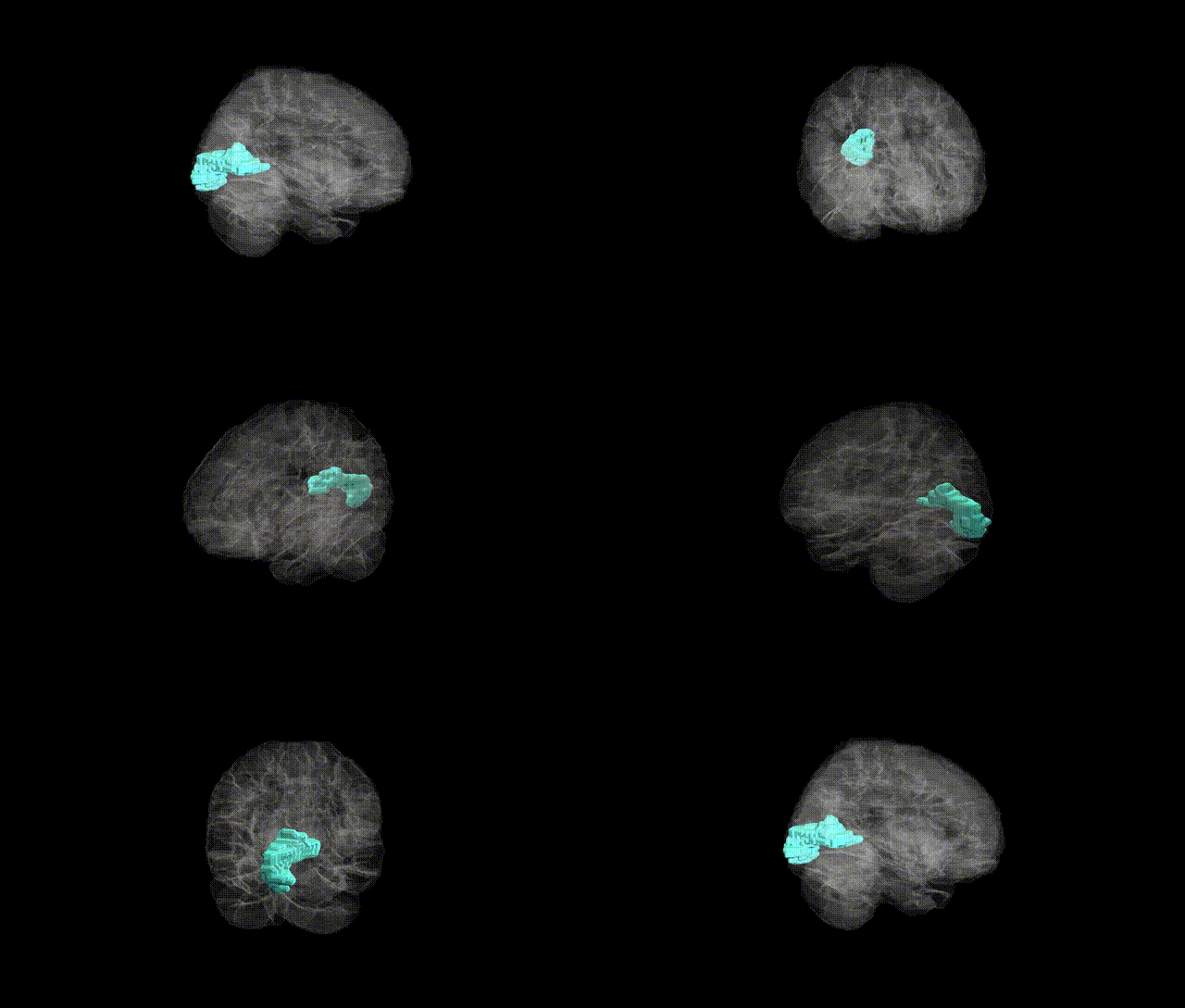
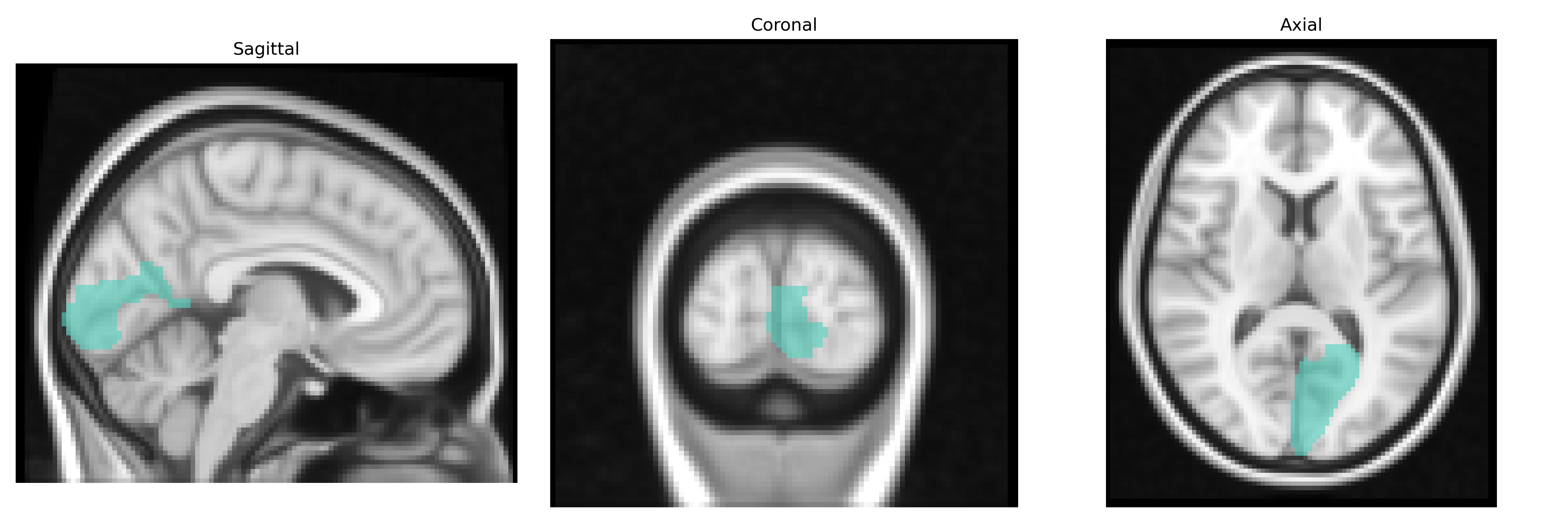
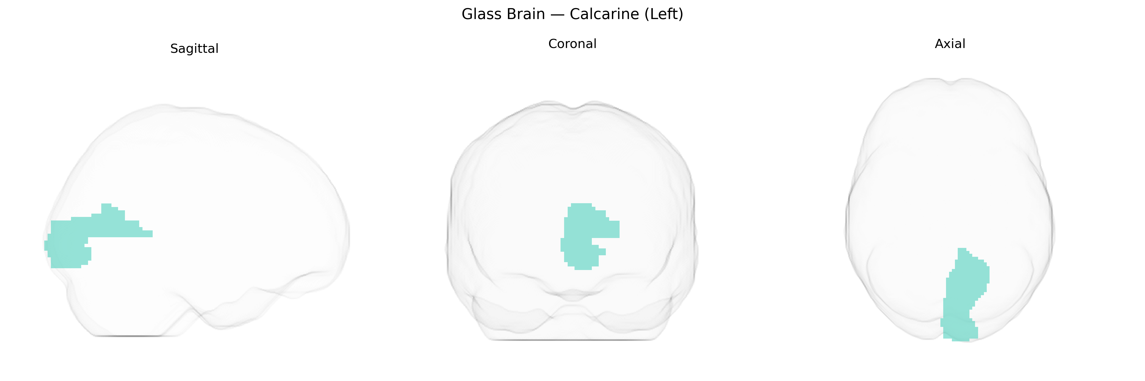
The left calcarine region, corresponding primarily to the medial occipital cortex surrounding the calcarine sulcus, encompasses the left primary visual cortex (V1, Brodmann area 17) as defined in the AAL2 atlas. This cortical area receives highly organized retinotopic input from the lateral geniculate nucleus of the thalamus via the optic radiations, and is crucial for the initial, high-fidelity processing of visual information such as orientation, spatial frequency, contrast, and basic motion signals. Neurons in this region are arranged in columnar and laminar patterns that support feature detection and integration, forming the foundation for subsequent visual processing in extrastriate areas. Lesions confined to the left calcarine cortex typically produce right homonymous visual field defects, reflecting the contralateral representation of the visual field. There is no direct Wikipedia page for “left calcarine” as a separate entry; a closely related structure is the Calcarine sulcus.
The left calcarine region, corresponding largely to the primary visual cortex (V1), has been implicated in several genetic and GWAS-based findings, though often at the level of occipital or visual cortex rather than the AAL2 label specifically. Neuroimaging GWAS consortia such as ENIGMA and UK Biobank have identified common variants (e.g., near genes involved in neurodevelopment and synaptic function like MEF2C, C4A, and others) associated with occipital cortical thickness, surface area, and volume, which encompass the calcarine sulcus. Polygenic risk for schizophrenia, bipolar disorder, and major depressive disorder has been linked to structural and functional alterations in calcarine/occipital regions, suggesting that risk variants for these disorders may partially exert effects through visual cortex circuitry. Calcarine volume and activity have also been associated with genetic risk scores for migraine and visual aura, and with variants influencing visual acuity and ocular traits, reflecting its central role in visual processing. In Alzheimer’s disease and other dementias, GWAS-identified risk loci (such as APOE) show downstream associations with occipital and calcarine atrophy patterns, while in autism spectrum conditions and dyslexia, common variants affecting cortical development and connectivity have been related to atypical activation and structural differences in calcarine and adjacent visual areas. Overall, genetic findings implicate the left calcarine region as a heritable, disease-relevant node in networks supporting visual perception, higher-order cognition, and psychopathology, although few studies have isolated this AAL2-defined parcel as a primary GWAS target.
Overview generated by GPT-4o (2026).
Region ID: 5001
Hemisphere: left
Atlas: AAL2
Full Quality Version: Download MP4
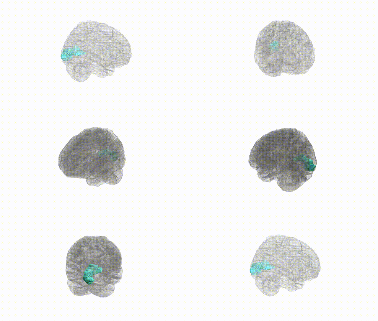
Full Quality Version: Download MP4
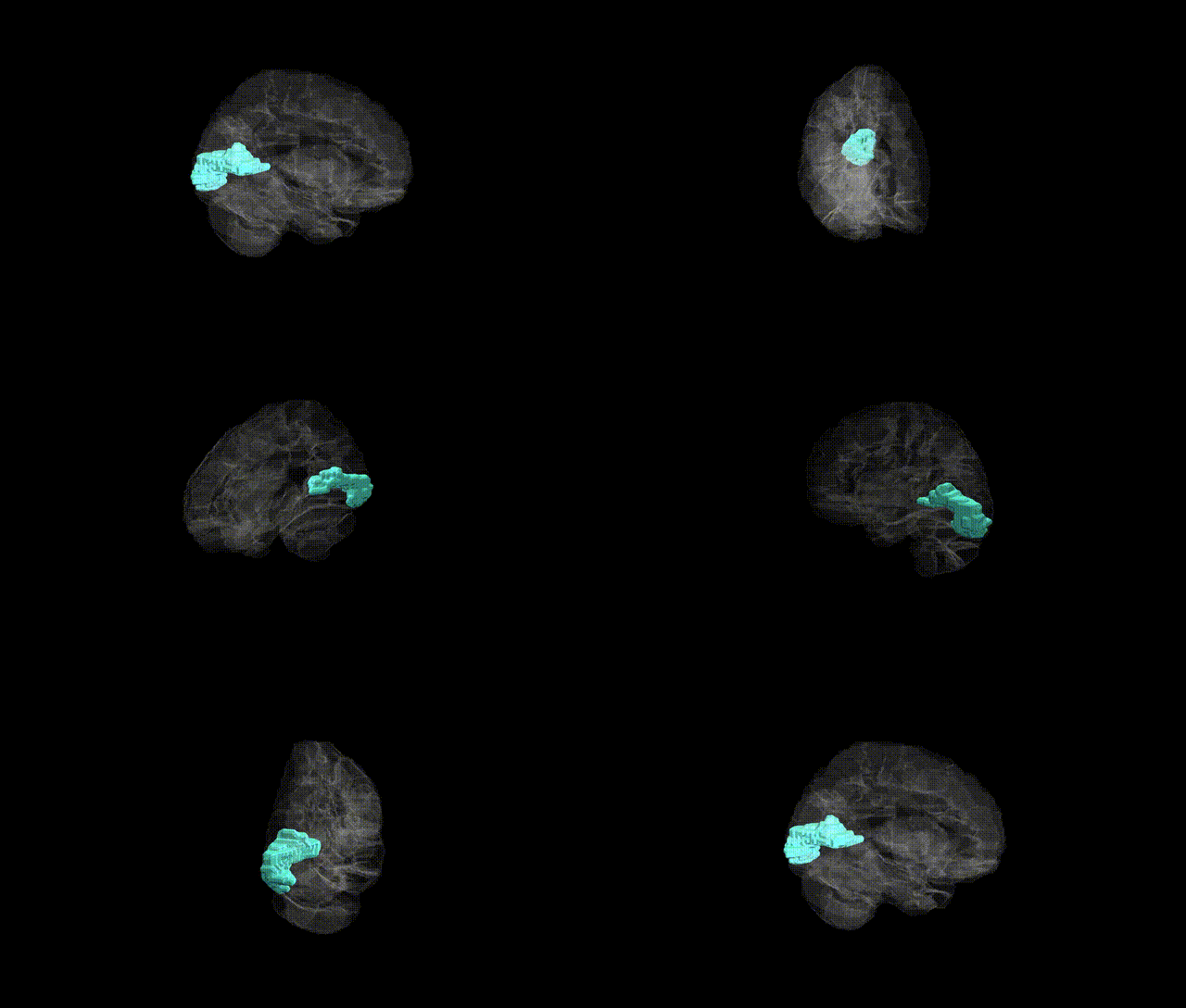
Full Quality Version: Download MP4
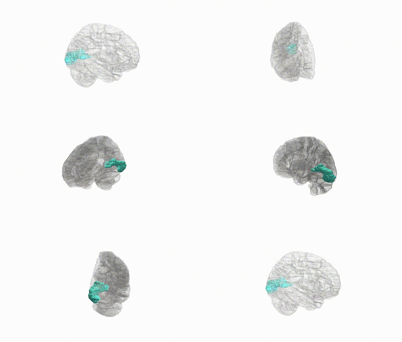
Full Quality Version: Download MP4
Wali Sidiqyar*, Gaurav Rudravaram*, Elyssa M. McMaster, Trent M. Schwartz, Adam M. Saunders, Kurt G. Schilling, Bennett A. Landman "Introducing SPINS: A Shared Public Visualization Library of Neuroanatomical Structures." Medical Imaging with Deep Learning- short paper
This resource is licensed under CC0 1.0 Universal (Public Domain).