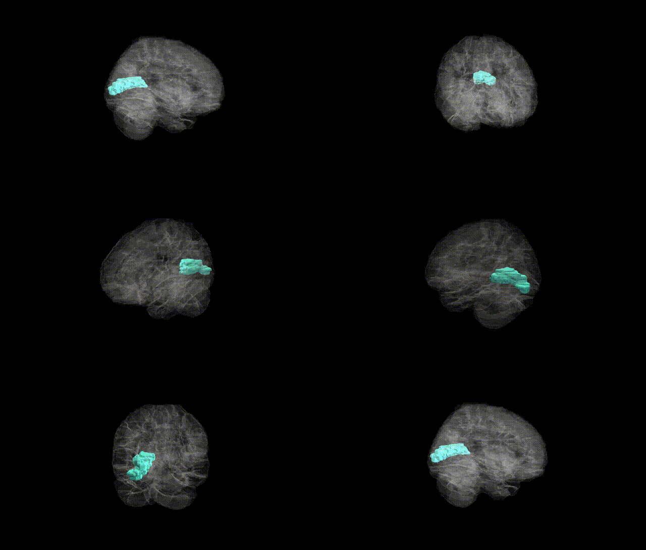
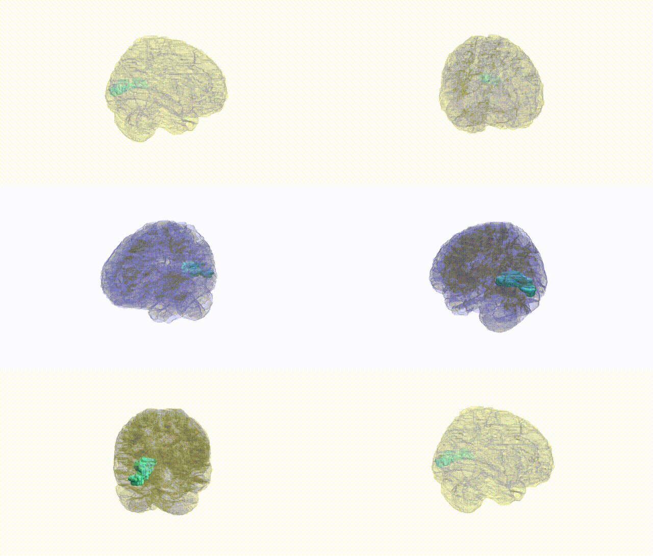
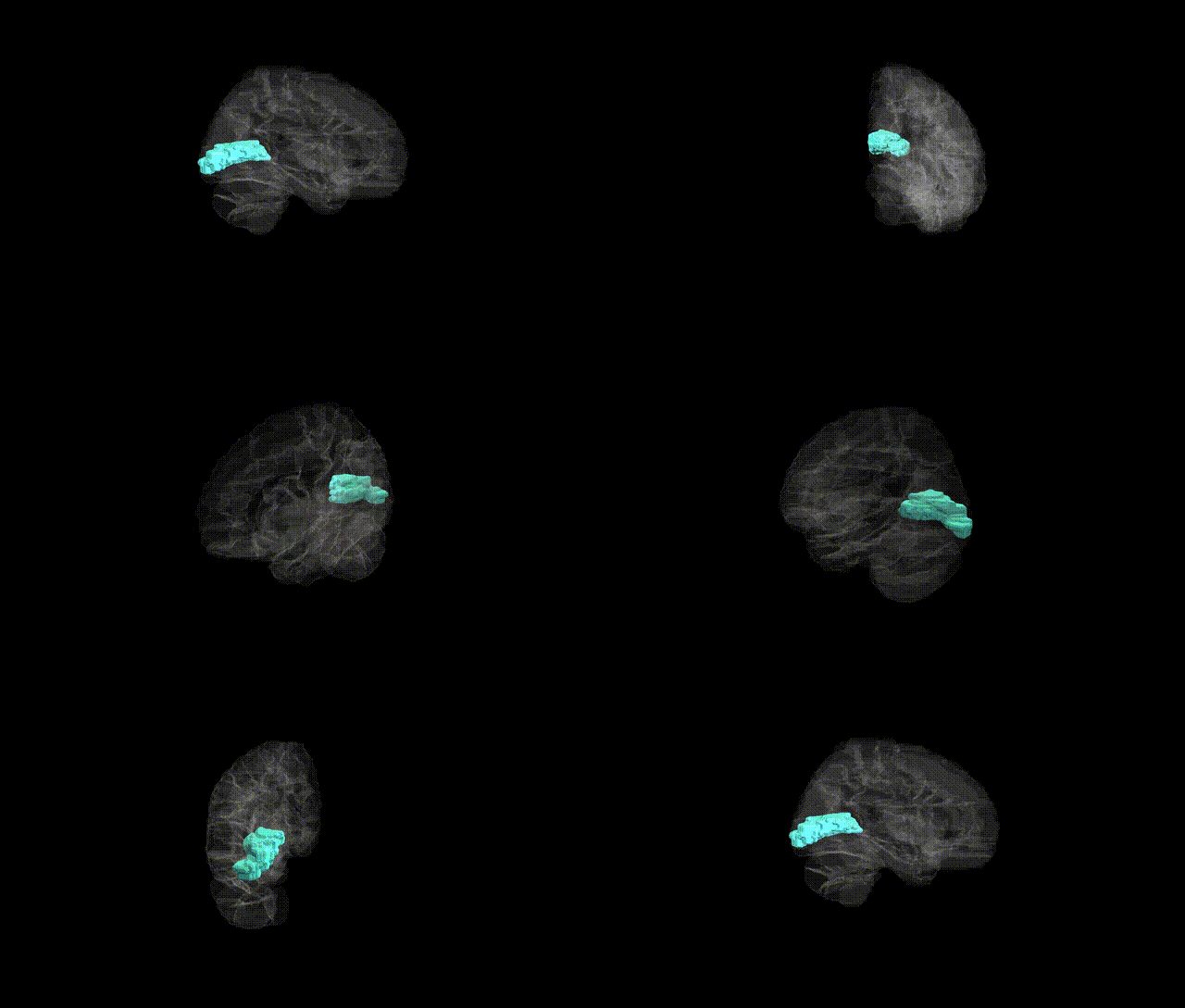
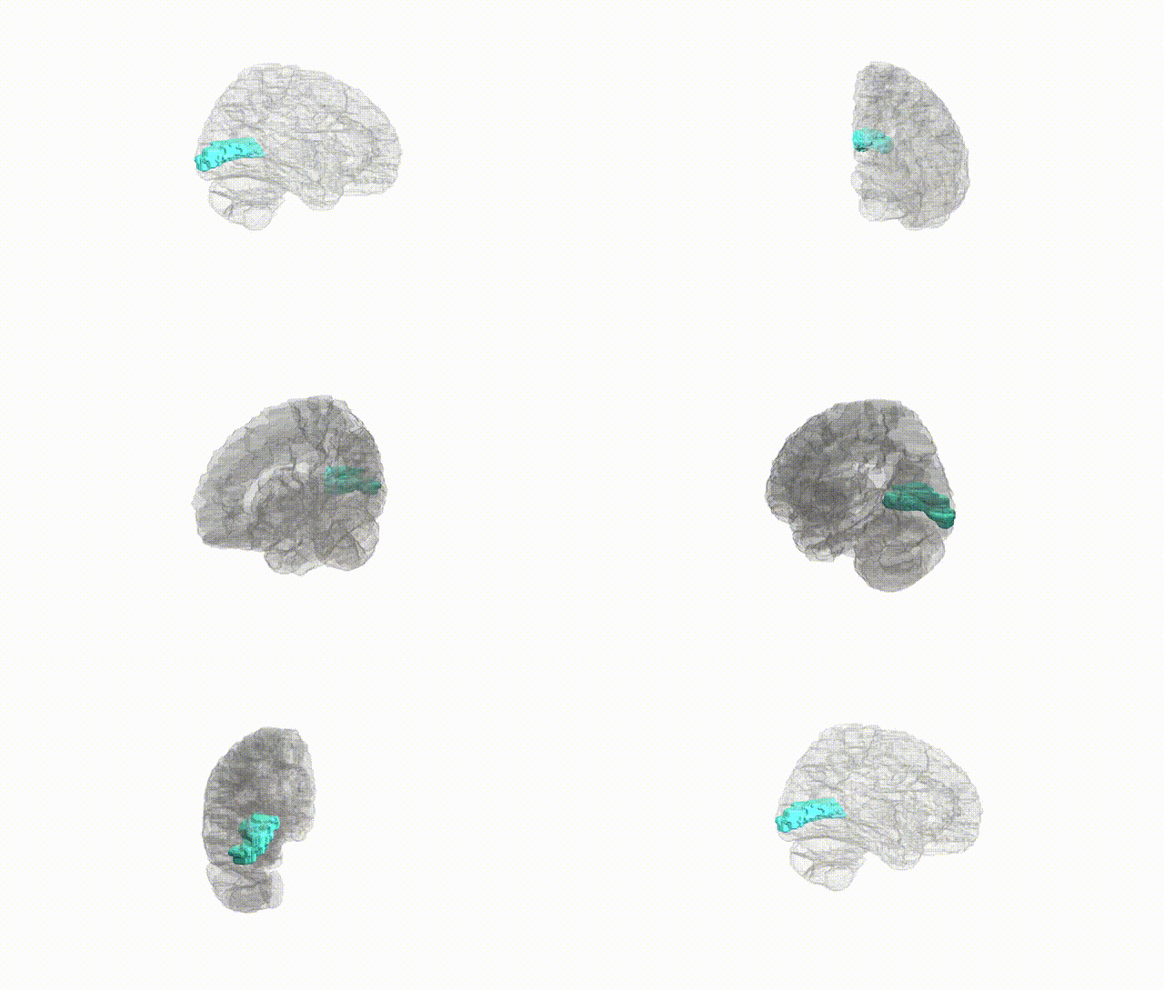
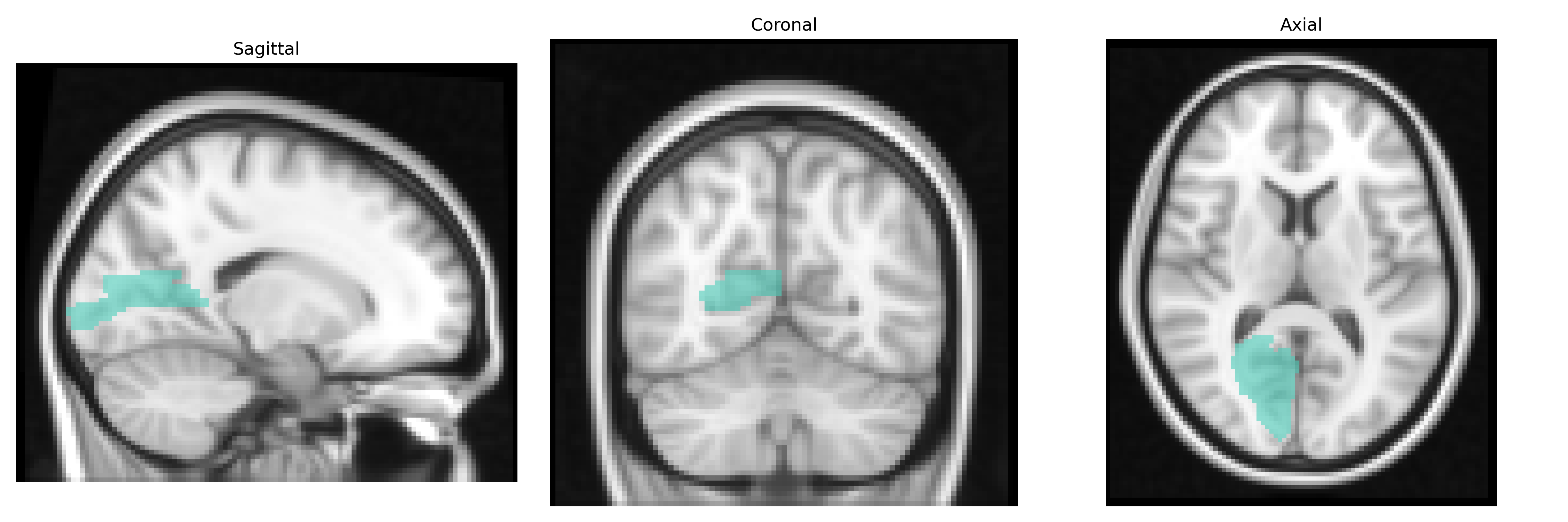
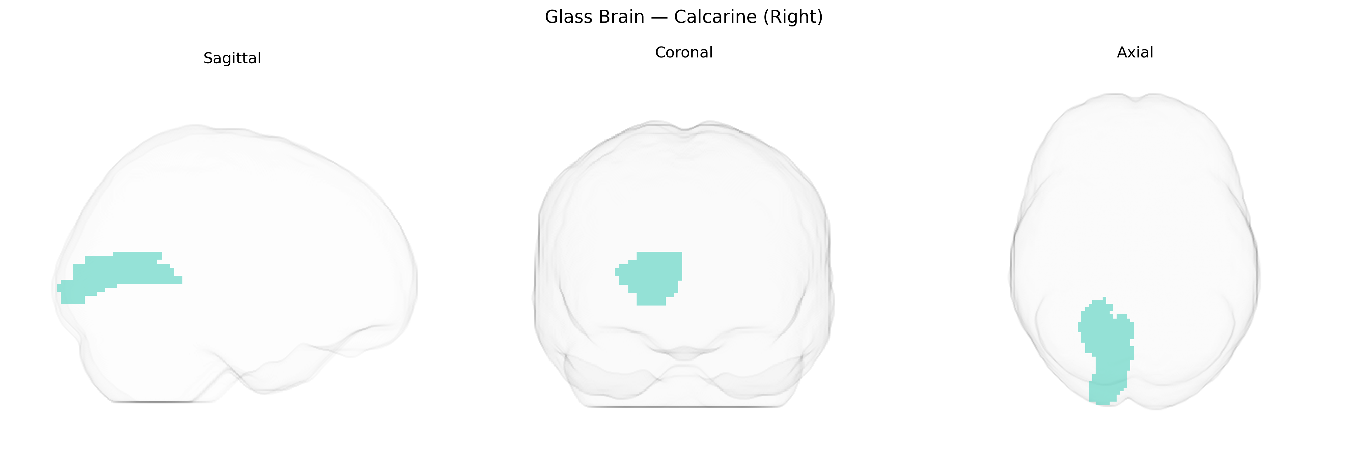
The right calcarine region, as defined in the AAL2 atlas, corresponds primarily to the right primary visual cortex (V1) located along the banks of the calcarine sulcus in the medial occipital lobe. It forms the initial cortical recipient of visual information from the lateral geniculate nucleus of the thalamus, preserving a retinotopic representation of the visual field with a strong emphasis on foveal input in its posterior portion and peripheral vision in its anterior portion. Neurons in this area are highly specialized for processing basic visual attributes such as orientation, spatial frequency, contrast, and motion direction, providing the foundational computations for higher-order visual processing in extrastriate areas. The right calcarine cortex is critical for contralateral (left) visual field perception, and lesions confined to this region often result in homonymous visual field defects. Related article: Primary visual cortex.
The right calcarine cortex, corresponding to primary visual cortex (V1) territory in the AAL2 atlas, has been implicated in several imaging-genetics and GWAS-derived associations, though often at the level of bilateral or generalized calcarine/occipital regions rather than strictly right-lateralized effects. Large neuroimaging GWAS (e.g., ENIGMA, UK Biobank) have identified SNPs in or near genes involved in neurodevelopment, synaptic function, and myelination (such as variants near PAX6, TCF4, and genes in Wnt and axon guidance pathways) associated with occipital/cortical thickness or surface area measures that include the calcarine cortex, and polygenic scores for overall brain structure frequently show loadings in this region. Structural and functional alterations of the calcarine cortex have been reported in genetic risk contexts for schizophrenia, major depression, bipolar disorder, and autism, where risk alleles (for example in CACNA1C, GRIN2A, and complement pathway genes) are associated with atypical visual processing and calcarine activation, though these effects are typically part of broader cortical patterns rather than region-specific loci. In migraine and visual aura, imaging-genetics work has linked calcarine excitability differences to variants in ion channel and glutamatergic genes identified in migraine GWAS, again without clear right-hemisphere specificity. Rare monogenic visual pathway disorders and albinism, involving genes such as OCA2 and others affecting retinal/optic tract development, show downstream structural and functional changes in the calcarine cortex, but these reflect developmental consequences rather than primary calcarine genetic loci. Overall, genetic associations involving the right calcarine region are predominantly inferred from broader occipital or bilateral calcarine findings in imaging-genetics studies, with no widely replicated GWAS reporting variants uniquely or specifically tied to right-sided calcarine anatomy or function.
Overview generated by GPT-4o (2026).
Region ID: 5002
Hemisphere: right
Atlas: AAL2
Full Quality Version: Download MP4
Full Quality Version: Download MP4
Full Quality Version: Download MP4
Full Quality Version: Download MP4
Wali Sidiqyar*, Gaurav Rudravaram*, Elyssa M. McMaster, Trent M. Schwartz, Adam M. Saunders, Kurt G. Schilling, Bennett A. Landman "Introducing SPINS: A Shared Public Visualization Library of Neuroanatomical Structures." Medical Imaging with Deep Learning- short paper
This resource is licensed under CC0 1.0 Universal (Public Domain).