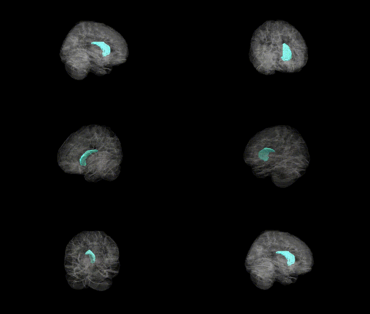
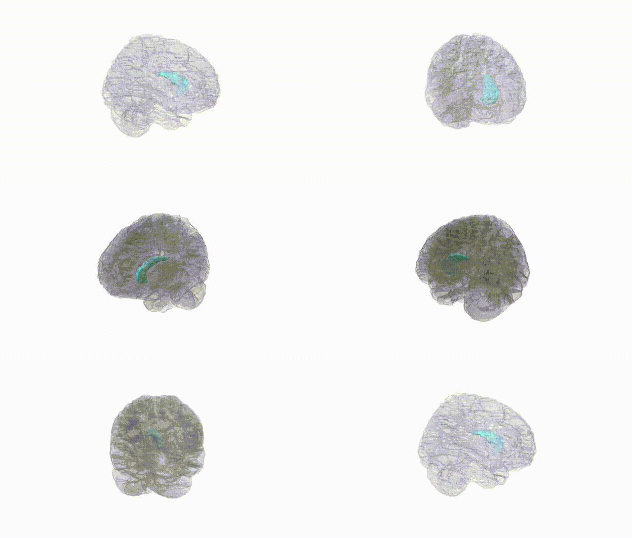
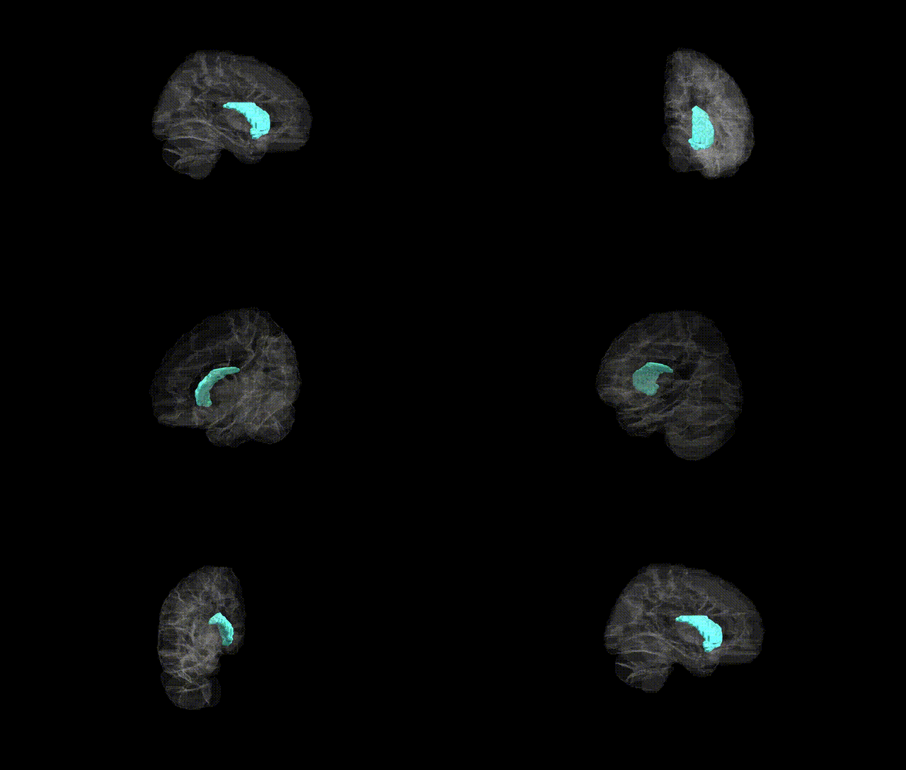
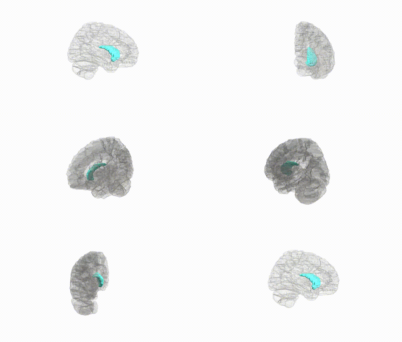
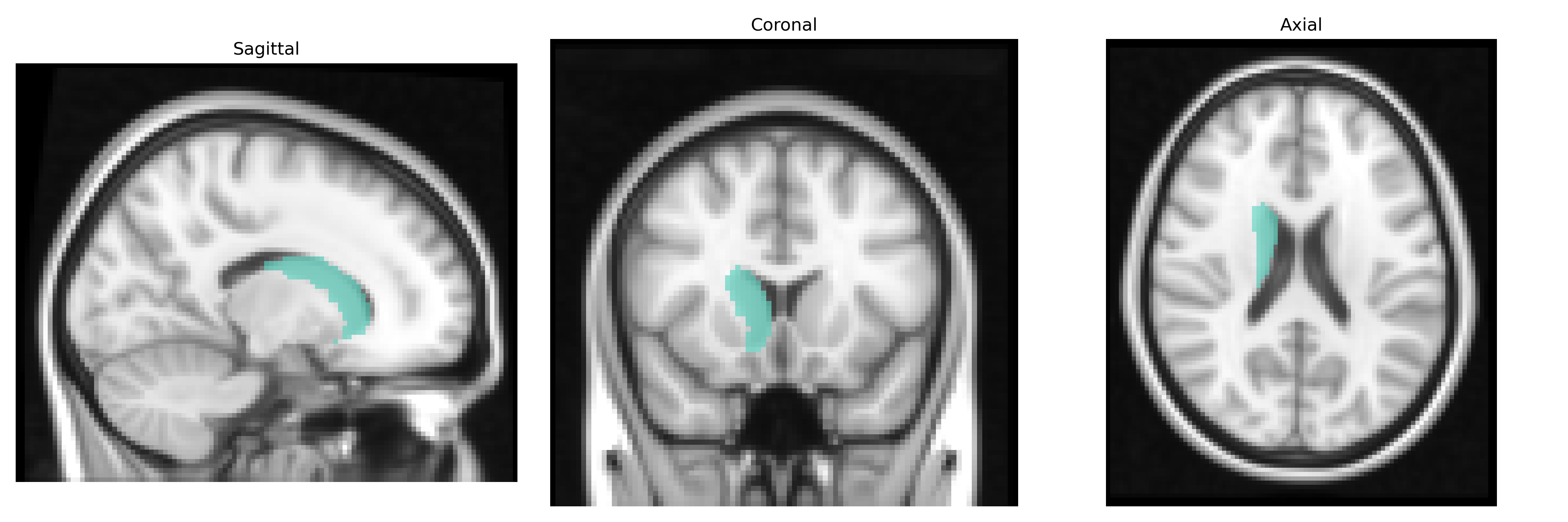
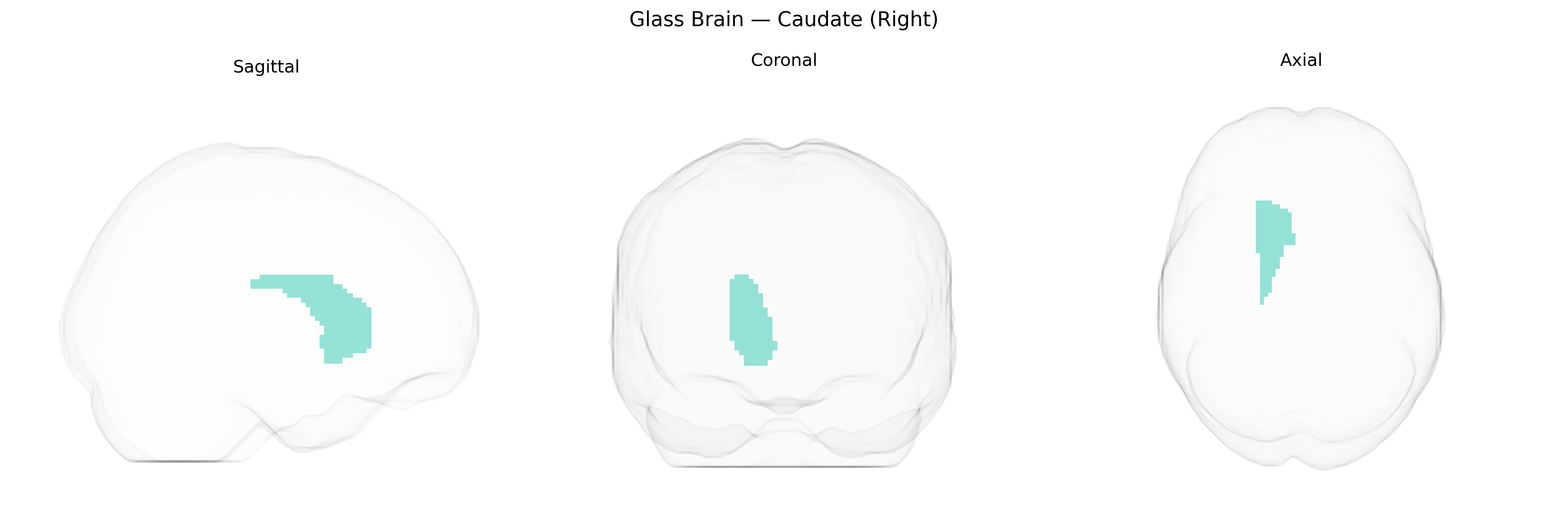
The right caudate nucleus is a C-shaped subcortical gray matter structure located in the right hemisphere of the brain, forming part of the dorsal striatum within the basal ganglia. It lies adjacent to the lateral ventricle and is composed of head, body, and tail segments that extend posteriorly and ventrally. Functionally, the caudate is heavily interconnected with frontal cortical regions via corticostriatal loops and plays key roles in motor control, procedural learning, goal-directed behavior, and various aspects of cognition such as executive function and action selection. It is also involved in reward processing and reinforcement learning through dopaminergic input from the substantia nigra. Structural or functional abnormalities of the caudate are implicated in movement disorders (e.g., Parkinson’s disease, Huntington’s disease), obsessive-compulsive disorder, attention-deficit/hyperactivity disorder, and other neuropsychiatric conditions. There is no direct link for “Right Caudate” as a separate entity; a closely related structure is the Caudate nucleus.
The right caudate, as defined in the AAL2 atlas, has been implicated in multiple genetic studies, particularly GWAS of subcortical brain volume and structure. Large-scale imaging–genetics consortia (e.g., ENIGMA) have identified common variants near KTN1, DCC, BDNF, and genes involved in neurodevelopment, synaptic plasticity, and axonal guidance that associate with caudate volume or morphology. Polygenic risk scores for schizophrenia, major depressive disorder, bipolar disorder, and attention-deficit/hyperactivity disorder show correlations with caudate size and functional measures, consistent with the caudate’s role in frontostriatal circuits underlying cognition, reward, and motor control. GWAS of Parkinson’s disease, obsessive–compulsive disorder, and Tourette syndrome highlight dopaminergic and glutamatergic genes whose effects partly manifest in striatal structures, including the caudate. In addition, genetic variants associated with general cognitive ability, educational attainment, and neuroticism have been linked to caudate volume or activation, suggesting that the right caudate serves as an anatomical substrate through which diverse neuropsychiatric and behavioral genetic influences converge. While many findings are bilateral or reported for total caudate volume, some imaging–genetics studies note lateralized effects, with right caudate associations particularly seen in reward-related and impulsivity traits, though this lateralization remains an active area of investigation rather than a fully established genetic pattern.
Overview generated by GPT-4o (2026).
Region ID: 7002
Hemisphere: right
Atlas: AAL2
Full Quality Version: Download MP4
Full Quality Version: Download MP4
Full Quality Version: Download MP4
Full Quality Version: Download MP4
Wali Sidiqyar*, Gaurav Rudravaram*, Elyssa M. McMaster, Trent M. Schwartz, Adam M. Saunders, Kurt G. Schilling, Bennett A. Landman "Introducing SPINS: A Shared Public Visualization Library of Neuroanatomical Structures." Medical Imaging with Deep Learning- short paper
This resource is licensed under CC0 1.0 Universal (Public Domain).