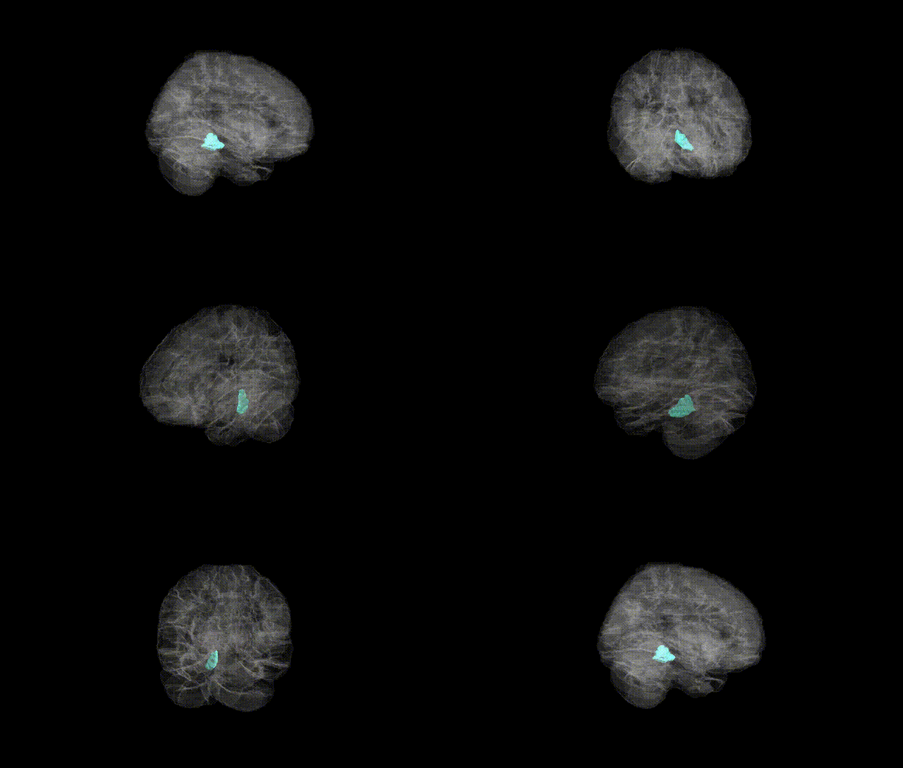
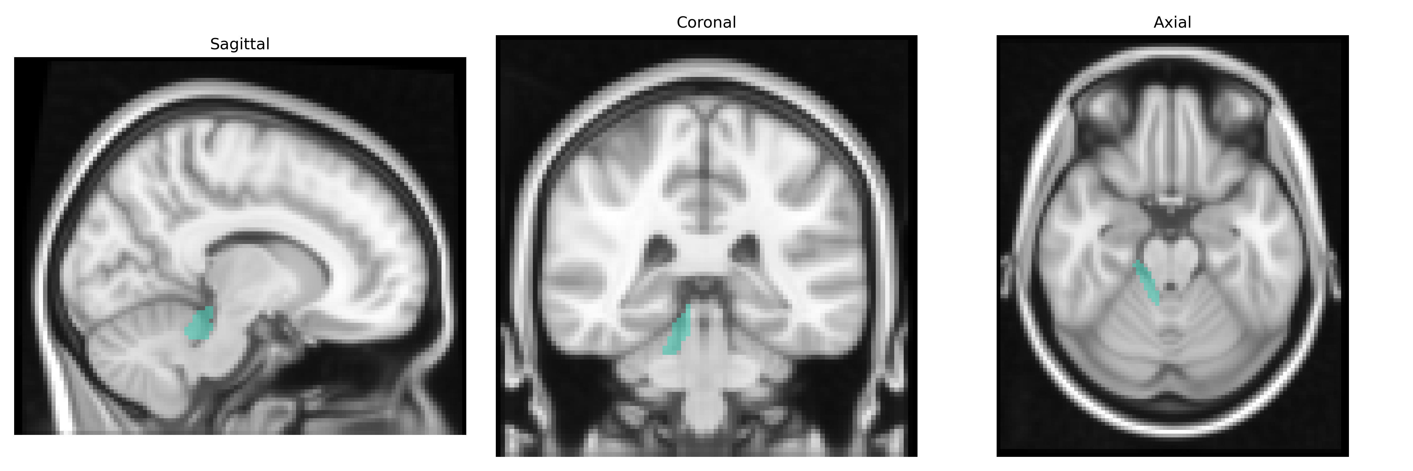
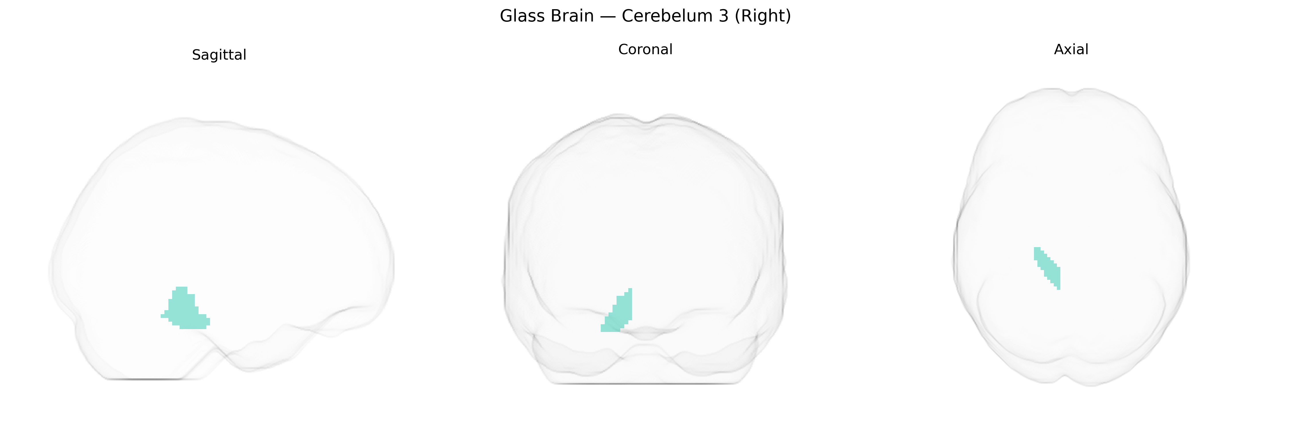
Cerebellum 3 (Right), as defined in the AAL2 atlas, corresponds approximately to lobule III of the right anterior cerebellar hemisphere, a small lobule situated anteriorly and superiorly near the primary fissure. Histologically, it is composed of the typical cerebellar cortical layers (molecular, Purkinje cell, and granular layers) overlying deep cerebellar white matter, and it projects primarily to the deep cerebellar nuclei, particularly the fastigial and interposed nuclei. Functionally, lobule III is implicated in fine-tuning limb and axial motor control, sensorimotor integration, and the timing and coordination of voluntary movements, contributing to error correction via cerebello-thalamo-cortical loops. Although there is no specific Wikipedia article for “Cerebellum 3 (Right),” it is part of the anterior cerebellar lobe: Anterior lobe of cerebellum.
The right cerebellar lobule III (AAL2 “Cerebelum_3_R”) has emerged in imaging genetics and GWAS as part of broader cerebellar and cerebellar–cortical circuits rather than as an isolated locus, with associations largely inferred from regional cerebellar volume, connectivity, or activation phenotypes. Large-scale brain structure GWAS (e.g., ENIGMA and UK Biobank cohorts) have identified multiple loci influencing cerebellar morphology—most consistently near genes involved in neurodevelopment, synaptic function, and axon guidance (such as KIAA0586, PAX6, and FOXP genes)—though these findings typically report global or lobular cerebellar measures and do not specifically single out right lobule III. Genetic correlations and polygenic scores for psychiatric and cognitive traits (schizophrenia, major depression, autism spectrum disorder, attention-deficit/hyperactivity disorder, and general cognitive ability) frequently show cerebellar involvement, with cerebellar regions including lobules I–VI implicated in motor control, timing, working memory, and affect regulation, and functional/structural alterations in these regions linked to risk variants in neurotransmission and synaptic plasticity genes (e.g., CACNA1C, GRM genes). GWAS of brain activation and functional connectivity have connected cerebellar regions to heritable differences in sensorimotor and executive networks, and cerebellar volume is genetically correlated with educational attainment and intelligence, suggesting pleiotropic influences on both motor and cognitive traits. However, current evidence remains coarse-grained: no robust, replicated GWAS has yet established a set of variants uniquely and specifically associated with Right Cerebelum_3_R, and genetic associations are best interpreted as affecting distributed cerebellar–cerebral systems in which this region participates rather than this single AAL2-defined parcel alone.
Overview generated by GPT-4o (2026).
Region ID: 9022
Hemisphere: right
Atlas: AAL2
Full Quality Version: Download MP4
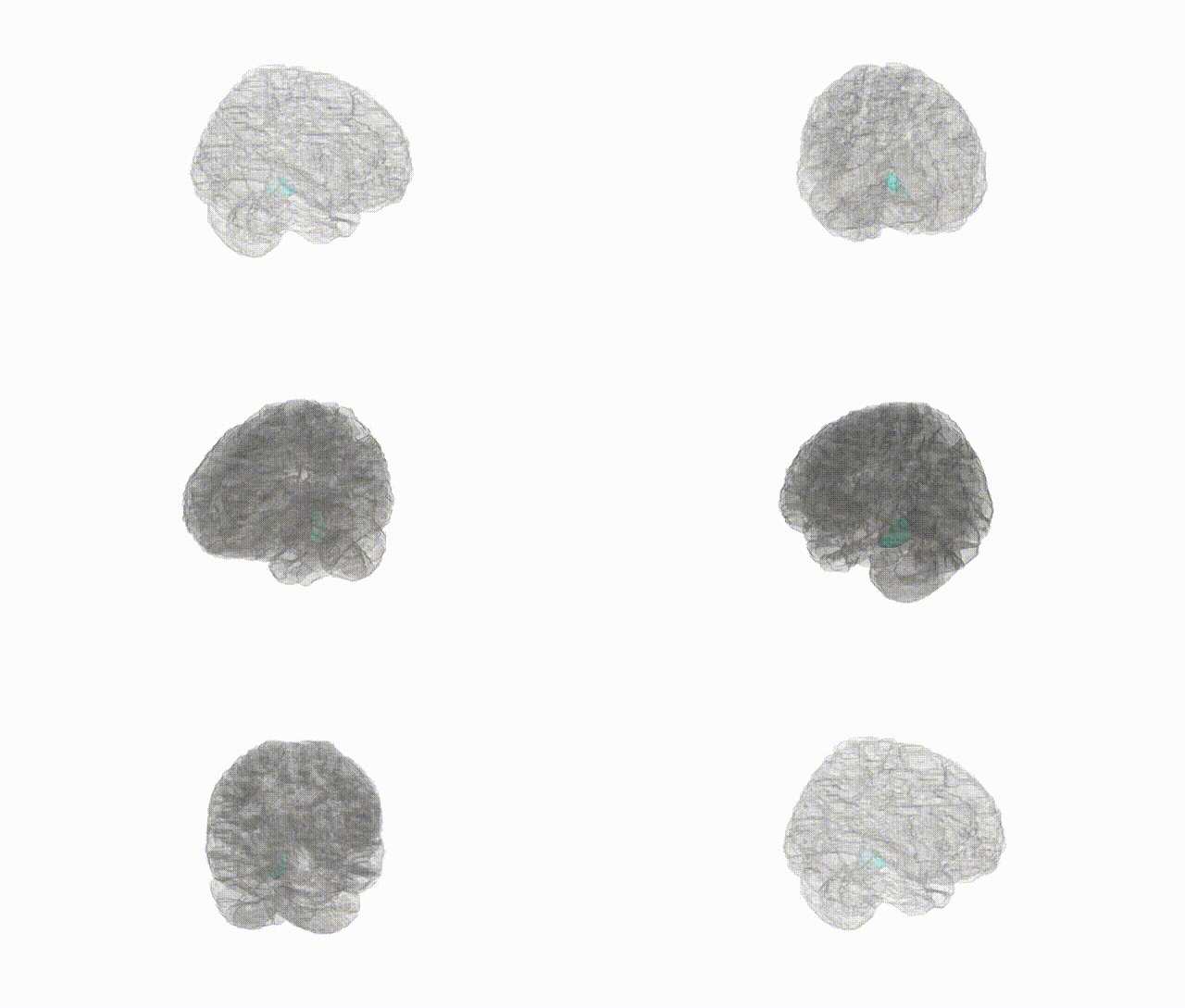
Full Quality Version: Download MP4
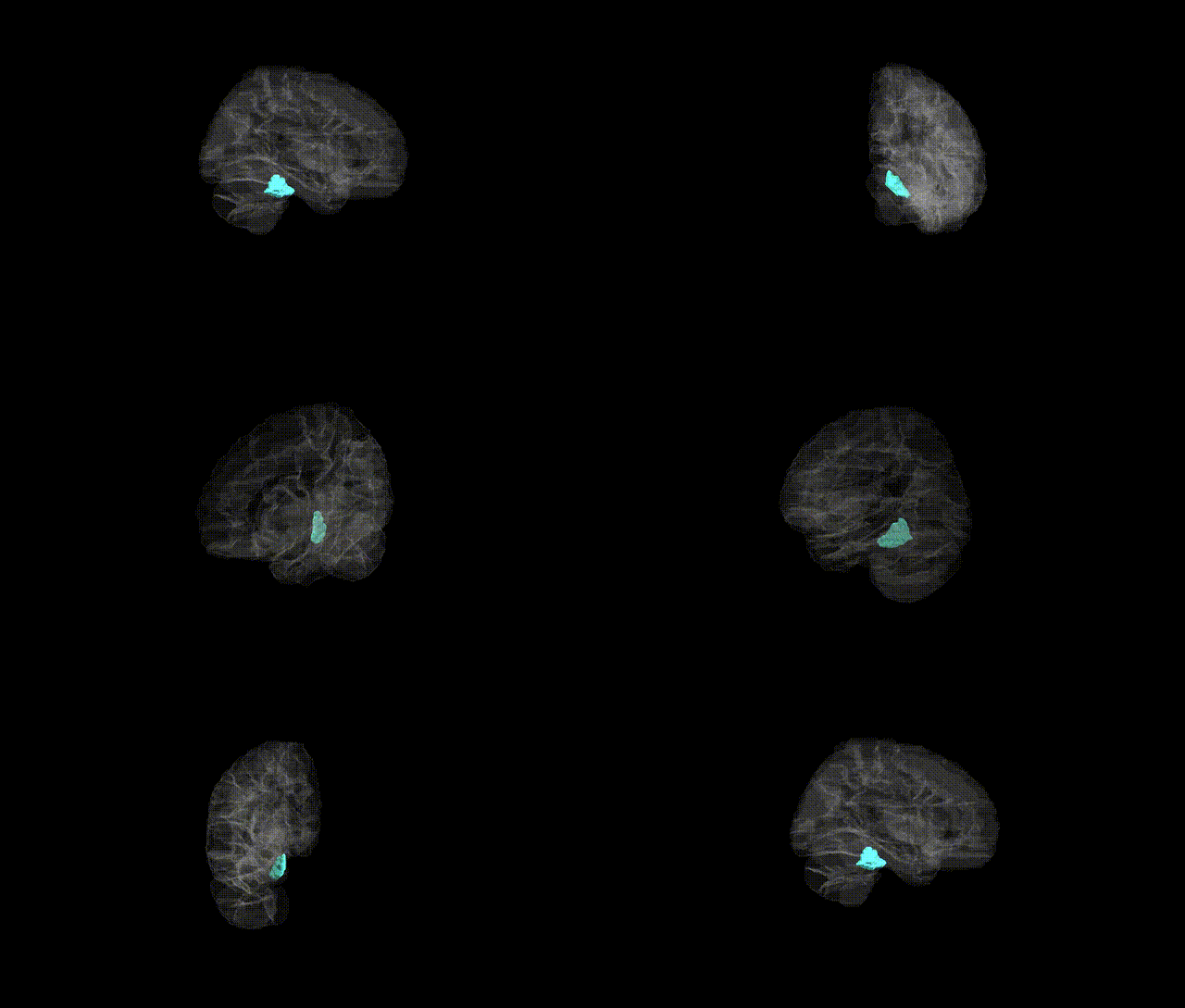
Full Quality Version: Download MP4
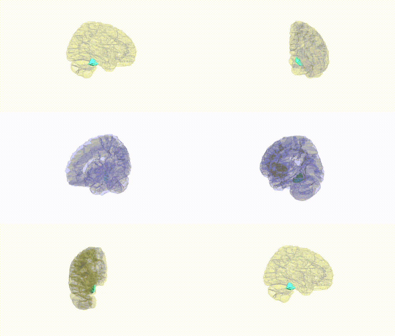
Full Quality Version: Download MP4
Wali Sidiqyar*, Gaurav Rudravaram*, Elyssa M. McMaster, Trent M. Schwartz, Adam M. Saunders, Kurt G. Schilling, Bennett A. Landman "Introducing SPINS: A Shared Public Visualization Library of Neuroanatomical Structures." Medical Imaging with Deep Learning- short paper
This resource is licensed under CC0 1.0 Universal (Public Domain).