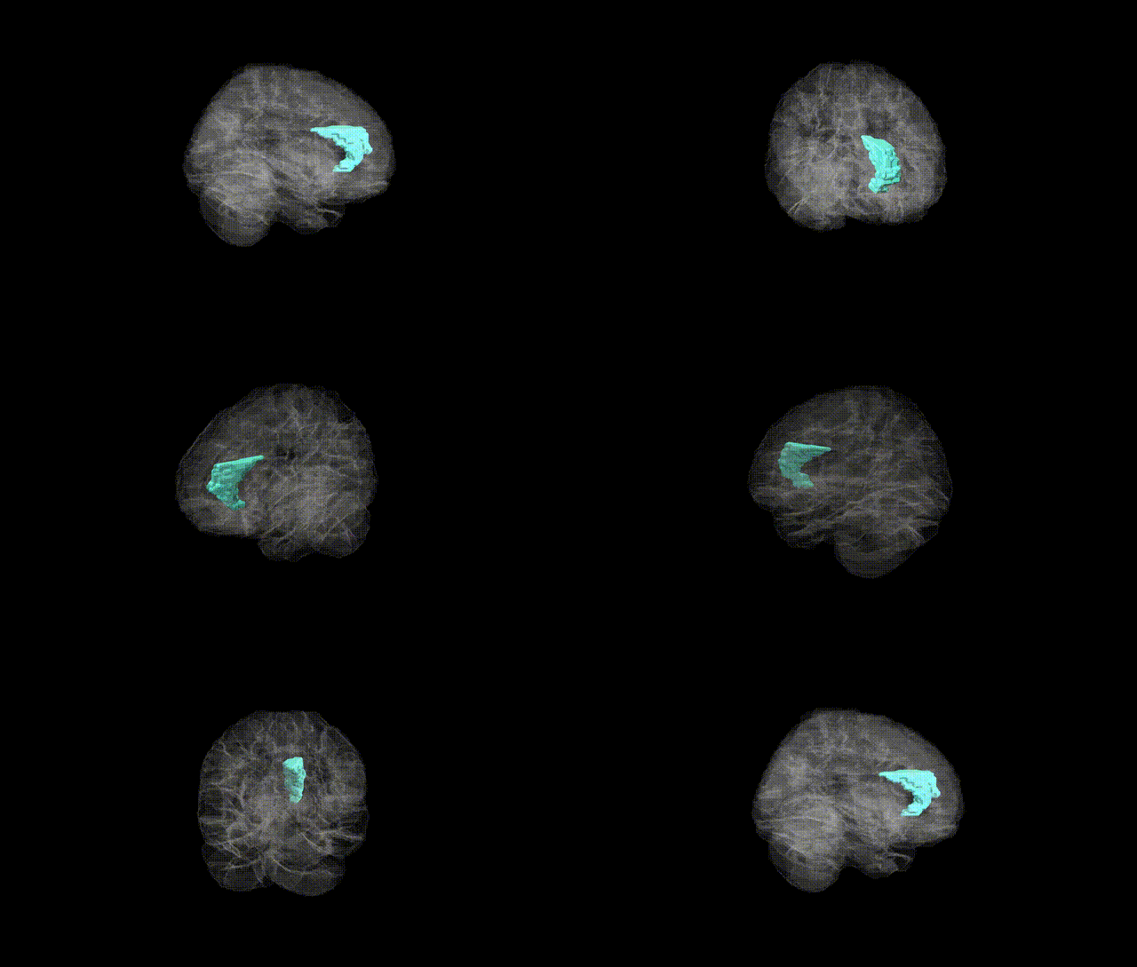
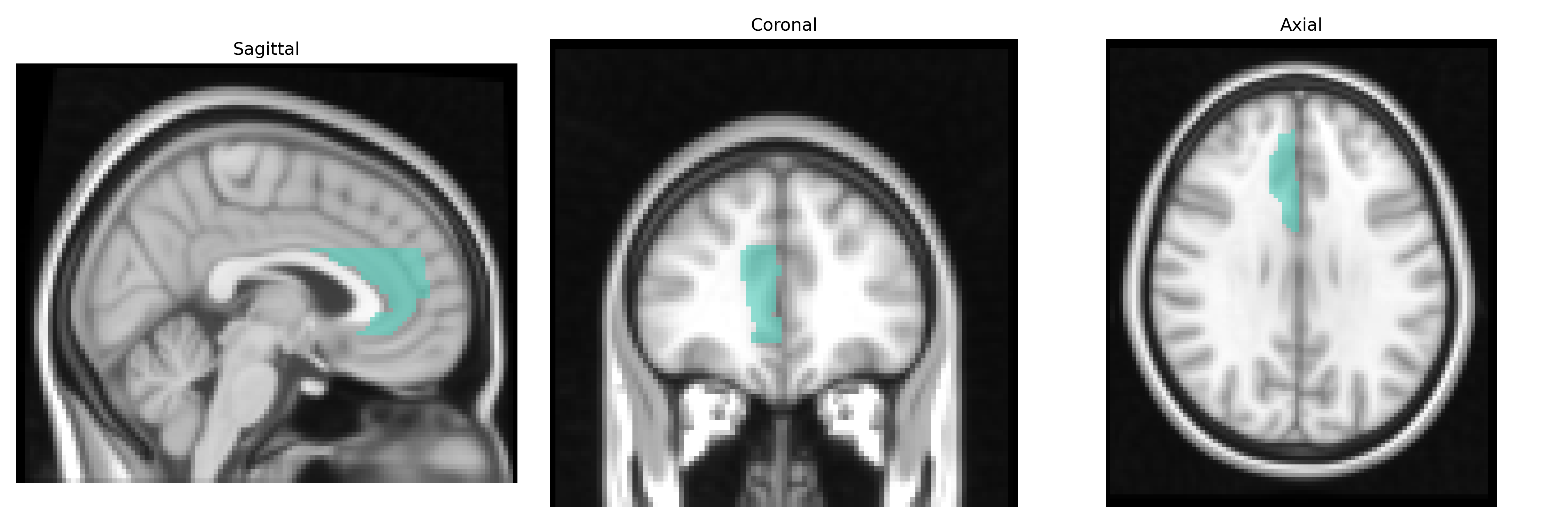
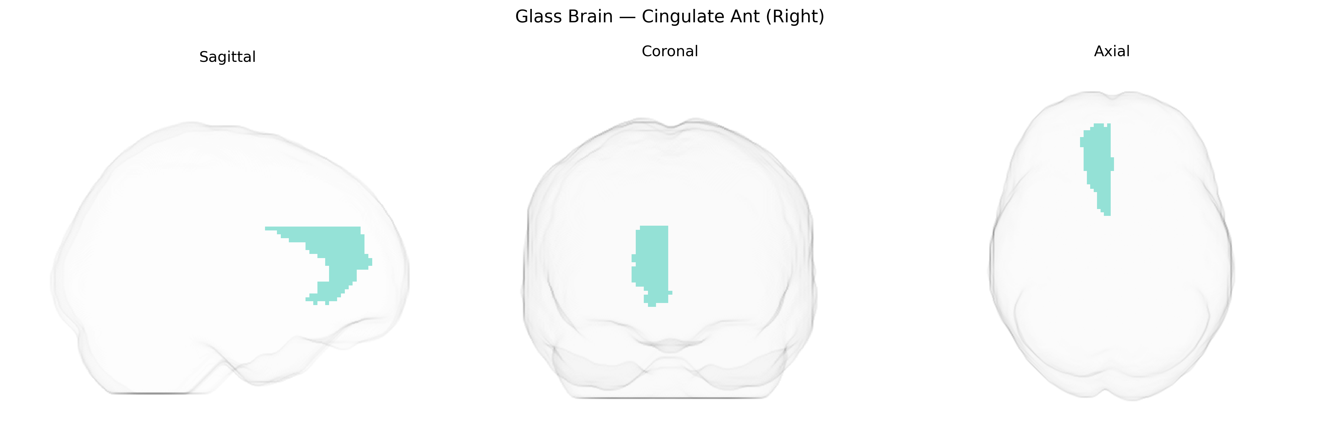
The right cingulate anterior region (Right Cingulate Ant) in the AAL2 atlas corresponds to the anterior portion of the cingulate gyrus in the right hemisphere, a key component of the limbic system implicated in emotion, motivation, autonomic regulation, and higher-order cognitive control. Anatomically, it lies on the medial surface of the cerebral hemisphere, superior to the corpus callosum and anterior to the midcingulate segment, with cytoarchitectonic fields overlapping Brodmann areas 24, 32, and 33. Functionally, this region contributes to conflict monitoring, error detection, decision-making, and regulation of affective responses, often showing activation during tasks involving attention, pain perception, and reward-based learning. It also maintains dense connections with prefrontal, parietal, limbic, and subcortical structures, supporting integration of cognitive and emotional information. Anterior cingulate cortex
The right anterior cingulate cortex (often mapped as “Cingulate Ant (Right)” in the AAL2 atlas) has been repeatedly implicated in imaging genetics and GWAS of brain structure and function, with variants in genes related to synaptic plasticity, neurodevelopment, and myelination (for example, CACNA1C, BDNF, NRG1, and NCAN) associated with its volume, thickness, and connectivity. Large-scale ENIGMA and UK Biobank–based studies show that common variants explaining individual differences in right anterior cingulate morphology overlap with loci implicated in major depressive disorder, bipolar disorder, schizophrenia, and attention-deficit/hyperactivity disorder, as well as in traits such as neuroticism, anxiety, and cognitive control. Polygenic risk for psychiatric disorders frequently correlates with altered right anterior cingulate volume or activation during tasks involving error monitoring, conflict processing, and emotion regulation, and several GWAS of cortical surface area and thickness identify this region as a key mediator between genetic risk and behavioral phenotypes, although specific loci often show hemisphere-symmetric rather than strictly right-lateralized effects.
Overview generated by GPT-4o (2026).
Region ID: 4002
Hemisphere: right
Atlas: AAL2
Full Quality Version: Download MP4
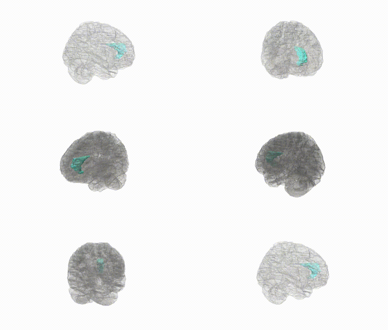
Full Quality Version: Download MP4
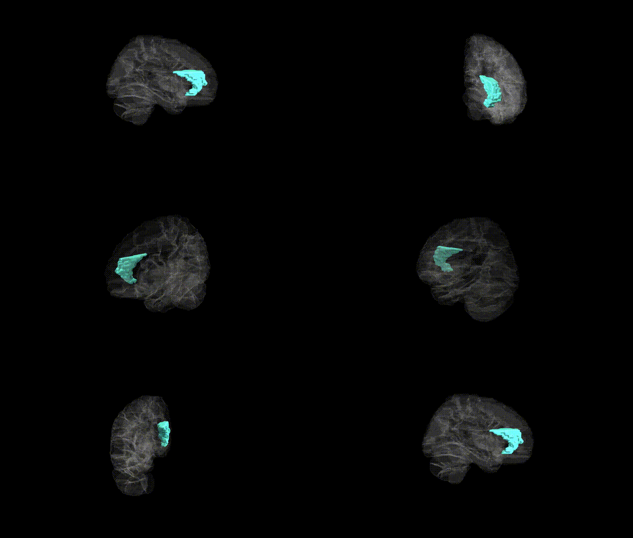
Full Quality Version: Download MP4
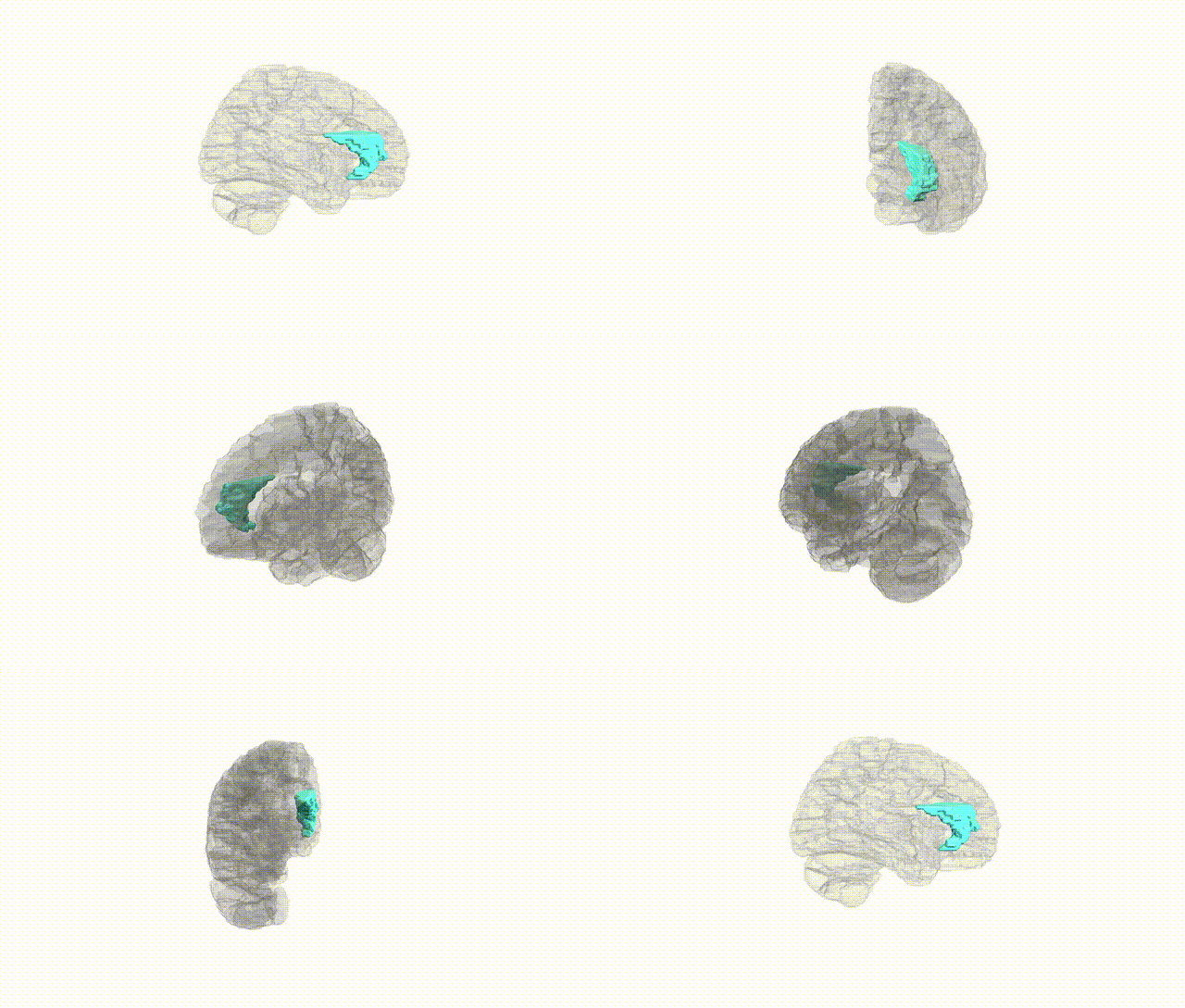
Full Quality Version: Download MP4
Wali Sidiqyar*, Gaurav Rudravaram*, Elyssa M. McMaster, Trent M. Schwartz, Adam M. Saunders, Kurt G. Schilling, Bennett A. Landman "Introducing SPINS: A Shared Public Visualization Library of Neuroanatomical Structures." Medical Imaging with Deep Learning- short paper
This resource is licensed under CC0 1.0 Universal (Public Domain).