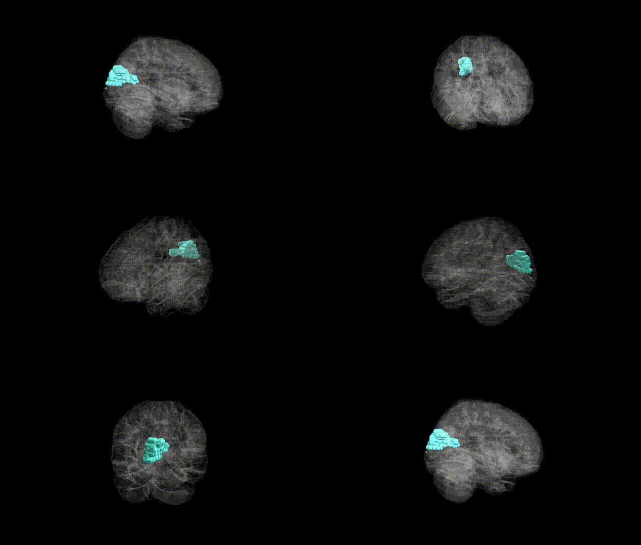
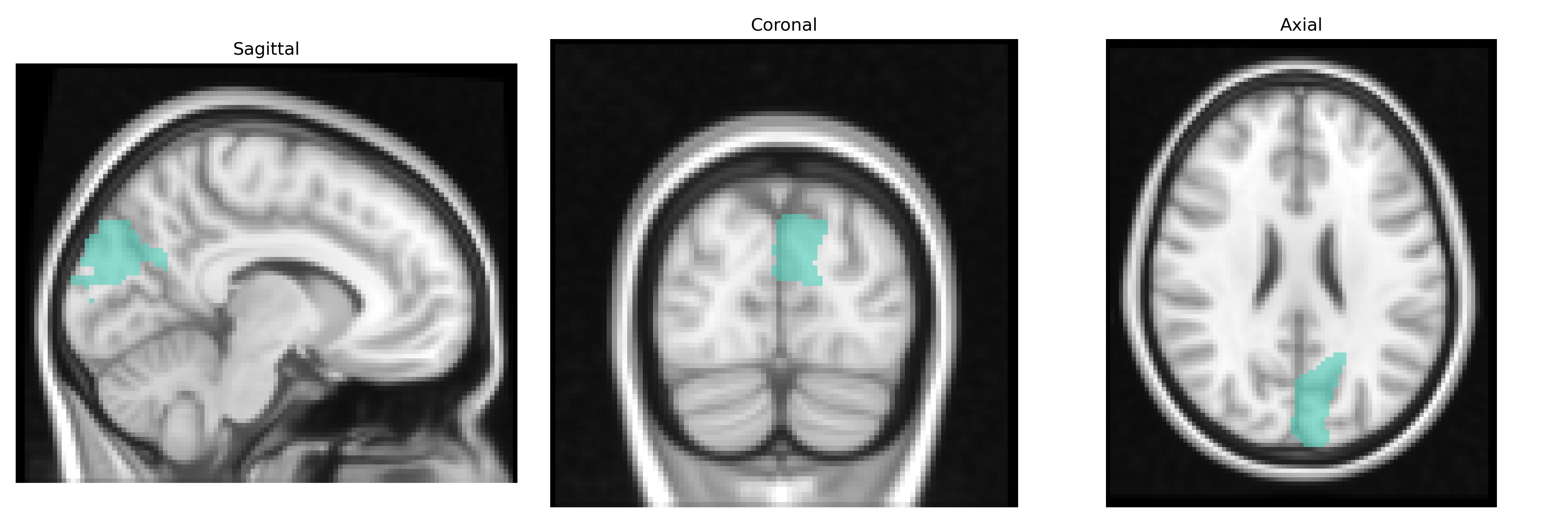
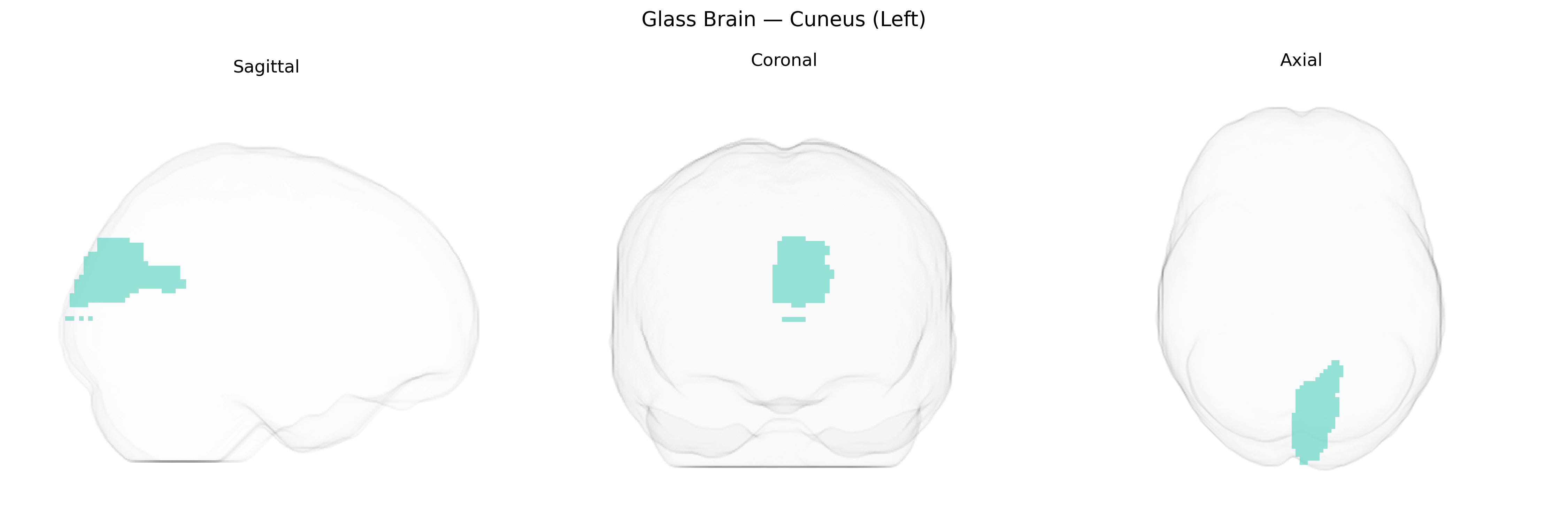
The left cuneus is a medial occipital lobe structure forming part of the dorsal visual cortex, located between the calcarine sulcus inferiorly and the parieto-occipital sulcus superiorly. It corresponds primarily to portions of Brodmann areas 17, 18, and 19, and is heavily involved in early-stage visual processing, including basic feature analysis, visuospatial integration, and aspects of visual attention. The cuneus receives major input from the lateral geniculate nucleus via the primary visual cortex and participates in integrating visual information with higher-order cognitive processes such as visual imagery and top-down modulation from parietal and frontal regions. In the AAL2 atlas, the left cuneus is delineated as a distinct cortical parcel within the occipital lobe, serving as a key node in the posterior visual network. Cuneus
The left cuneus, a primary visual and higher-order visual association region defined in the AAL2 atlas, shows genetic associations largely through imaging-genetics and GWAS of cortical structure and function rather than direct locus-specific studies. Large-scale ENIGMA and UK Biobank analyses have identified common variants in genes related to neurodevelopment, synaptic function, and myelination (for example, in or near MAPT, HMGA2, and loci on chromosomes 3p, 6p, and 17q) that influence cuneus cortical thickness, surface area, and volume, often as part of broader occipital or visual-network measures. Polygenic risk scores for schizophrenia, major depressive disorder, autism spectrum disorder, and attention-deficit/hyperactivity disorder have been associated with altered cuneus morphology or activation, implicating this region in psychiatric liability. GWAS of visual processing traits and resting-state functional connectivity have also linked variants in sensory and visual pathway genes to cuneus activity patterns. In neurodegenerative conditions, Alzheimer’s disease and Parkinson’s disease risk variants show downstream effects on cuneus structure and metabolism as part of posterior cortical and default-mode–adjacent networks, while migraine and photophobia-related genetic studies occasionally report cuneus involvement within broader visual cortical phenotypes rather than as a primary locus.
Overview generated by GPT-4o (2026).
Region ID: 5011
Hemisphere: left
Atlas: AAL2
Full Quality Version: Download MP4
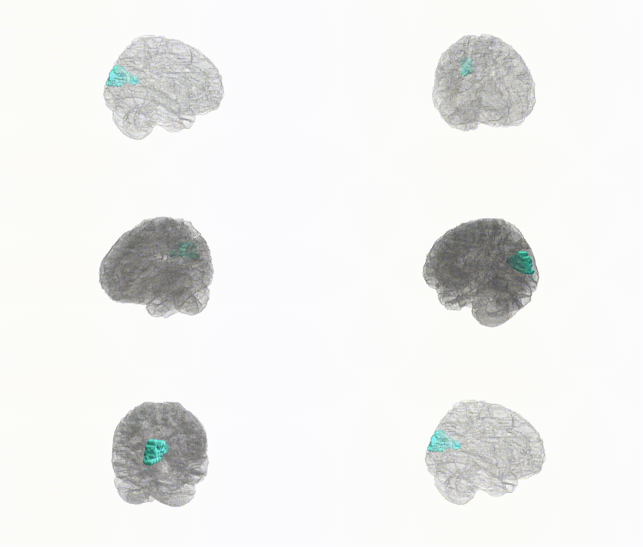
Full Quality Version: Download MP4
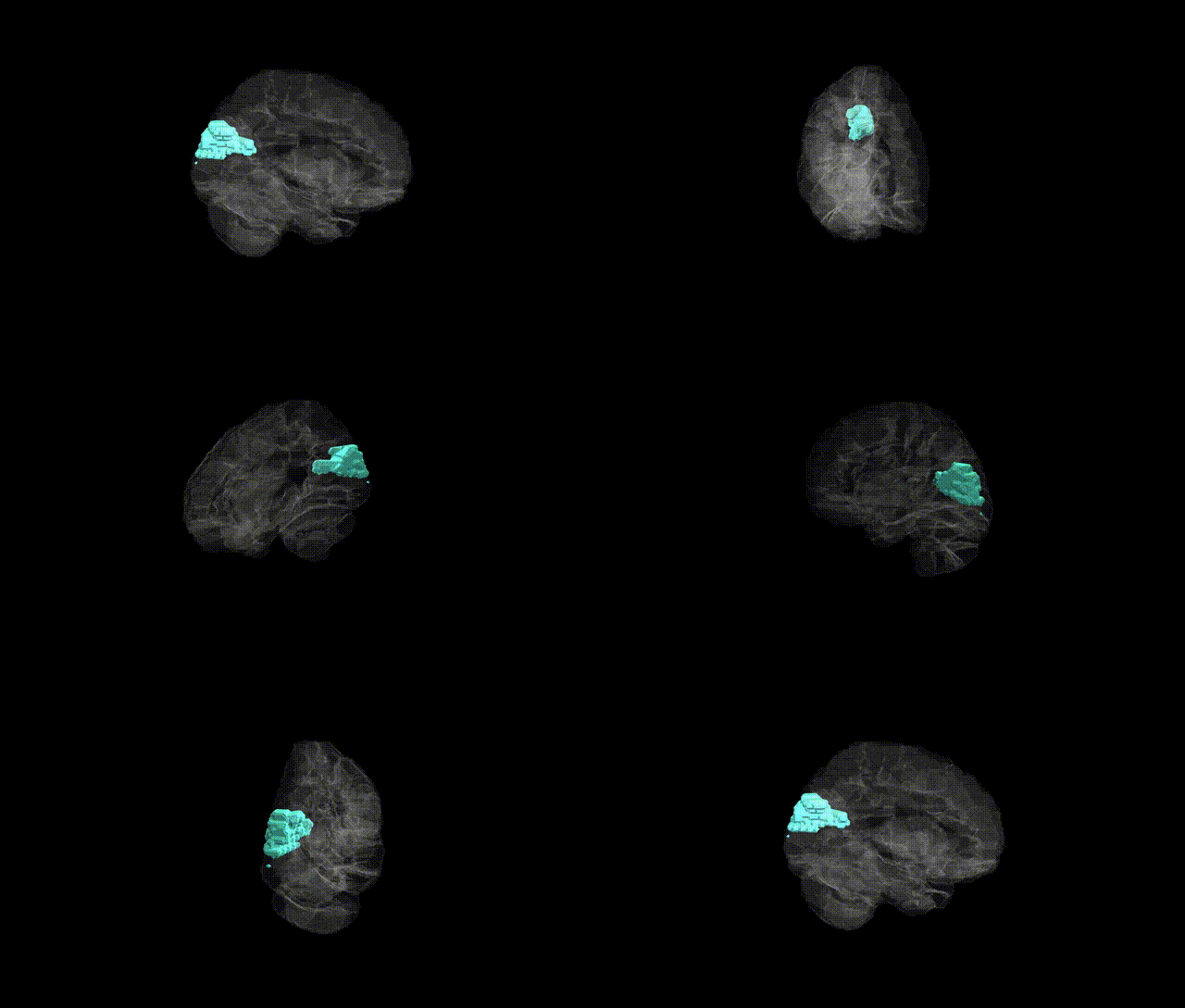
Full Quality Version: Download MP4
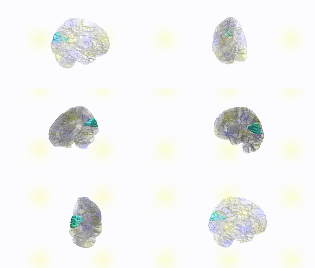
Full Quality Version: Download MP4
Wali Sidiqyar*, Gaurav Rudravaram*, Elyssa M. McMaster, Trent M. Schwartz, Adam M. Saunders, Kurt G. Schilling, Bennett A. Landman "Introducing SPINS: A Shared Public Visualization Library of Neuroanatomical Structures." Medical Imaging with Deep Learning- short paper
This resource is licensed under CC0 1.0 Universal (Public Domain).