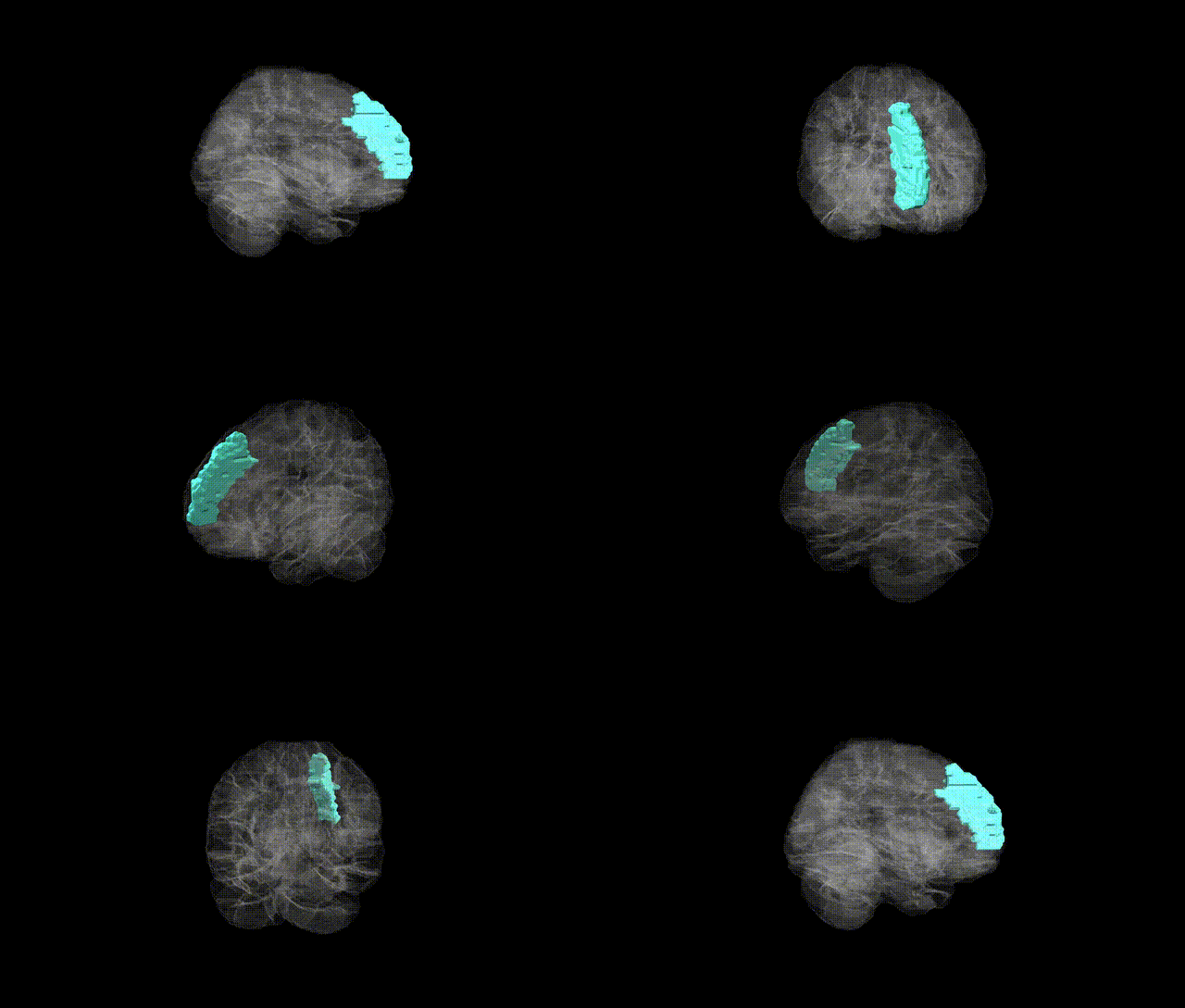
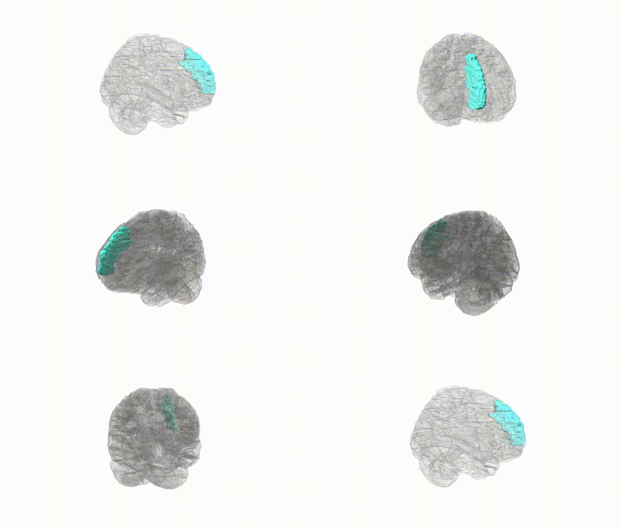
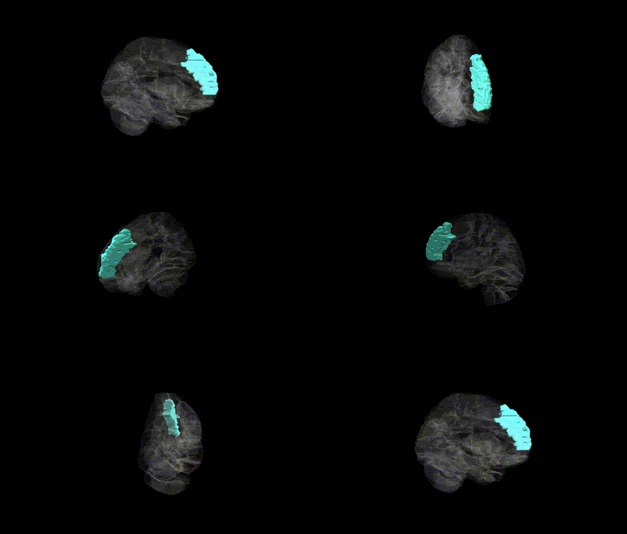
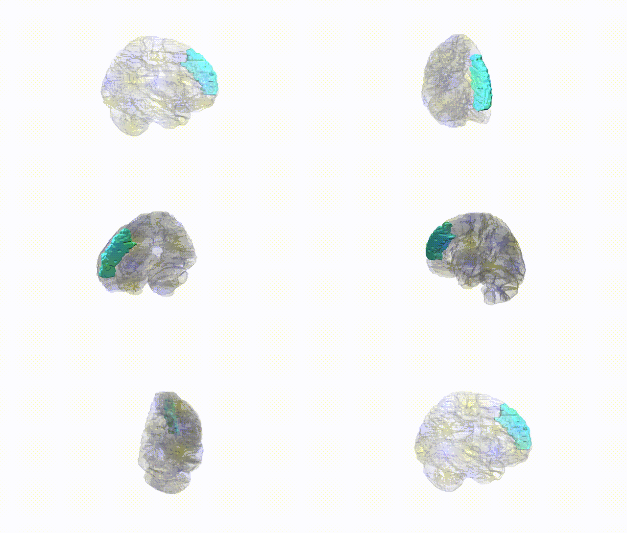
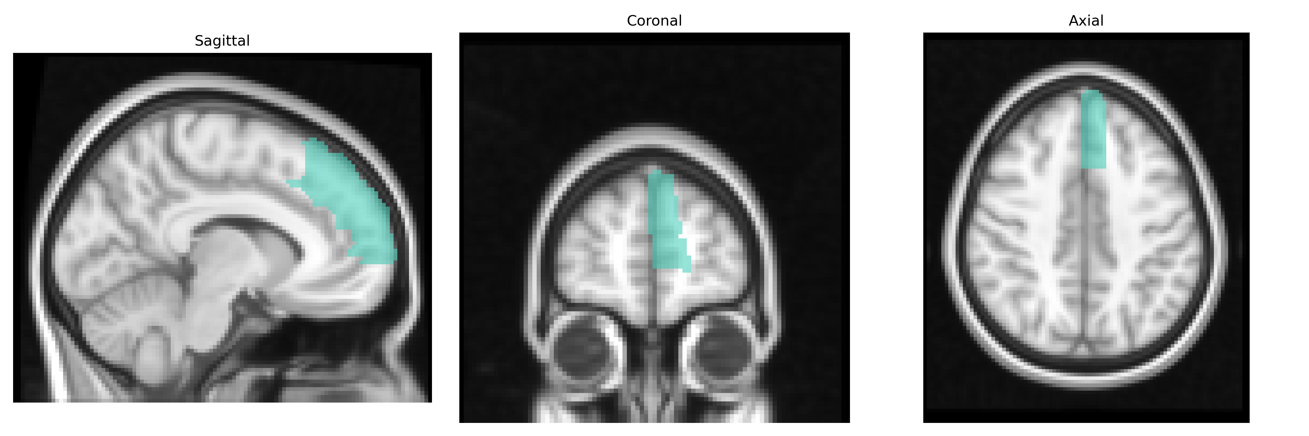
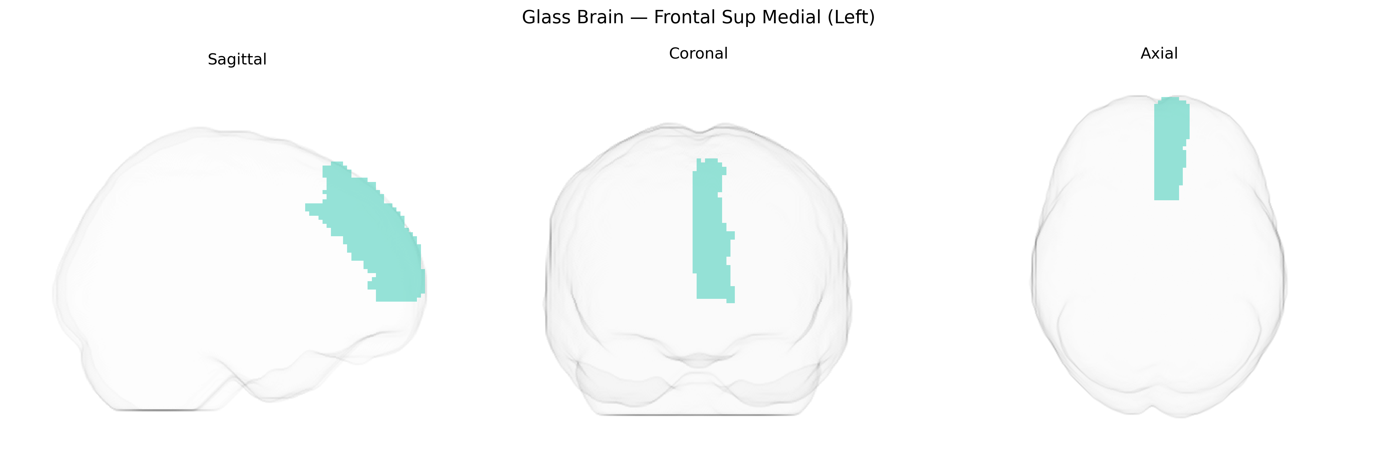
The left Frontal Superior Medial region (AAL2: Frontal_Sup_Medial_L) corresponds largely to the medial portion of the superior frontal gyrus on the left hemisphere, encompassing parts of the medial prefrontal cortex and bordering anterior cingulate territories. This area is implicated in high-level executive functions, self-referential processing, decision-making, and aspects of social cognition, including evaluation of internal states and regulation of behavior. Functionally, it participates in large-scale networks such as the default mode and frontoparietal control networks, integrating cognitive, affective, and motivational information. There is no direct link for the AAL2 label itself; a closely related structure is the medial prefrontal cortex: Medial prefrontal cortex.
The left medial superior frontal region (often encompassing medial prefrontal and dorsomedial prefrontal cortex in the AAL2 atlas) shows genetic associations in several neuroimaging GWAS and disorder-focused studies, though findings are typically reported at the level of cortical thickness, surface area, or functional networks rather than the exact “Frontal Sup Medial (Left)” label. Large imaging-genetics consortia such as ENIGMA and UK Biobank–based GWAS have identified common variants in genes related to neurodevelopment (e.g., microtubule-associated and synaptic genes) influencing medial prefrontal thickness and volume, including loci near genes like MAPT, HMGA2, and NRXN1, among others, though associations are generally distributed and polygenic. Medial superior frontal cortex volume and thickness show significant SNP-based heritability and share polygenic architecture with cognitive ability and educational attainment, as well as with psychiatric traits such as major depressive disorder, schizophrenia, and bipolar disorder, in which case-control and imaging-genetics studies frequently report altered structure or function in this region. Functional and structural variation in this medial frontal area, linked through GWAS and polygenic risk score analyses, has also been associated with traits such as neuroticism, impulsivity, and risk-taking, and with disorders including attention-deficit/hyperactivity disorder and anxiety disorders, particularly within default-mode and cognitive control networks where medial frontal nodes figure prominently. Overall, genetic influences on the left medial superior frontal cortex appear highly polygenic, with overlapping risk architecture across multiple neuropsychiatric disorders and cognitive traits, rather than being driven by a small number of region-specific loci.
Overview generated by GPT-4o (2026).
Region ID: 2601
Hemisphere: left
Atlas: AAL2
Full Quality Version: Download MP4
Full Quality Version: Download MP4
Full Quality Version: Download MP4
Full Quality Version: Download MP4
Wali Sidiqyar*, Gaurav Rudravaram*, Elyssa M. McMaster, Trent M. Schwartz, Adam M. Saunders, Kurt G. Schilling, Bennett A. Landman "Introducing SPINS: A Shared Public Visualization Library of Neuroanatomical Structures." Medical Imaging with Deep Learning- short paper
This resource is licensed under CC0 1.0 Universal (Public Domain).