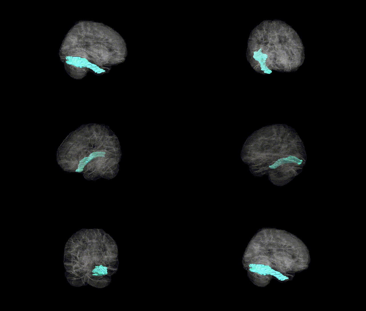
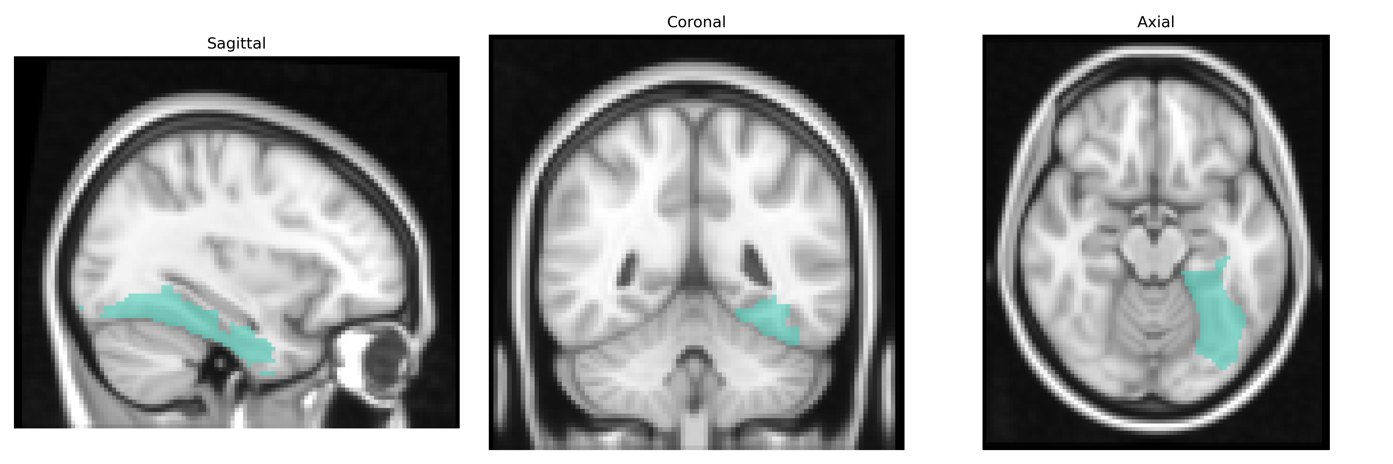
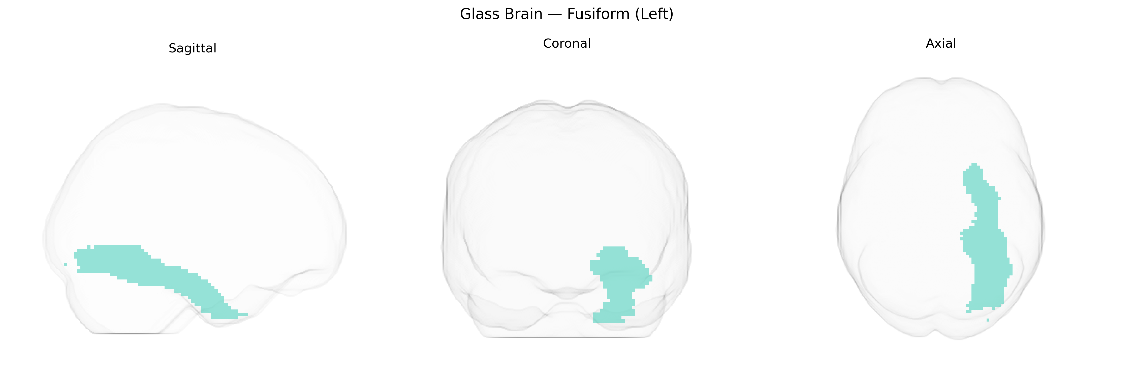
The left fusiform gyrus is a ventral temporal lobe structure extending along the basal surface of the cerebral hemisphere between the inferior temporal gyrus laterally and the parahippocampal gyrus medially, supplied primarily by branches of the posterior cerebral artery. Cytoarchitectonically, it encompasses multiple visual association areas (e.g., Brodmann areas 37 and adjacent regions) and is characterized by columnar organization supporting high-level visual processing. Functionally, the left fusiform gyrus is critically involved in orthographic and lexical processing for reading, visual word recognition, and complex object and face perception, including contributions to category-selective regions such as the visual word form area. It participates in distributed networks for language, memory, and visual perception, with strong reciprocal connections to occipital visual cortices, inferior temporal cortex, and perisylvian language regions. Lesions of the left fusiform gyrus can result in pure alexia, deficits in visual word recognition, and category-specific agnosias. Fusiform gyrus
The left fusiform gyrus, as defined in the AAL2 atlas, has been implicated in multiple genetic and GWAS-based findings, particularly in relation to language, reading, and neuropsychiatric traits. Twin and imaging-genetics studies indicate moderate heritability of fusiform volume and activation patterns, with variants in KIAA0319, DCDC2, and other dyslexia-associated genes linked to altered left fusiform structure and function, supporting its role in orthographic and phonological processing. Large neuroimaging GWAS consortia (e.g., ENIGMA) have reported associations between common variants in genes related to neural development and synaptic function (such as BDNF and HMGA2) and fusiform surface area or cortical thickness, though findings are often regionally broader rather than strictly fusiform-specific. In autism spectrum disorder, de novo and rare variants in synaptic and chromatin-remodeling genes (e.g., SHANK3, CHD8) have been associated with atypical fusiform activation and morphology, consistent with impaired face processing circuits. Schizophrenia and major depressive disorder GWAS, while not pinpointing the fusiform directly, show polygenic risk scores that correlate with fusiform volume and connectivity changes in imaging-genetics analyses. Additionally, GWAS of facial recognition and social cognition traits have reported associations with loci near genes involved in visual processing and white matter integrity that modulate fusiform responses, underscoring this region as a genetically influenced hub for high-level visual and socio-emotional processing.
Overview generated by GPT-4o (2026).
Region ID: 5401
Hemisphere: left
Atlas: AAL2
Full Quality Version: Download MP4
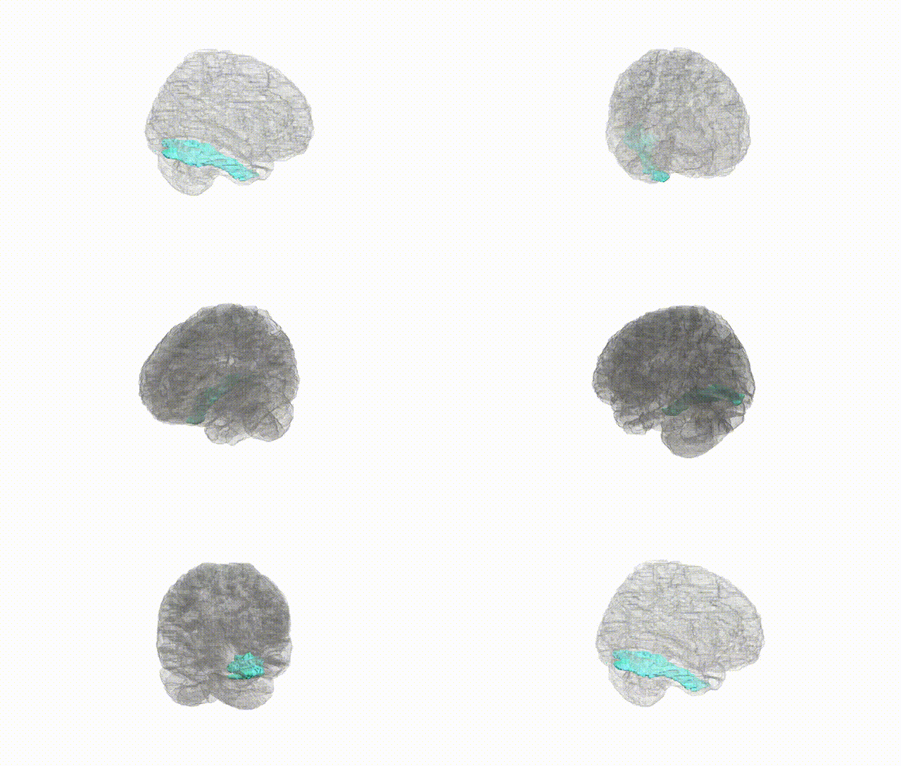
Full Quality Version: Download MP4
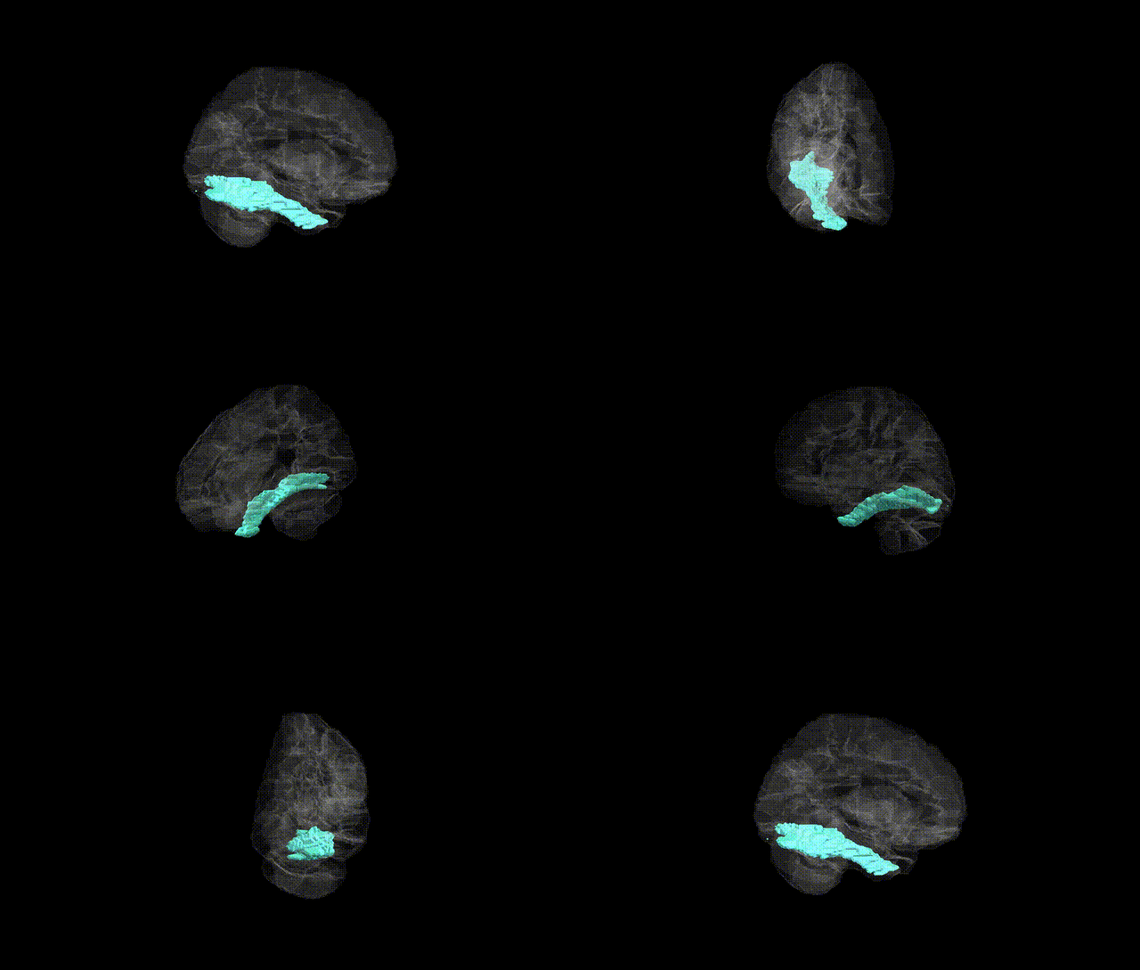
Full Quality Version: Download MP4
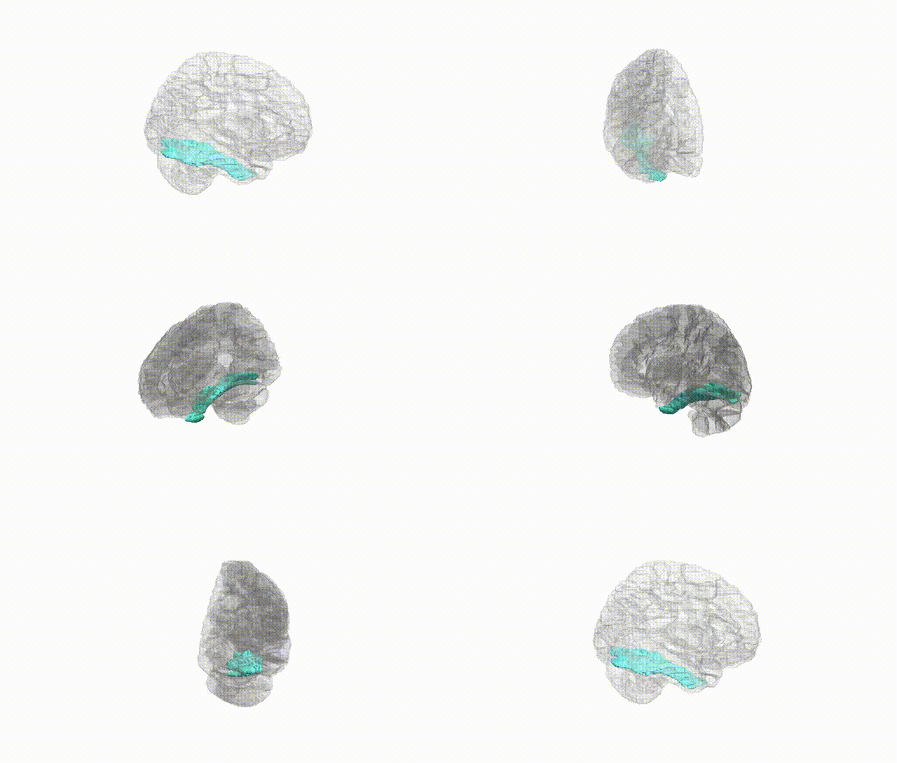
Full Quality Version: Download MP4
Wali Sidiqyar*, Gaurav Rudravaram*, Elyssa M. McMaster, Trent M. Schwartz, Adam M. Saunders, Kurt G. Schilling, Bennett A. Landman "Introducing SPINS: A Shared Public Visualization Library of Neuroanatomical Structures." Medical Imaging with Deep Learning- short paper
This resource is licensed under CC0 1.0 Universal (Public Domain).