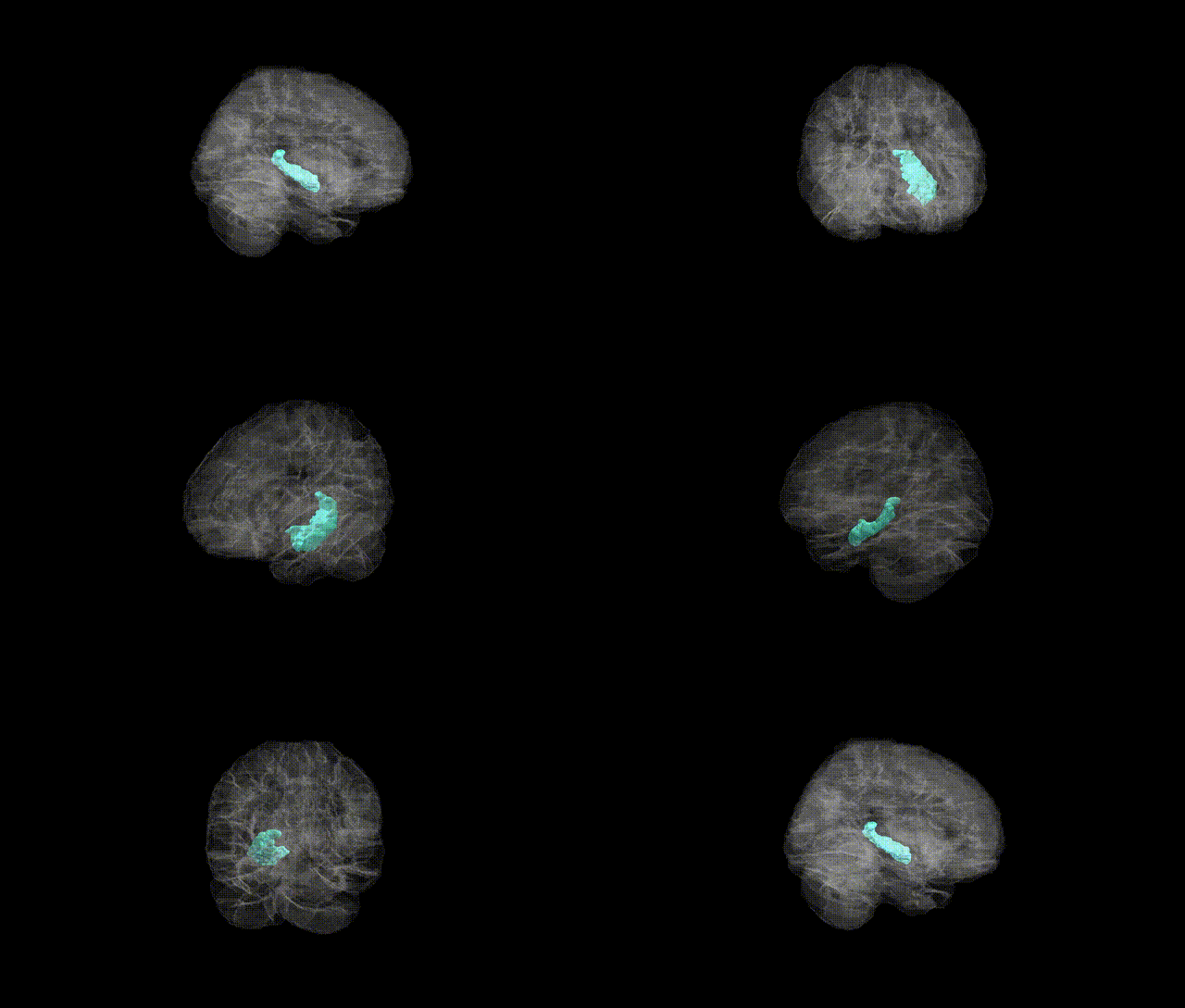
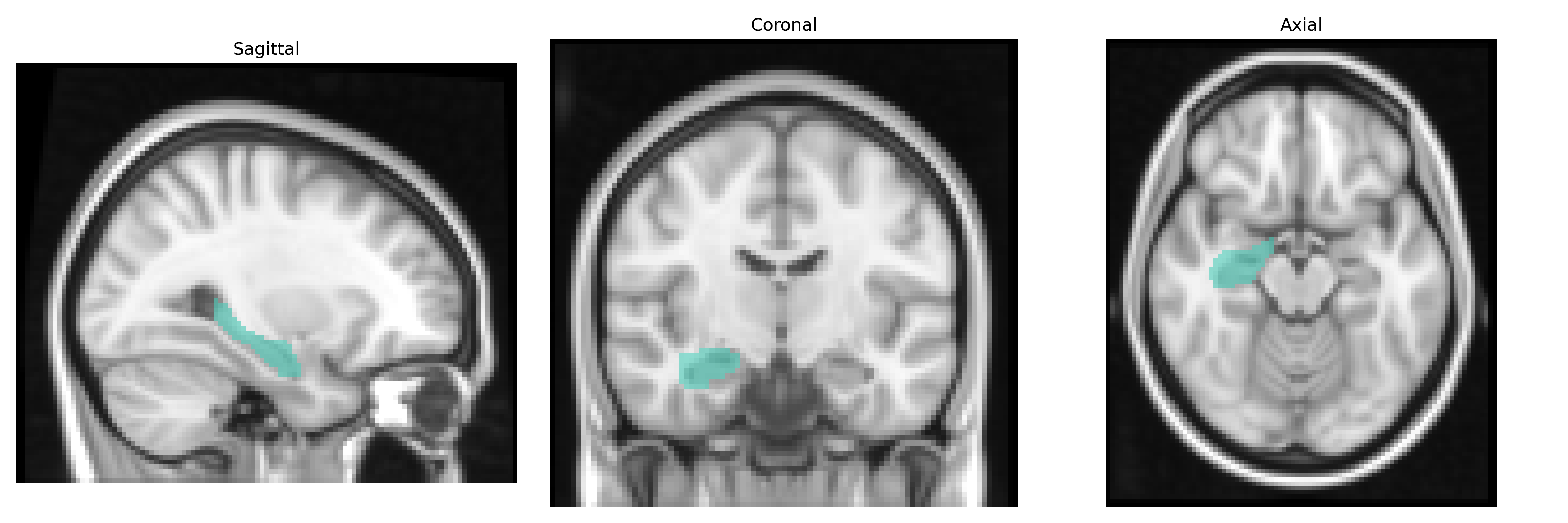
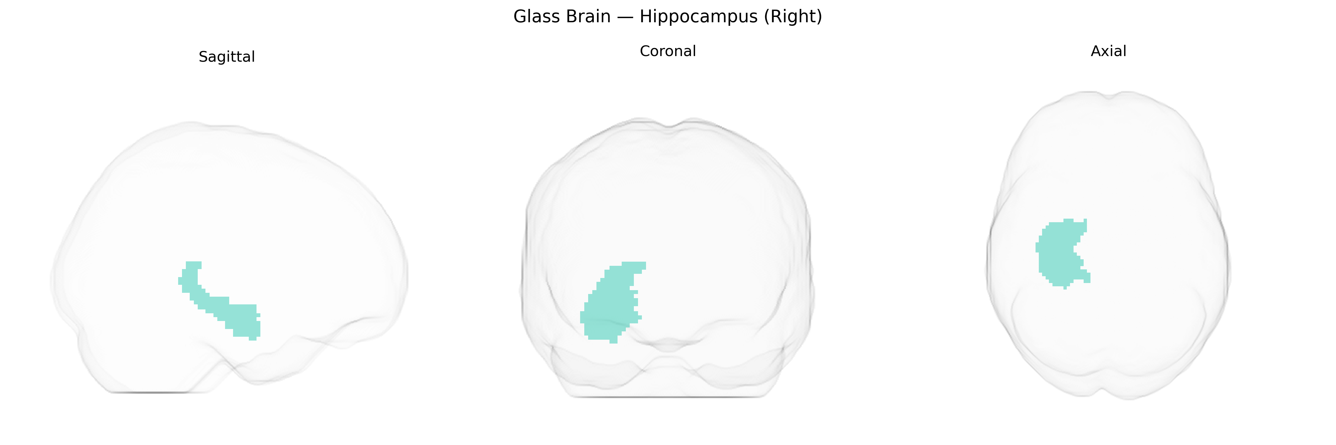
The right hippocampus is a bilateral medial temporal lobe structure critically involved in episodic memory formation, spatial navigation, and contextual processing, with a particular emphasis—relative to the left hippocampus—on spatial and visuospatial memory functions. Cytoarchitectonically, it comprises the cornu ammonis (CA fields), dentate gyrus, and subiculum, interconnected in a trisynaptic circuit that supports long-term potentiation and memory consolidation. The right hippocampus receives multimodal input via the entorhinal cortex and projects through the fornix to diencephalic and limbic structures, forming a key node within the hippocampal–diencephalic–cortical memory network. It is highly plastic and vulnerable to hypoxia, neurodegenerative processes, and stress-related pathology, with volumetric changes often observed in conditions such as temporal lobe epilepsy, Alzheimer’s disease, and post-traumatic stress disorder.
Hippocampus
The right hippocampus, as defined in the AAL2 atlas, has been repeatedly implicated in genetic studies of brain structure and function, with numerous GWAS identifying variants that influence its volume, microstructure, and connectivity. Large-scale imaging genetics consortia (e.g., ENIGMA, UK Biobank) have highlighted loci near or within genes such as HMGA2, DPP4, WNT3, ASTN2, APOE, KIBRA (WWC1), BDNF, and CLU, among others, as associated with hippocampal volume, often with hemisphere-specific or asymmetric effects. APOE ε4 and related Alzheimer’s disease risk variants, including in BIN1, CR1, and ABCA7, have been linked to reduced right hippocampal volume and accelerated atrophy, and similar structural associations have been reported for schizophrenia and bipolar disorder risk loci in genes such as CACNA1C, ZNF804A, and MIR137. Genetic risk scores for major depressive disorder, PTSD, and anxiety traits show correlations with right hippocampal morphology and activity, reflecting the region’s role in stress regulation and episodic memory. Variants affecting neurogenesis and synaptic plasticity, including BDNF Val66Met and KIBRA alleles, have been associated with memory performance and functional MRI activation in the right hippocampus, while polygenic scores for educational attainment and general cognitive ability show modest associations with its volume and connectivity. Overall, GWAS and candidate-gene studies converge on the right hippocampus as a key heritable endophenotype linking genetic variation to neurodegenerative disease, psychosis, affective disorders, memory traits, and cognitive performance.
Overview generated by GPT-4o (2026).
Region ID: 4102
Hemisphere: right
Atlas: AAL2
Full Quality Version: Download MP4
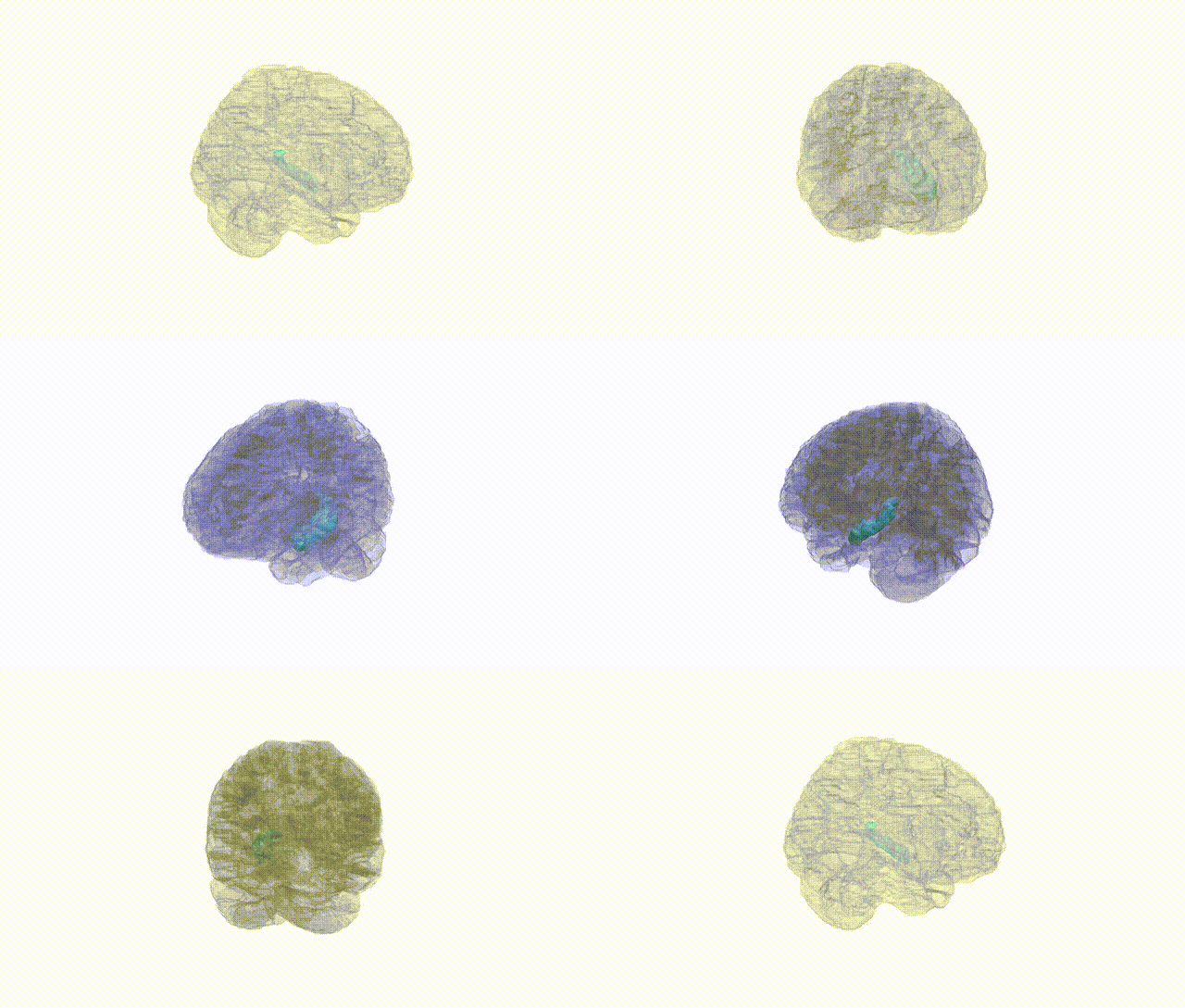
Full Quality Version: Download MP4
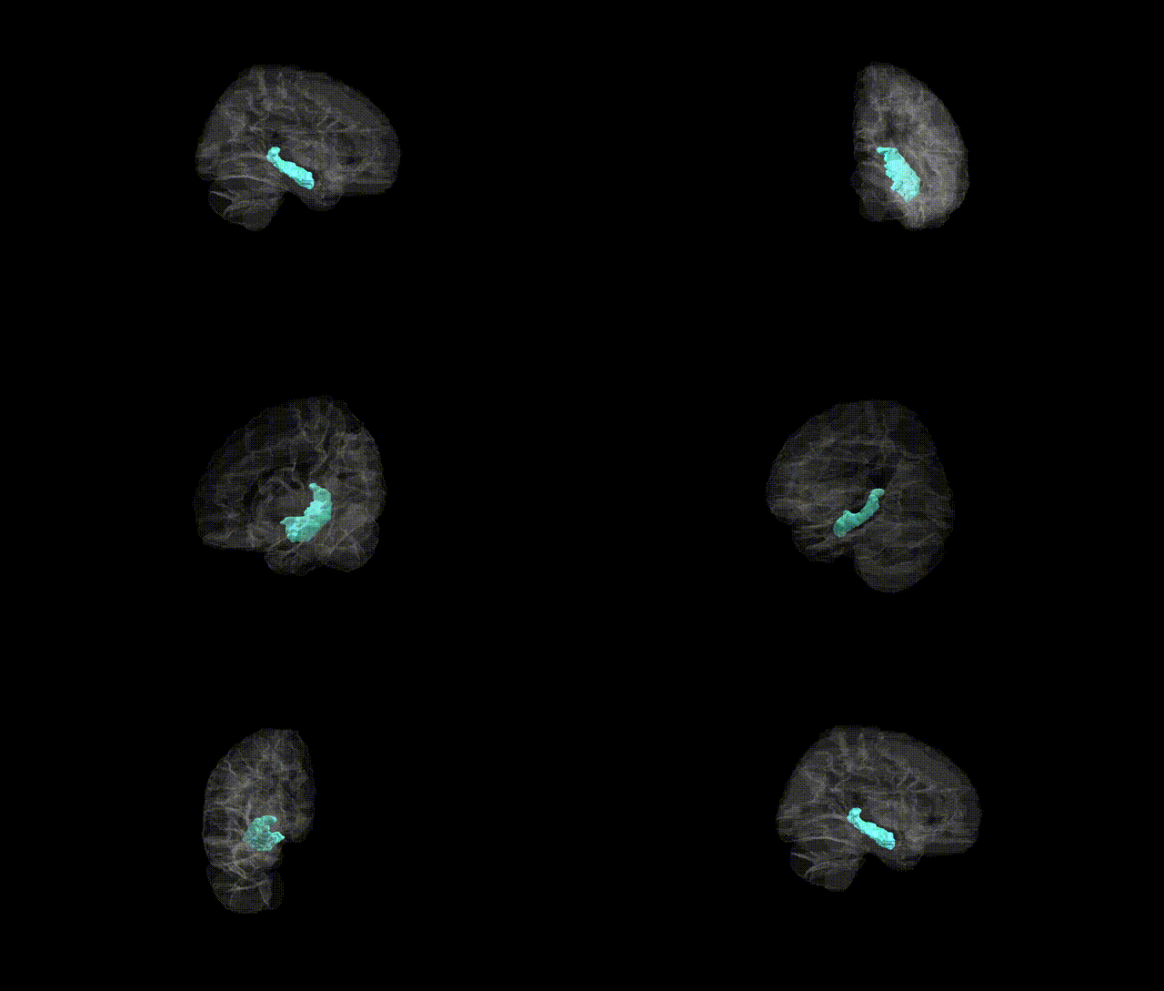
Full Quality Version: Download MP4
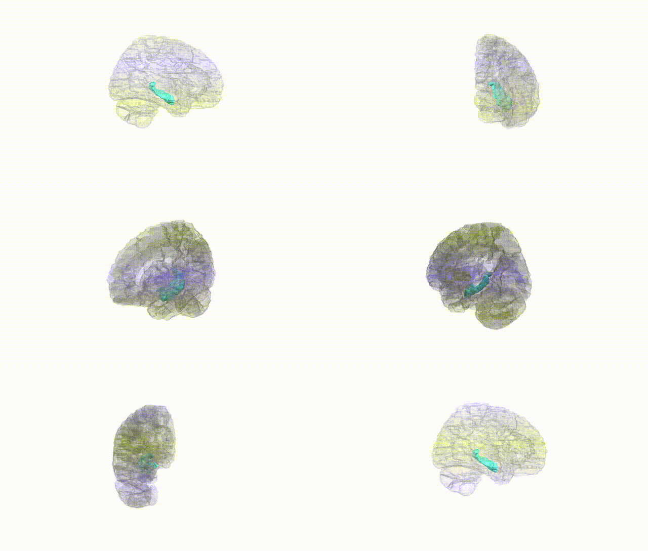
Full Quality Version: Download MP4
Wali Sidiqyar*, Gaurav Rudravaram*, Elyssa M. McMaster, Trent M. Schwartz, Adam M. Saunders, Kurt G. Schilling, Bennett A. Landman "Introducing SPINS: A Shared Public Visualization Library of Neuroanatomical Structures." Medical Imaging with Deep Learning- short paper
This resource is licensed under CC0 1.0 Universal (Public Domain).