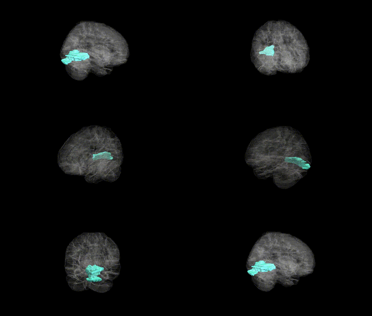
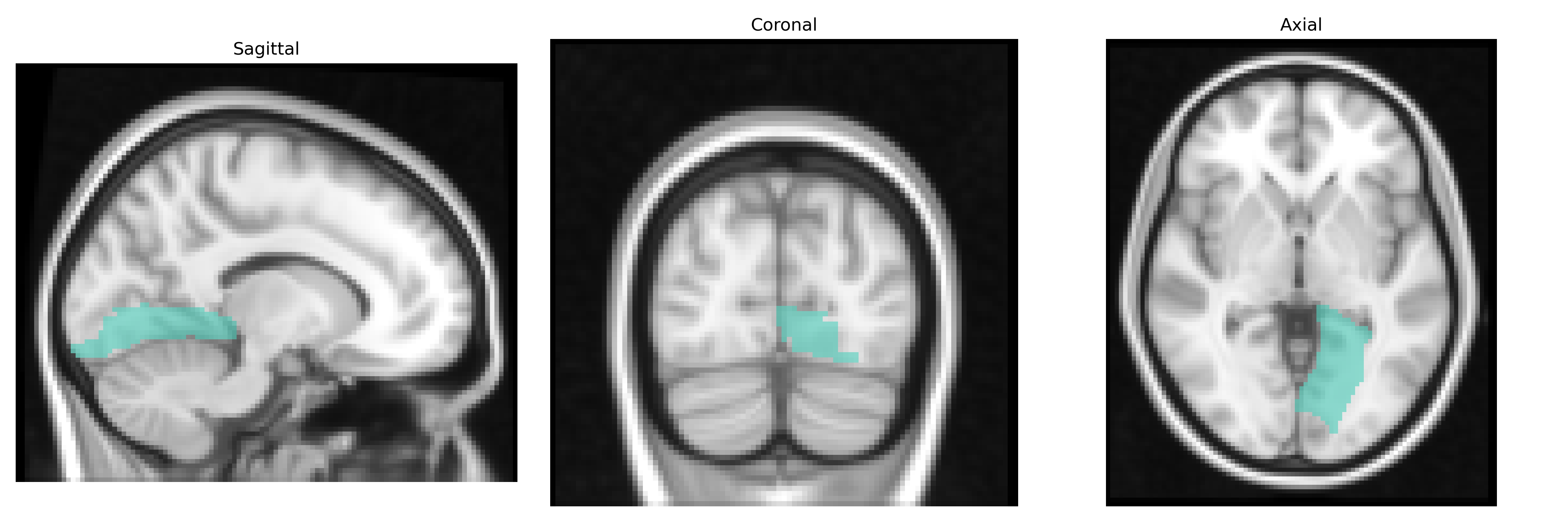
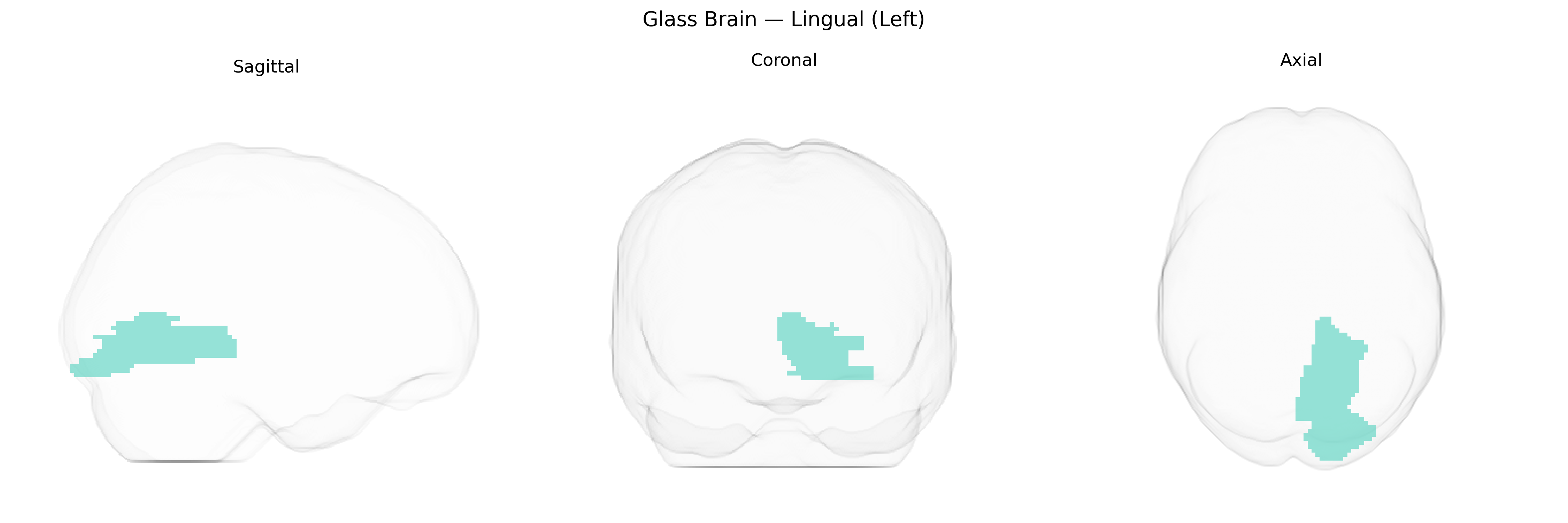
The left lingual gyrus, as defined in the AAL2 Atlas, is a medial occipital cortical region located inferior to the calcarine sulcus and extending into the posterior part of the temporal lobe. It is primarily involved in visual processing, including the analysis of complex visual patterns, letter and word recognition, and contributes to early stages of visual memory encoding. This region participates in networks supporting reading, visual imagery, and visuospatial attention, and is supplied by branches of the posterior cerebral artery. Functionally, it integrates input from primary visual areas and relays processed information to higher-order visual and language-related cortices, playing a role in transforming visual stimuli into more abstract representations. There is no direct Wikipedia article for “left lingual (left) brain region”; a related structure is the Lingual gyrus.
The left lingual gyrus, an occipital lobe region implicated in visual processing and reading, has been highlighted in neuroimaging genetics studies as a locus where common variants influence both structure and function. Large-scale GWAS of brain morphology (e.g., ENIGMA, UK Biobank) have identified associations between variants in genes involved in synaptic development, axon guidance, and neurogenesis—such as MEF2C, DLG2, and genes near the 15q14 and 6p21 loci—and cortical surface area, thickness, or volume in occipital regions that include the lingual gyrus, although these findings are often reported at regional or lobar rather than AAL2-level specificity. Functional imaging genetics has linked polymorphisms in genes like BDNF (e.g., Val66Met), COMT, and DISC1 to altered activation of the lingual gyrus during visual, memory, and language tasks. In clinical GWAS and imaging–genetics studies, lingual gyrus structure and connectivity have been reported as intermediate phenotypes associated with risk alleles for major depressive disorder, schizophrenia, bipolar disorder, autism spectrum disorder, and dyslexia, including overlap with loci implicated in general cognitive ability and educational attainment. Additionally, migraine and visual aura GWAS, as well as studies of photophobia and visual hallucinations, have pointed to genetic influences on occipital regions encompassing the lingual gyrus, suggesting that common variants affecting glutamatergic and GABAergic signaling contribute to individual differences in visual and emotional processing mediated by this area.
Overview generated by GPT-4o (2026).
Region ID: 5021
Hemisphere: left
Atlas: AAL2
Full Quality Version: Download MP4
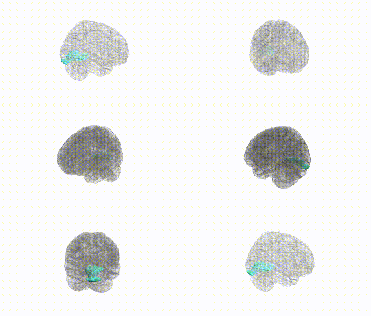
Full Quality Version: Download MP4
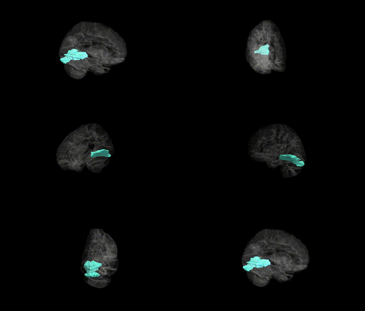
Full Quality Version: Download MP4
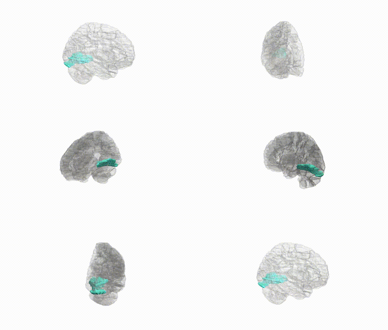
Full Quality Version: Download MP4
Wali Sidiqyar*, Gaurav Rudravaram*, Elyssa M. McMaster, Trent M. Schwartz, Adam M. Saunders, Kurt G. Schilling, Bennett A. Landman "Introducing SPINS: A Shared Public Visualization Library of Neuroanatomical Structures." Medical Imaging with Deep Learning- short paper
This resource is licensed under CC0 1.0 Universal (Public Domain).