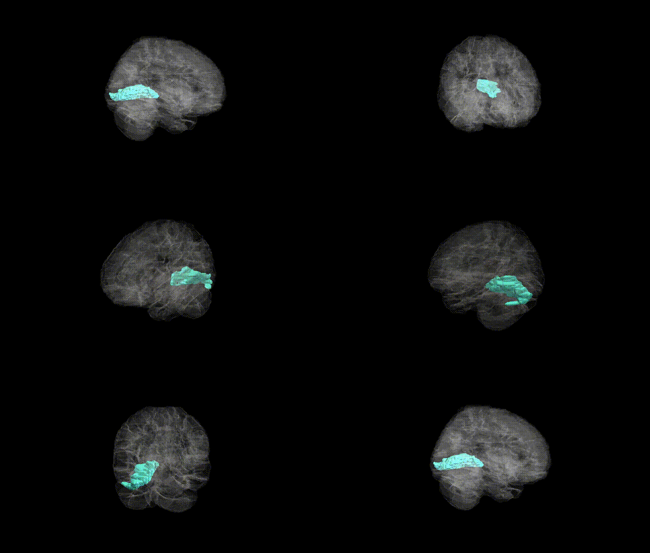
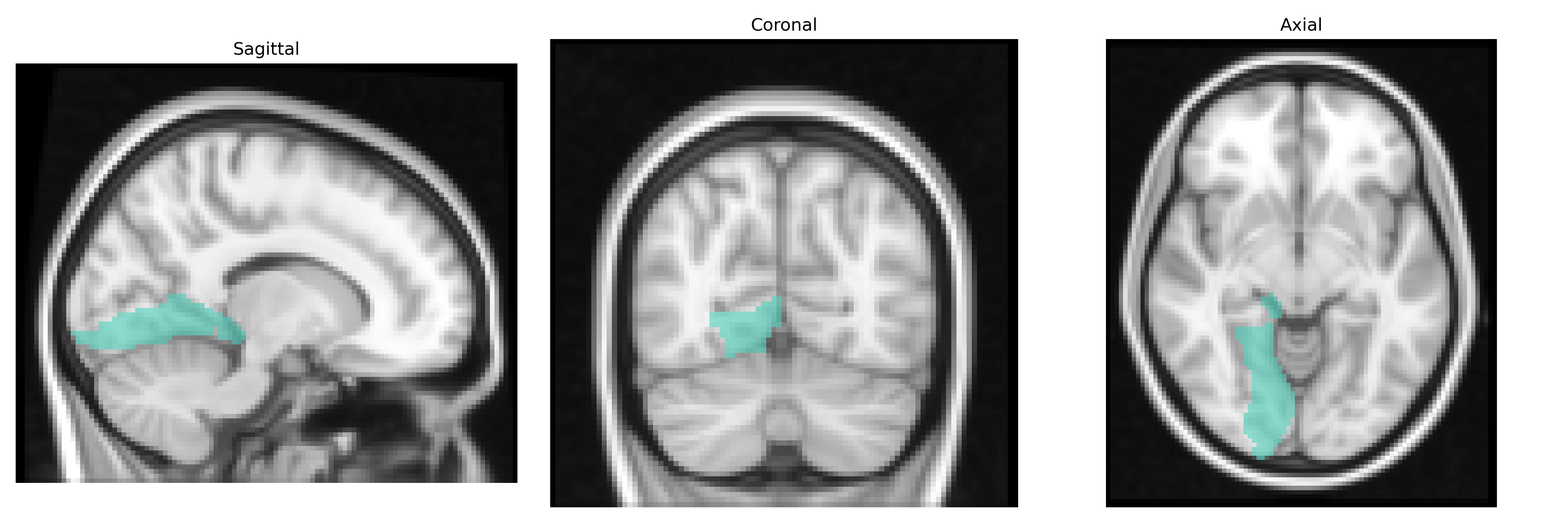
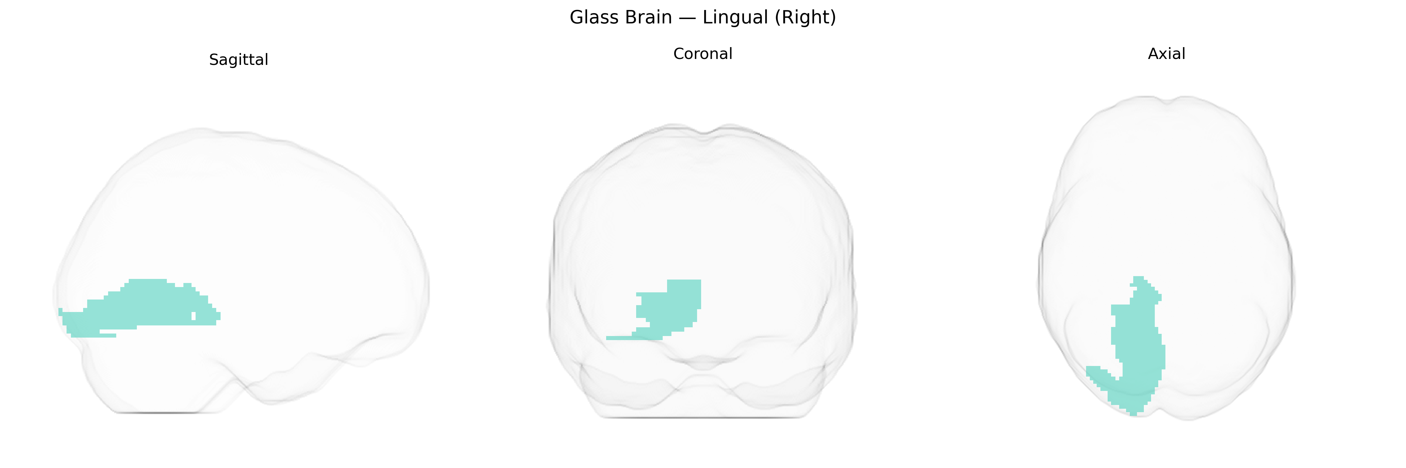
The right lingual gyrus, corresponding to the Right Lingual region in the AAL2 atlas, is a medial occipital cortical area located on the ventral surface of the occipital lobe, extending into the posterior part of the temporal lobe. It lies inferior to the calcarine sulcus and is bordered by the fusiform gyrus laterally and the cuneus superiorly. Cytoarchitectonically, it is part of the visual association cortex and is heavily involved in processing complex visual information, including form, color, and visual memory, and contributes to visuospatial analysis and word-form recognition. The right hemisphere lingual gyrus is particularly implicated in visuospatial and scene processing and participates in networks subserving visual imagery and memory encoding. There is no direct Wikipedia article specifically for the “Right Lingual” AAL2 region; the related structure is the Lingual gyrus.
The right lingual gyrus, as defined in the AAL2 atlas, has been implicated in several genetic and neuroimaging-genetic studies, particularly in the context of visual processing, psychiatric and neurodevelopmental disorders, and brain-structure GWAS. Large-scale imaging genetics consortia (e.g., ENIGMA) and UK Biobank analyses have identified common variants in genes involved in neurodevelopment and synaptic signaling (such as those in or near MAPT, HMGA2, and genes in Wnt and glutamatergic pathways) that influence occipital and lingual cortical thickness, surface area, or gray matter volume, with some effects showing hemispheric specificity or stronger associations on the right. The right lingual region has also appeared in polygenic risk–brain structure correlation analyses for schizophrenia, major depressive disorder, bipolar disorder, and autism spectrum disorder, where higher disorder-related polygenic scores associate with altered lingual gyrus volume or activation during visual, emotional, or language tasks. In addition, GWAS of functional MRI patterns and visual cortex activation have linked variation in occipital/lingual activation (including right-lateralized effects) to loci affecting neuronal excitability and myelination, while studies of disorders such as migraine, epilepsy, and reading or visual processing difficulties have reported lingual gyrus involvement in imaging endophenotypes that share genetic risk with these traits, although the mapping to specific right-lingual–restricted loci remains indirect and often reflects broader occipital or visual network genetic influences.
Overview generated by GPT-4o (2026).
Region ID: 5022
Hemisphere: right
Atlas: AAL2
Full Quality Version: Download MP4
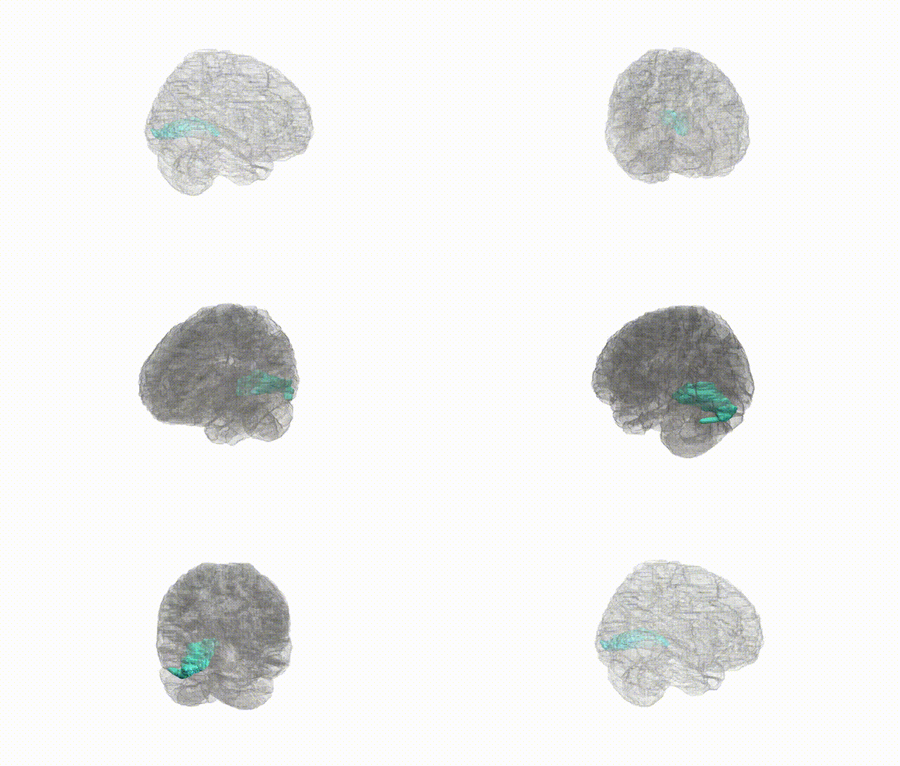
Full Quality Version: Download MP4
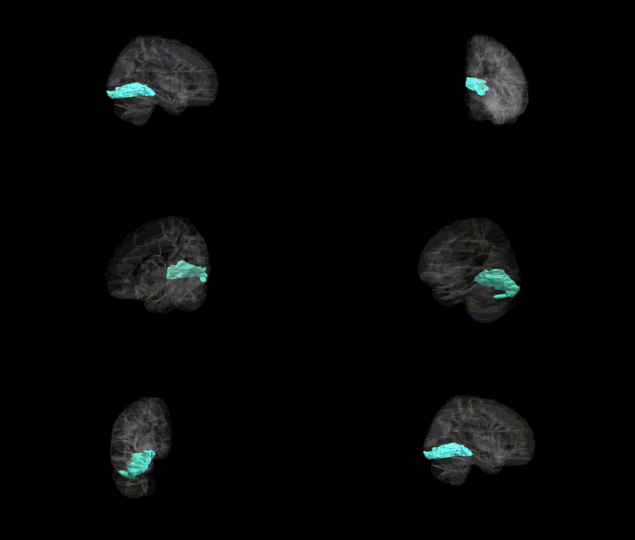
Full Quality Version: Download MP4
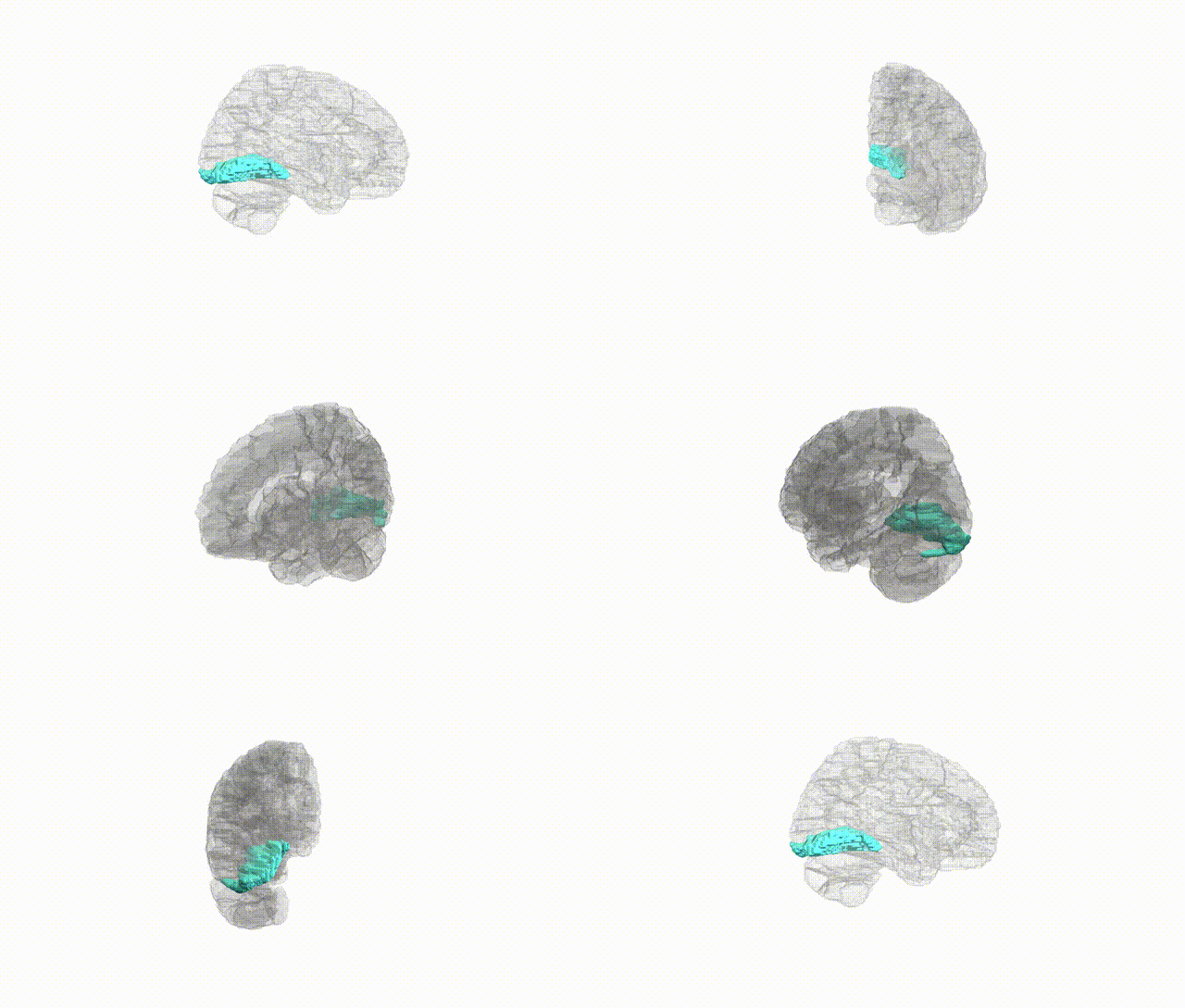
Full Quality Version: Download MP4
Wali Sidiqyar*, Gaurav Rudravaram*, Elyssa M. McMaster, Trent M. Schwartz, Adam M. Saunders, Kurt G. Schilling, Bennett A. Landman "Introducing SPINS: A Shared Public Visualization Library of Neuroanatomical Structures." Medical Imaging with Deep Learning- short paper
This resource is licensed under CC0 1.0 Universal (Public Domain).