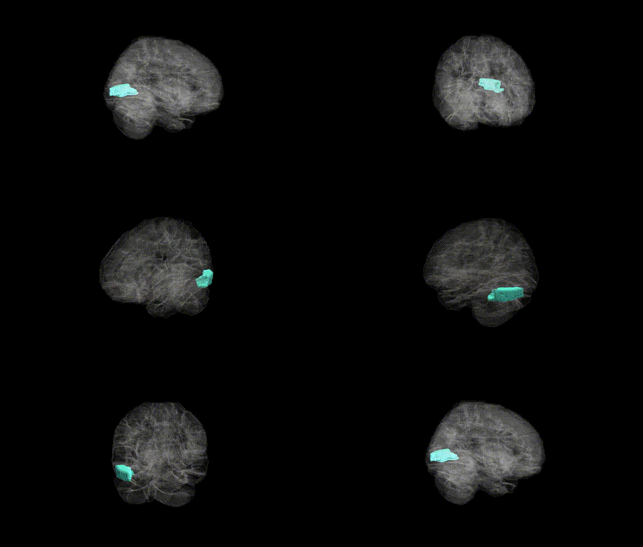
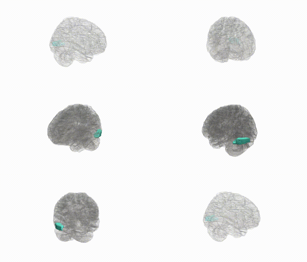
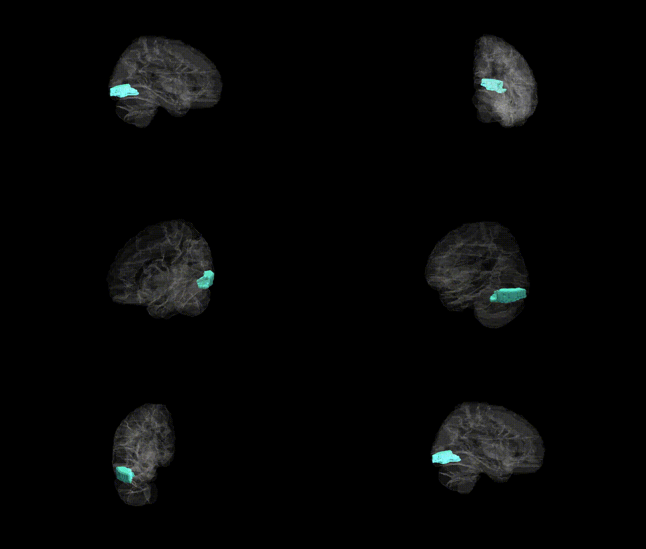
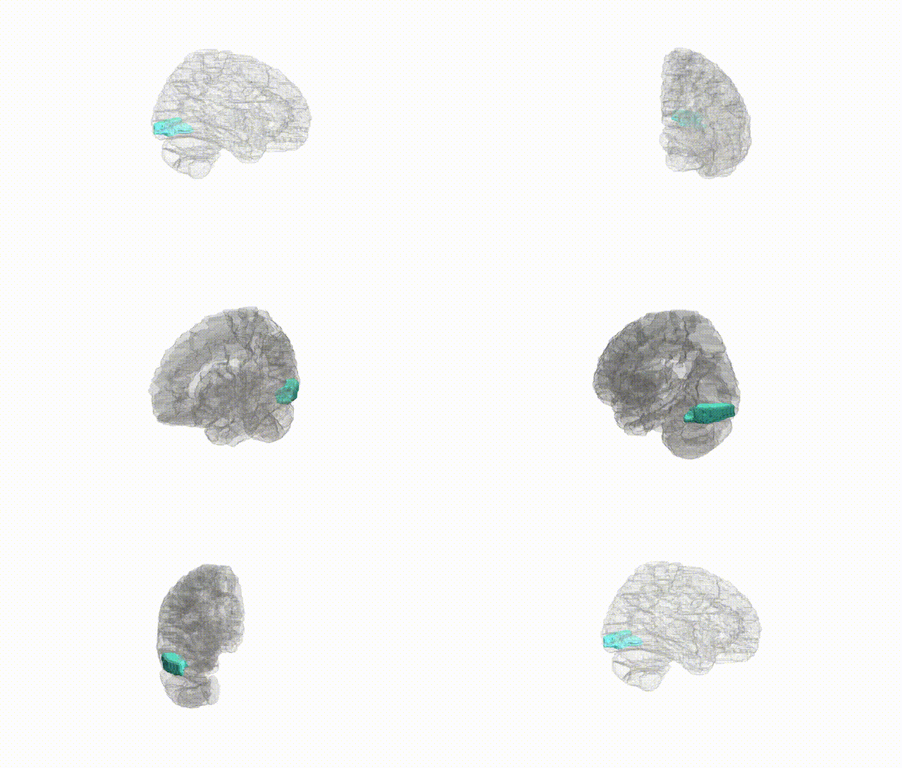
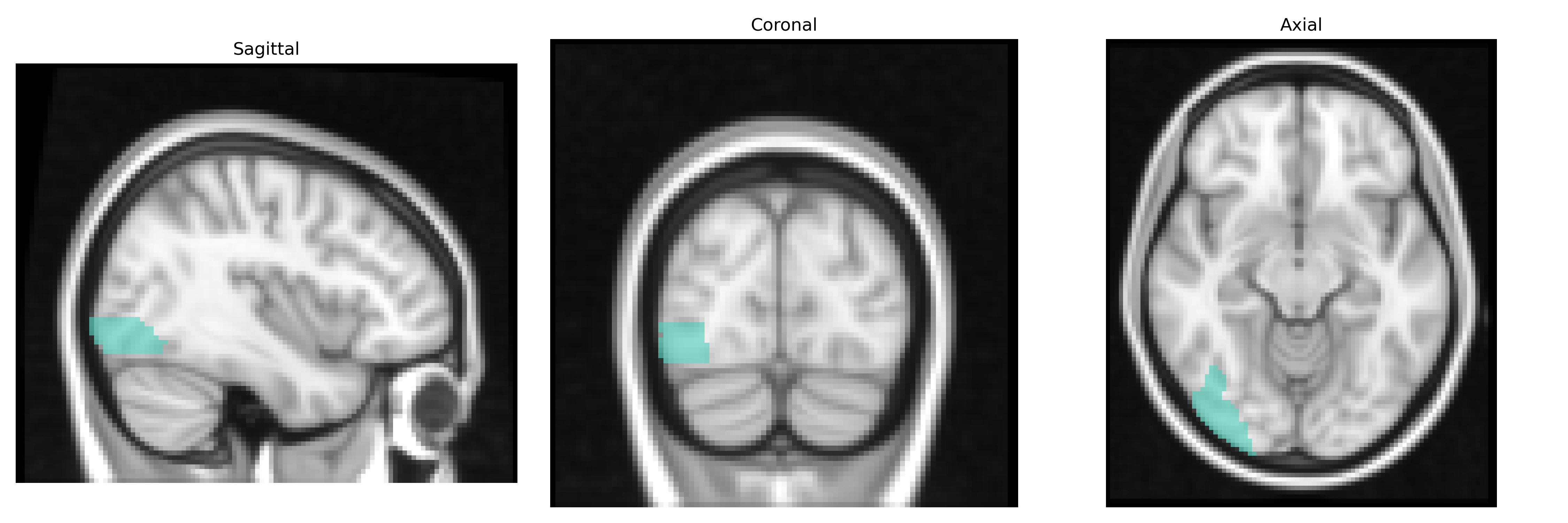
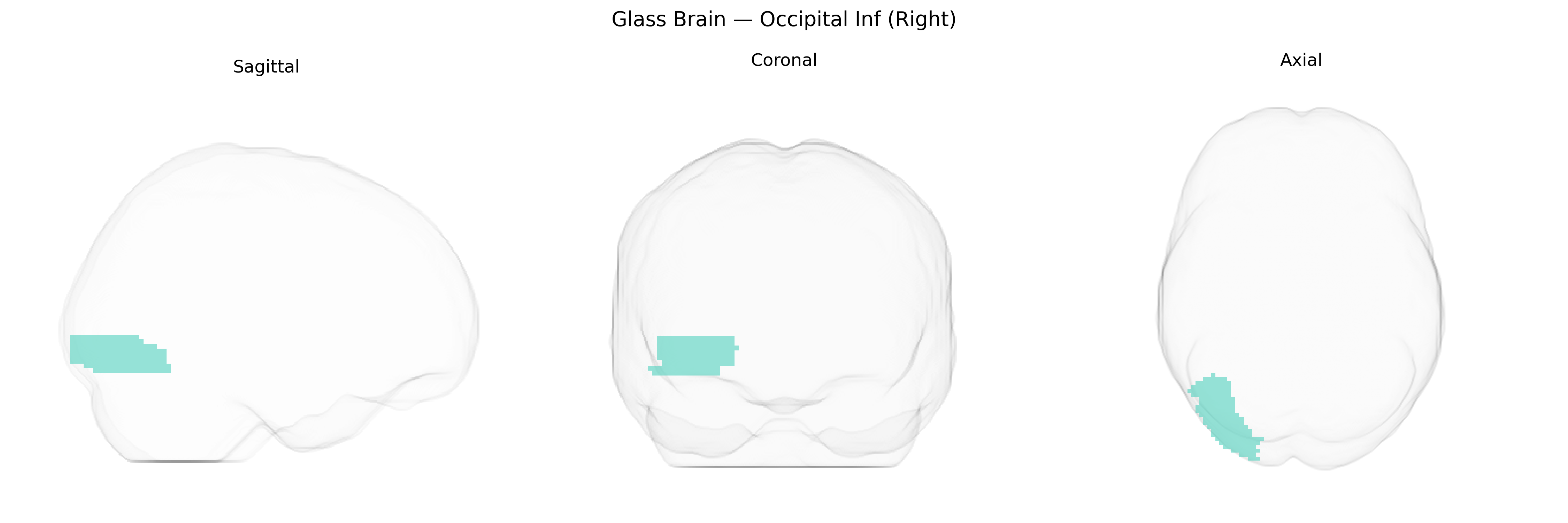
The right Inferior Occipital (Occipital Inf Right) region in the AAL2 atlas corresponds primarily to the inferior occipital gyrus on the right hemisphere, a ventral portion of the occipital lobe located along the lateral and basal surface near the occipitotemporal junction. This region is part of the early and intermediate visual processing network, receiving inputs from primary and secondary visual cortices and contributing to the analysis of complex visual features such as shape, contours, and aspects of object form that support higher-order recognition in ventral visual stream areas (e.g., fusiform and inferior temporal cortices). It participates in pathways crucial for object perception and may be involved in processing visually driven information relevant to reading, face perception, and scene analysis. Inferior temporal gyrus
The right inferior occipital gyrus (Occipital Inf Right in the AAL2 atlas) has been implicated in several imaging genetics and GWAS findings, largely through vertex- or parcel-wise analyses of cortical thickness, surface area, and volume rather than region-specific candidate genes. Large-scale consortia such as ENIGMA and UK Biobank–based studies have identified common variants in loci including 14q23 (e.g., DACT1), 15q14 (e.g., THBS1), and 6q22 (e.g., RSPO3) associated with occipital cortical morphology, with some effects overlapping the inferior occipital cortex. Polygenic influences related to general brain size, visual cortical development, and synaptic/axon guidance pathways contribute to structural variation in this region, and these variants often show pleiotropy with cognitive performance, educational attainment, and intracranial volume. Occipital regions, including inferior occipital cortex, appear in genetic correlation analyses linking visual cortical measures with neurodevelopmental and psychiatric traits (e.g., schizophrenia, autism spectrum traits, ADHD), though these associations are typically modest and distributed across many regions rather than specific to the inferior occipital gyrus. In neurodegenerative and neuropsychiatric GWAS-imaging studies, genetically driven atrophy or thinning in occipital areas has been reported in Alzheimer’s disease, dementia with Lewy bodies, and major depressive disorder, again reflecting widespread polygenic effects rather than a single disorder-specific locus for right inferior occipital cortex.
Overview generated by GPT-4o (2026).
Region ID: 5302
Hemisphere: right
Atlas: AAL2
Full Quality Version: Download MP4
Full Quality Version: Download MP4
Full Quality Version: Download MP4
Full Quality Version: Download MP4
Wali Sidiqyar*, Gaurav Rudravaram*, Elyssa M. McMaster, Trent M. Schwartz, Adam M. Saunders, Kurt G. Schilling, Bennett A. Landman "Introducing SPINS: A Shared Public Visualization Library of Neuroanatomical Structures." Medical Imaging with Deep Learning- short paper
This resource is licensed under CC0 1.0 Universal (Public Domain).