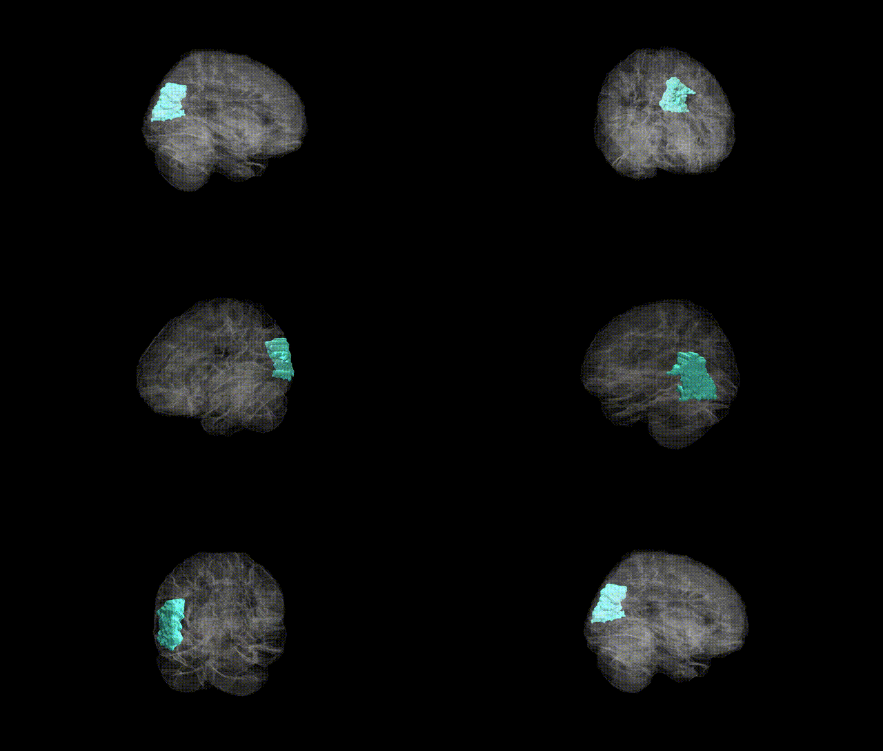
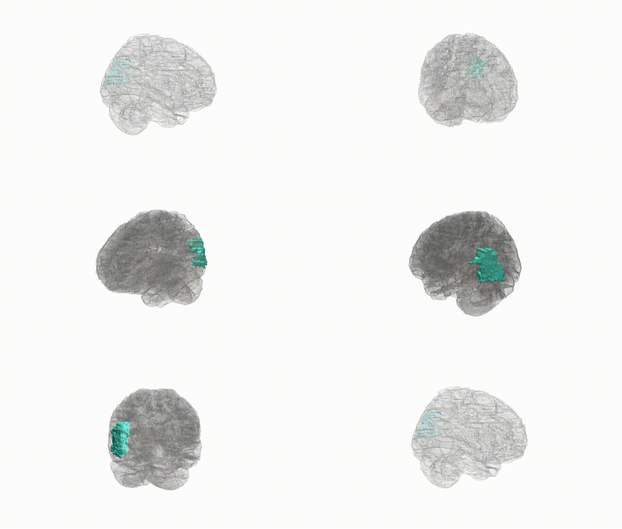
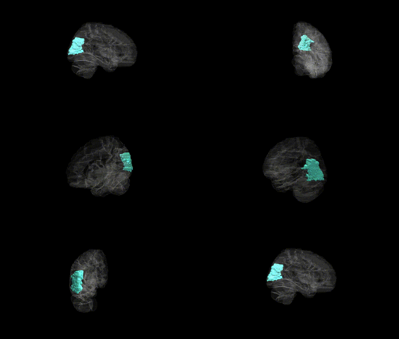
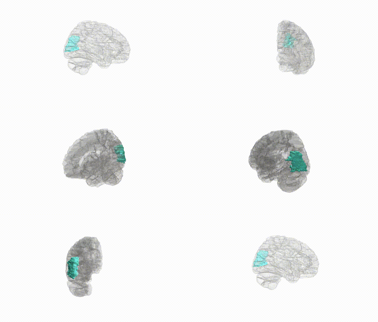
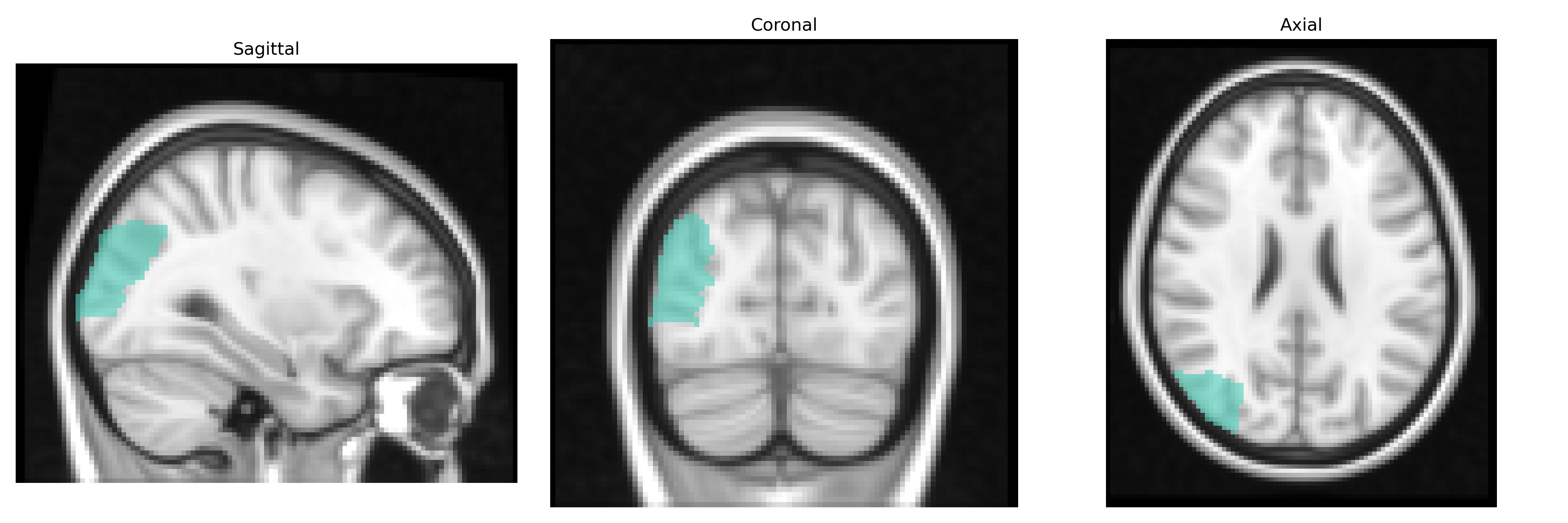
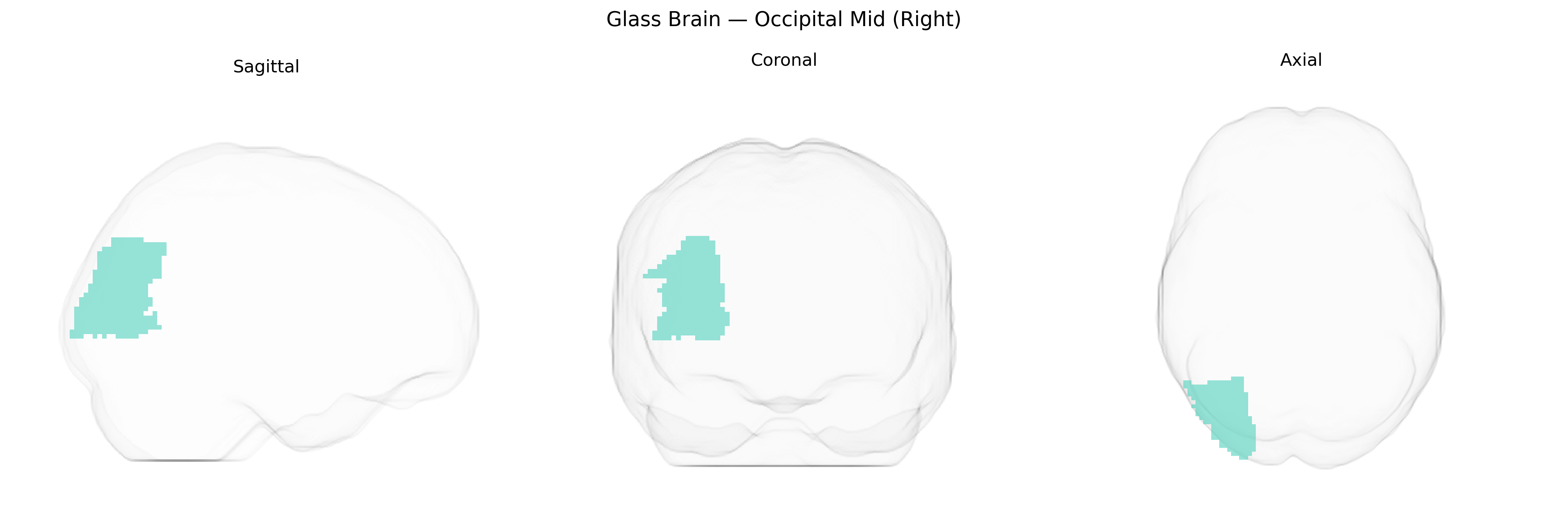
The right Occipital Mid (Right) region in the AAL2 atlas corresponds primarily to the middle occipital gyrus of the right cerebral hemisphere, a portion of the occipital lobe implicated in intermediate-level visual processing. This region participates in analysis of complex visual features, including object form, motion, and spatial relationships, and contributes to the integration of visual information from primary visual cortex (V1) and higher-order associative areas in the dorsal and ventral visual streams. It receives feedforward and feedback projections from surrounding occipital cortices and parietal and temporal visual association areas, supporting functions such as visuospatial attention, visual scene perception, and recognition. Lesions affecting the right middle occipital region can produce deficits in visual perception and visuospatial processing, potentially contributing to impairments in object recognition and spatial orientation. There is no direct link for the AAL2 label “Occipital Mid (Right)”; a closely related anatomical structure is the middle occipital gyrus: Middle occipital gyrus.
Genetic association findings specifically targeting the right middle occipital gyrus (Occipital Mid Right in AAL2) are sparse, and most evidence is indirect, coming from GWAS of brain structure and function that report occipital or visual-association regions more broadly. Large neuroimaging-genetics consortia (e.g., ENIGMA, UK Biobank) have identified common variants near genes such as HMGA2, IGF1, and genes involved in neurodevelopment and synaptic signaling that influence global and regional cortical surface area and thickness, including occipital cortex measures that encompass the middle occipital region. Occipital structural variation linked to polygenic risk scores for schizophrenia, major depressive disorder, and autism spectrum disorder points to shared genetic architectures affecting visual-association cortex, while visual-processing GWAS (e.g., for visual acuity, visual field deficits, or migraine with aura) implicate pathways in axon guidance, myelination, and vascular regulation that may influence occipital lobe function. Moreover, imaging-genetic studies of face and motion perception, reading, and dyslexia sometimes report associations in lateral occipital areas overlapping or adjacent to the right middle occipital gyrus, driven by variants in genes related to cortical patterning (such as those in Wnt and Notch signaling) and synaptic plasticity; however, precise, consistently replicated gene-level associations uniquely and specifically assigned to the right Occipital Mid (Right) parcel in AAL2 remain limited and are typically inferred from broader occipital or visual-network findings rather than region-exclusive GWAS.
Overview generated by GPT-4o (2026).
Region ID: 5202
Hemisphere: right
Atlas: AAL2
Full Quality Version: Download MP4
Full Quality Version: Download MP4
Full Quality Version: Download MP4
Full Quality Version: Download MP4
Wali Sidiqyar*, Gaurav Rudravaram*, Elyssa M. McMaster, Trent M. Schwartz, Adam M. Saunders, Kurt G. Schilling, Bennett A. Landman "Introducing SPINS: A Shared Public Visualization Library of Neuroanatomical Structures." Medical Imaging with Deep Learning- short paper
This resource is licensed under CC0 1.0 Universal (Public Domain).