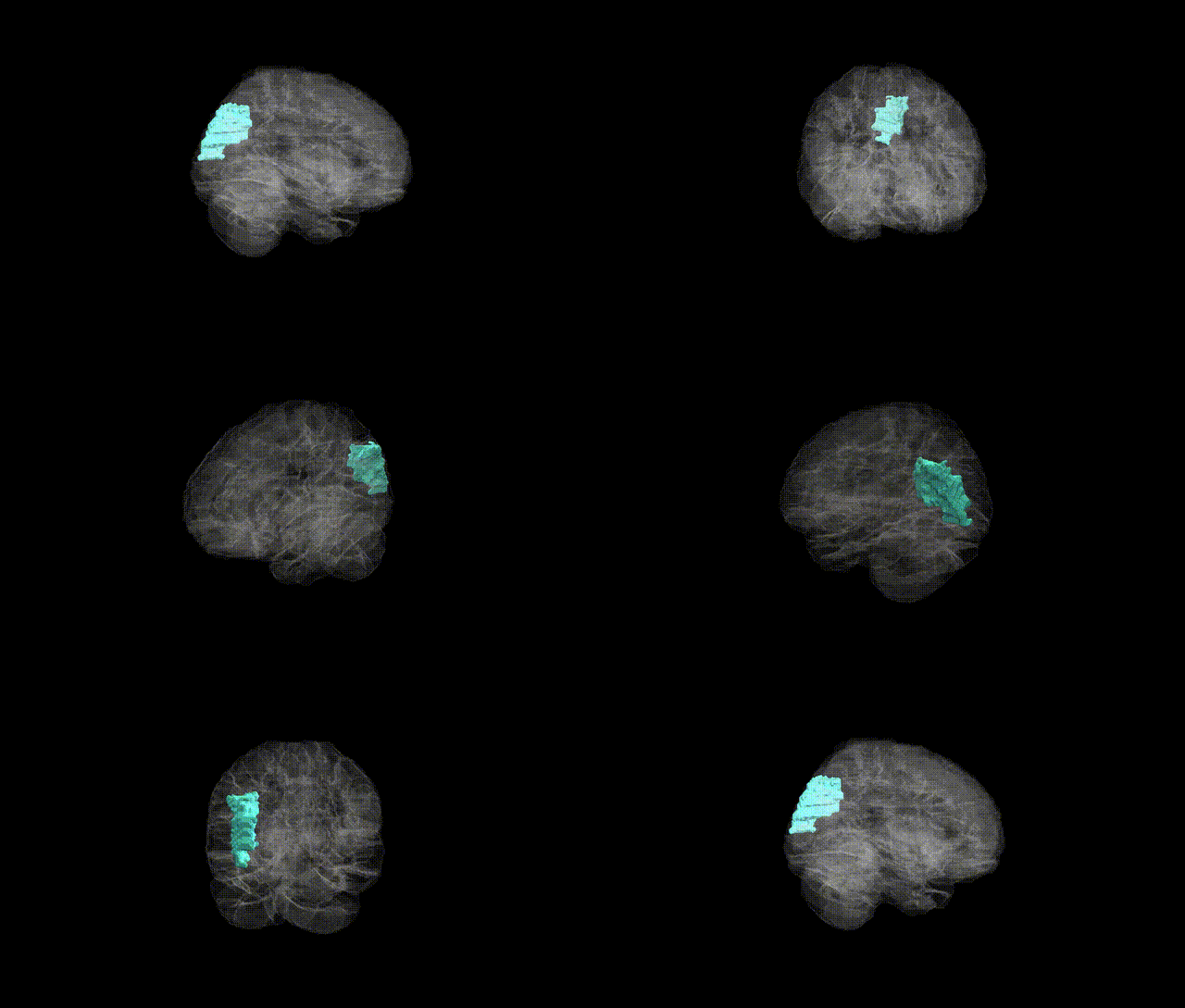
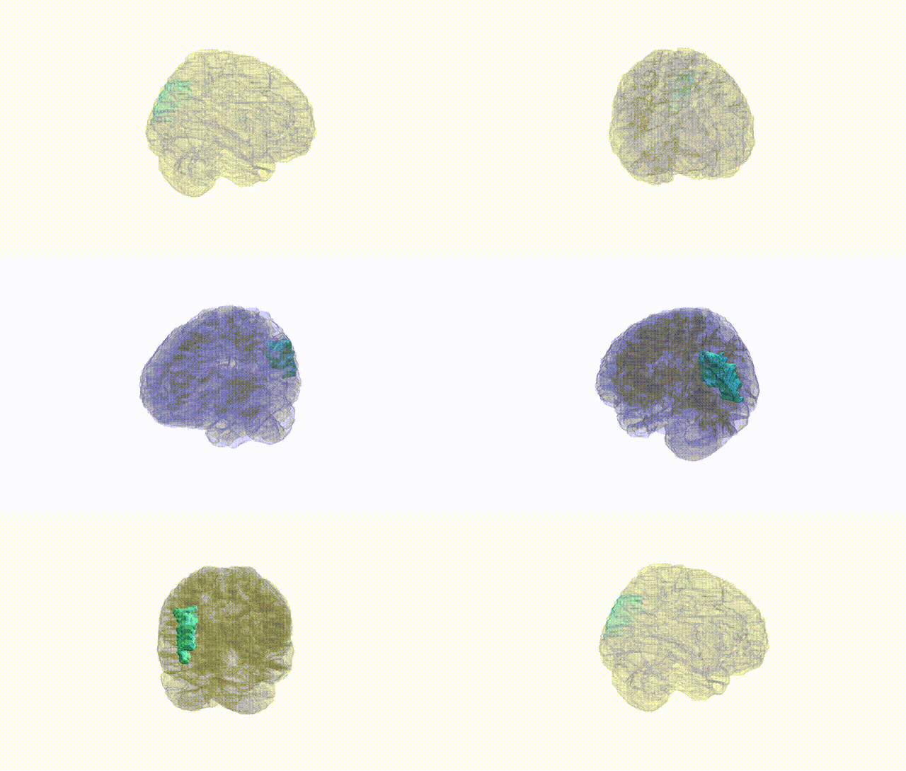
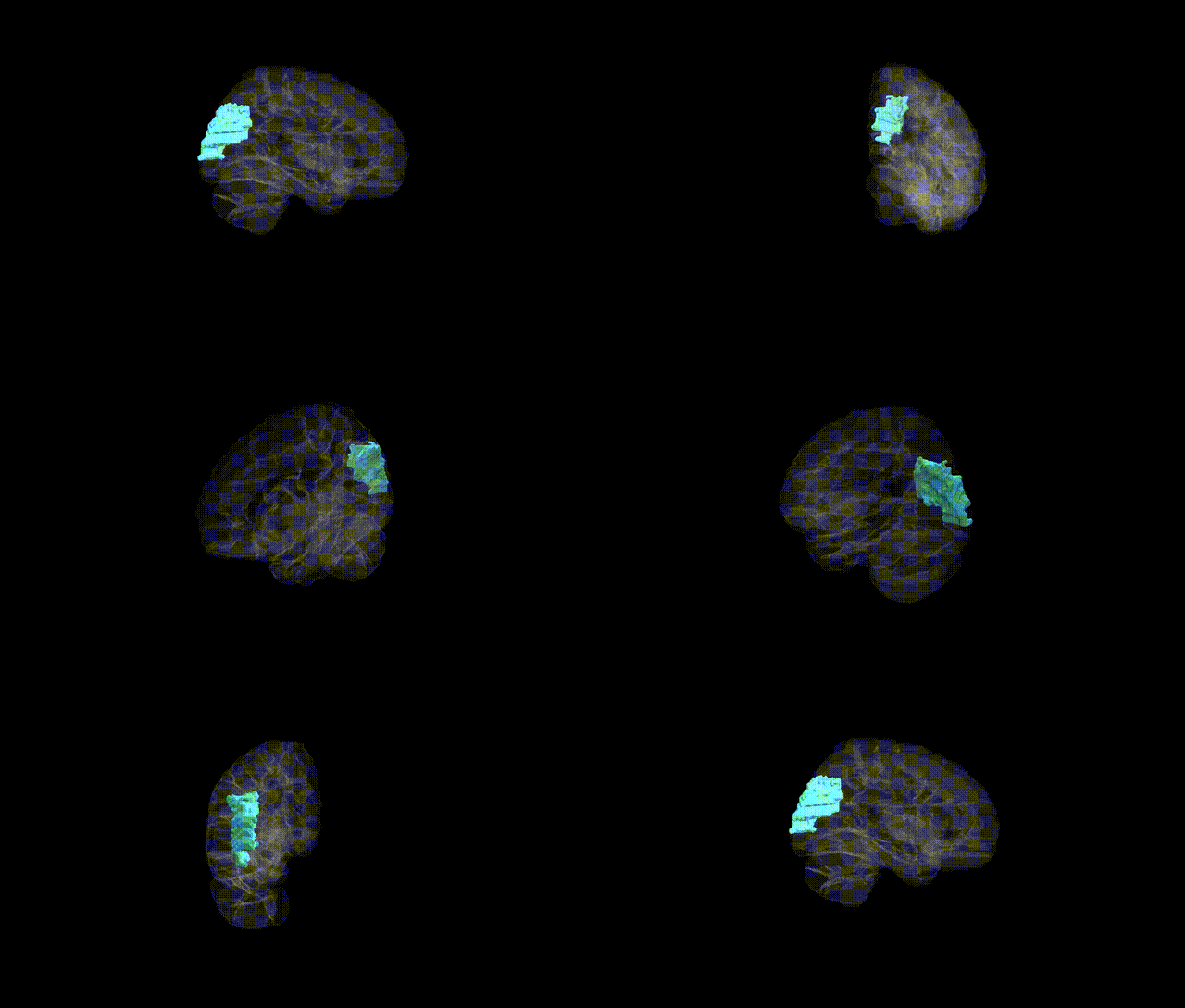
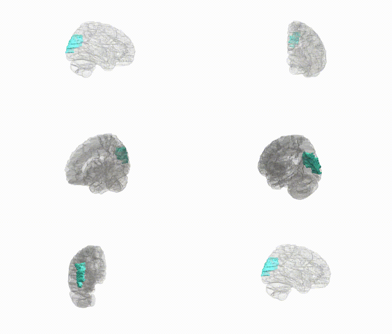
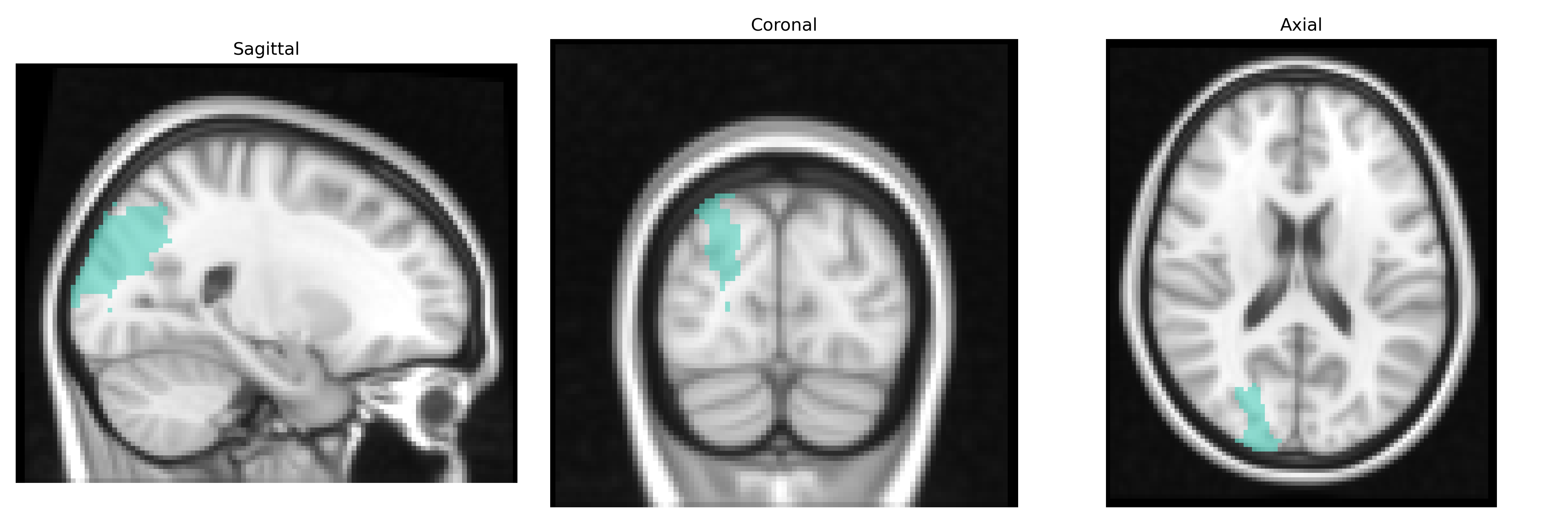
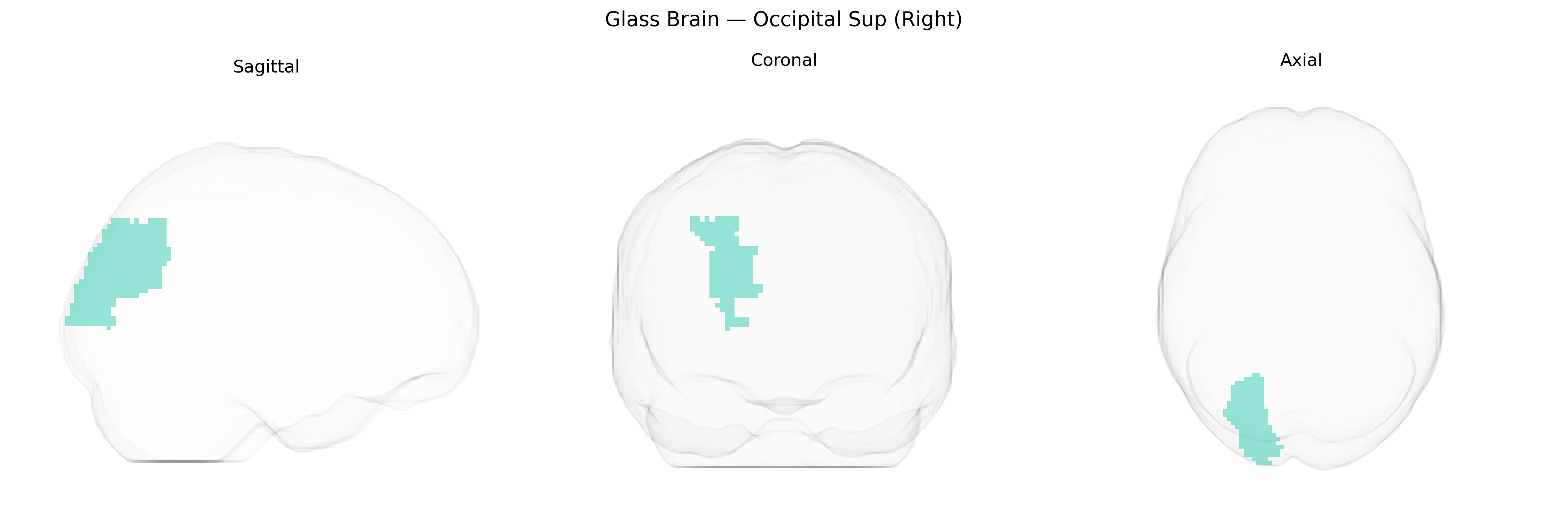
The right superior occipital gyrus (Occipital Sup Right in the AAL2 atlas) is a dorsal region of the occipital lobe located posteriorly and superiorly along the medial-lateral convexity, bordered by other occipital gyri and parietal regions. It is composed predominantly of visual association cortex (mainly Brodmann areas 19 and adjacent 18), participating in higher-order processing of visual information, including integration of motion, spatial orientation, and complex form analysis within dorsal “where/how” visual pathways. This region receives afferent input from primary and secondary visual cortices and projects to parietal and frontal regions involved in visuospatial attention, eye-movement control, and visually guided action. The right hemisphere dominance in many individuals contributes to its role in global visual processing and spatial awareness. There is no direct link for this specific gyrus; see the related structure Occipital lobe.
The right superior occipital gyrus (Occipital Sup Right in AAL2) is part of dorsal visual association cortex and, while not typically the primary focus of region-of-interest genetic studies, emerges repeatedly in imaging‑genetics and GWAS analyses of brain structure and function. Large-scale neuroimaging GWAS consortia (e.g., ENIGMA, UK Biobank) have identified common variants in genes involved in neurodevelopment, axon guidance, and synaptic signaling (such as MIR137, CNTNAP2, and genes in the 22q11.2 region) that associate with occipital cortical thickness, surface area, or functional connectivity, including in superior occipital regions. Polygenic risk for schizophrenia, autism spectrum disorder, and major depression has been linked to structural and functional alterations in occipital association cortex, with several studies reporting reduced cortical thickness or altered activation in right superior occipital areas in carriers of higher psychiatric genetic risk. Variants near or within visual pathway and myelination-related genes (e.g., NRG1/ERBB signaling components and oligodendrocyte-related loci) have been associated with white-matter microstructure affecting occipital tracts that project to superior occipital cortex, while GWAS of visual perception and reading-related traits occasionally implicate occipital association regions, including superior occipital parcels, in polygenic architectures for visuospatial and reading performance. Overall, genetic associations with this specific AAL2 parcel are typically indirect, arising from broader occipital or cortical measures, psychiatric polygenic risk, or visual-network traits rather than from region-exclusive GWAS signals.
Overview generated by GPT-4o (2026).
Region ID: 5102
Hemisphere: right
Atlas: AAL2
Full Quality Version: Download MP4
Full Quality Version: Download MP4
Full Quality Version: Download MP4
Full Quality Version: Download MP4
Wali Sidiqyar*, Gaurav Rudravaram*, Elyssa M. McMaster, Trent M. Schwartz, Adam M. Saunders, Kurt G. Schilling, Bennett A. Landman "Introducing SPINS: A Shared Public Visualization Library of Neuroanatomical Structures." Medical Imaging with Deep Learning- short paper
This resource is licensed under CC0 1.0 Universal (Public Domain).