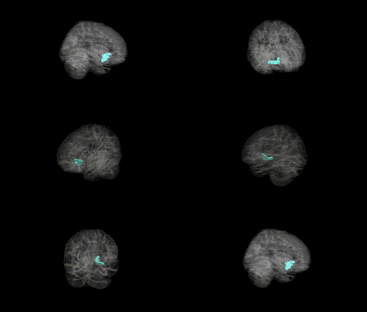
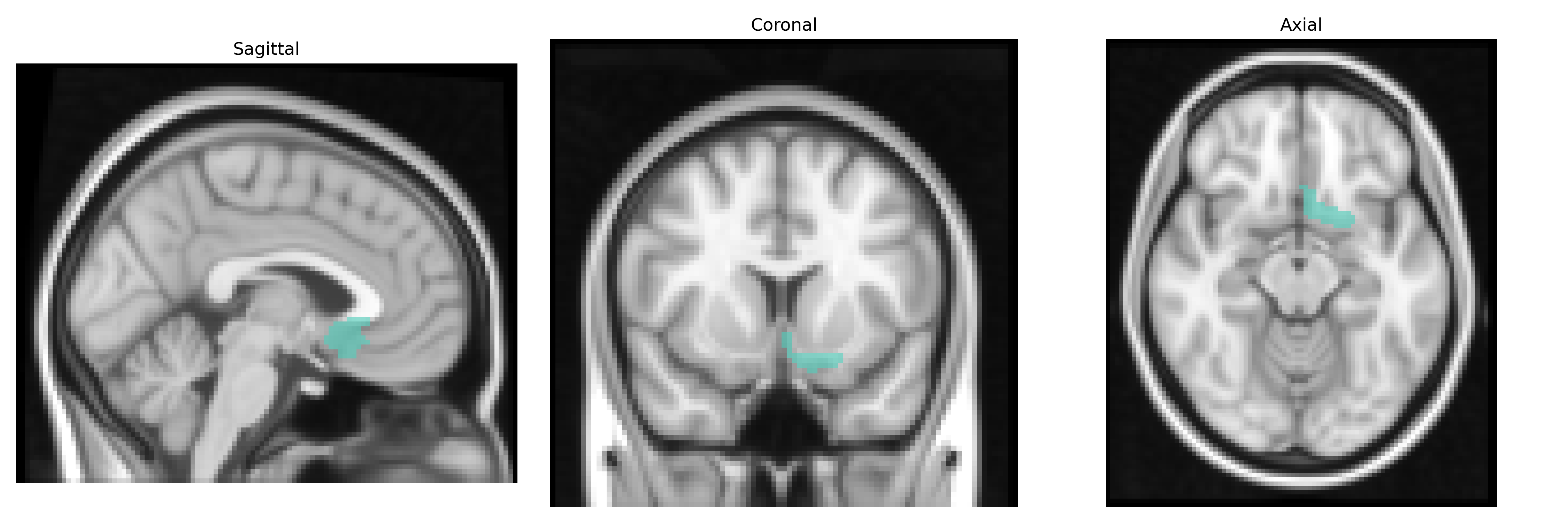
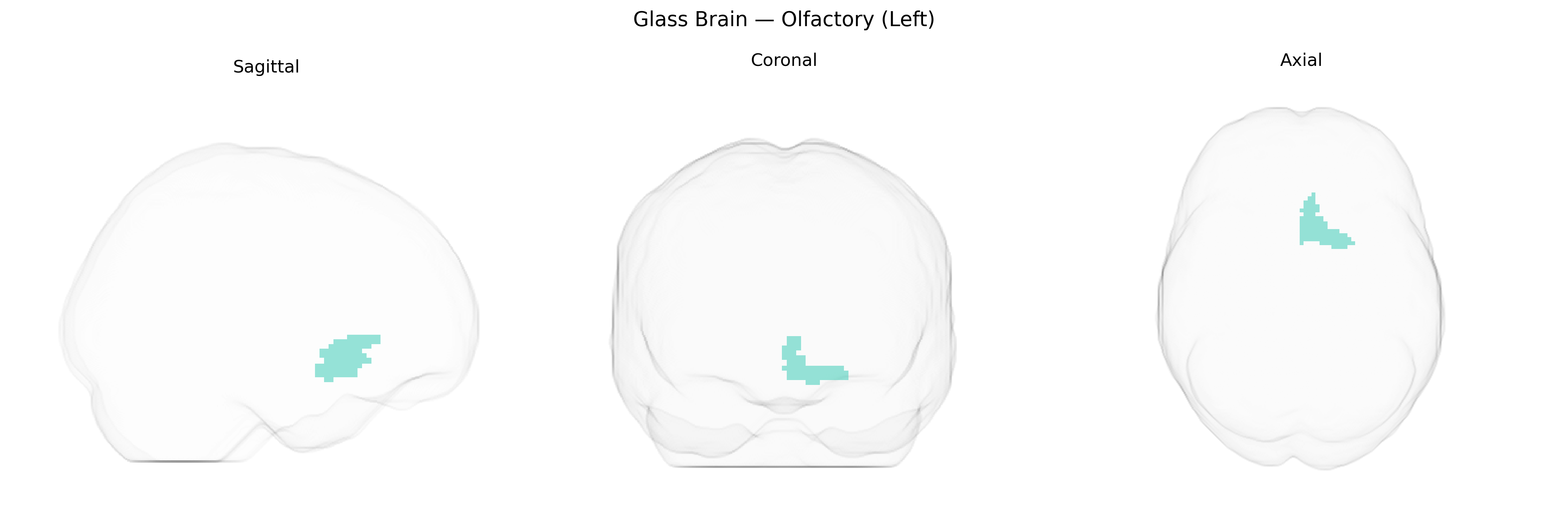
The left Olfactory (Olfactory_L) region in the AAL2 atlas corresponds to the left olfactory cortex, a primary cortical area involved in the processing of odor information received from the olfactory bulb via the olfactory tract. This region includes parts of the piriform cortex and adjacent anterior temporal and frontal areas that participate in odor detection, discrimination, and the integration of olfactory signals with emotion and memory, often through strong connections with the amygdala and hippocampus. As a key component of the limbic system, it contributes to associative learning and the affective valence of smells, influencing behaviors such as feeding, social interaction, and hazard avoidance. There is no direct Wikipedia article for “Olfactory_L” from the AAL2 atlas; a closely related structure is the Piriform cortex.
The left olfactory region, as defined in the AAL2 atlas, shows relatively limited but growing genetic association evidence, primarily through imaging genetics and olfactory-function GWAS rather than region-specific structural GWAS. Variants in genes central to olfactory signal transduction and axon guidance, including large clusters of olfactory receptor (OR) genes (e.g., OR4, OR5, and OR2 families), KAL1 (ANOS1), PROKR2, FGFR1, and other loci implicated in Kallmann syndrome and isolated congenital anosmia, are linked to altered development or function of olfactory bulbs and tracts, with imaging studies frequently demonstrating hypoplastic or absent olfactory structures predominantly on one or both sides rather than strictly lateralized effects. Common-variant GWAS of smell identification and sensitivity have repeatedly implicated OR gene clusters on chromosomes 11 and 17, as well as genes involved in synaptic function and neurodevelopment, which in turn are associated with variation in olfactory bulb/tract volume and functional activation in MRI studies. Neurodegenerative and psychiatric disorder GWAS (including those for Parkinson’s disease, Alzheimer’s disease, and schizophrenia) have identified risk loci in genes such as APOE, GBA, SNCA, and others that correlate with early olfactory dysfunction and structural or functional changes in olfactory pathways, often including asymmetric or left-dominant alterations in volumetric or connectivity measures. Overall, however, no robust, widely replicated GWAS signal is currently specific to the left olfactory AAL2 parcel; instead, genetic associations are inferred indirectly from olfactory bulb/tract imaging phenotypes, congenital or acquired anosmia, and disorders where olfactory impairment and related structural changes constitute an early or prominent feature.
Overview generated by GPT-4o (2026).
Region ID: 2501
Hemisphere: left
Atlas: AAL2
Full Quality Version: Download MP4
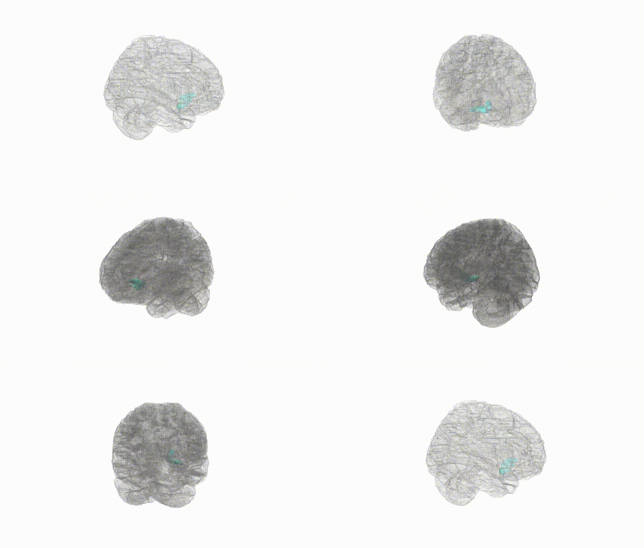
Full Quality Version: Download MP4
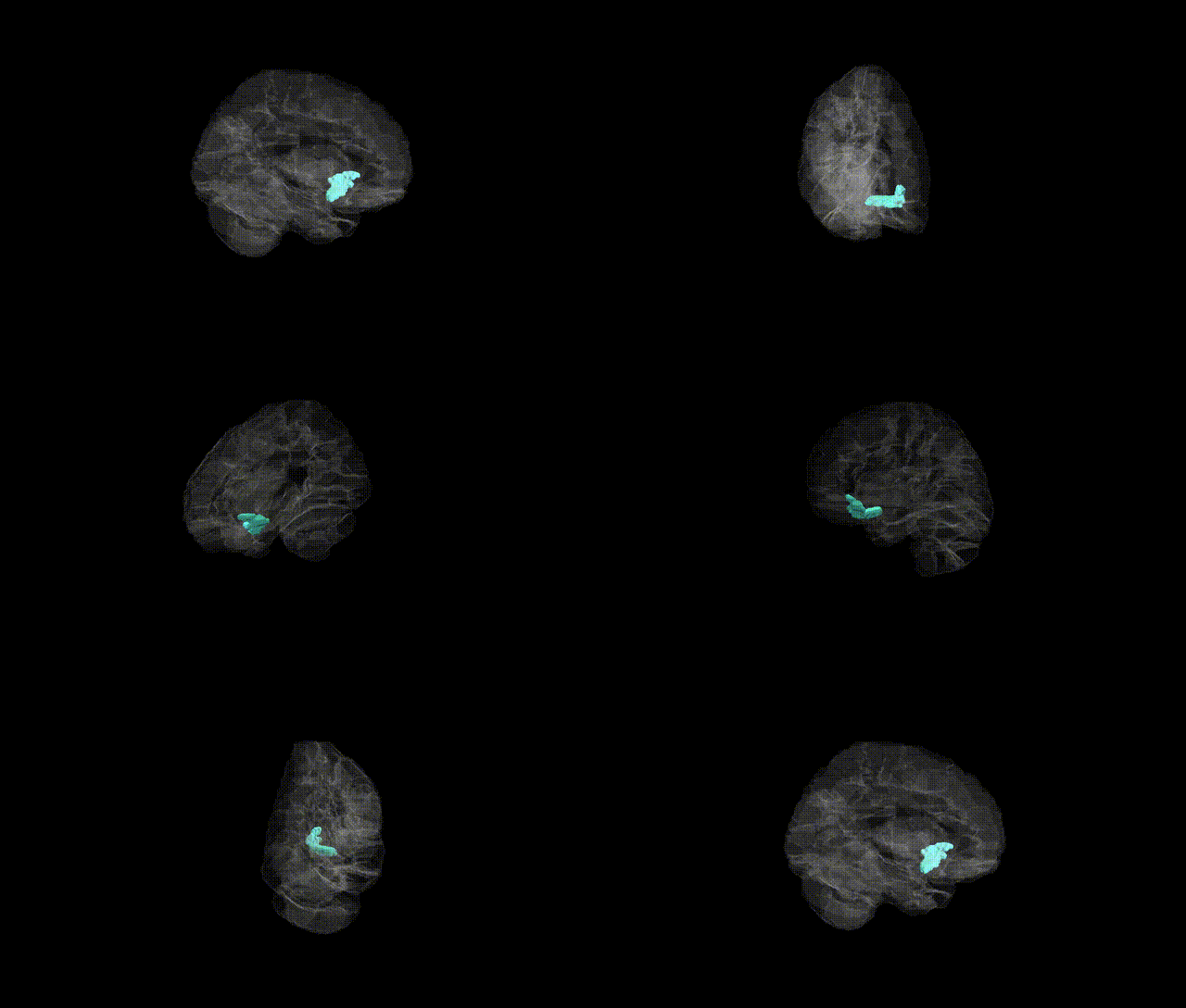
Full Quality Version: Download MP4
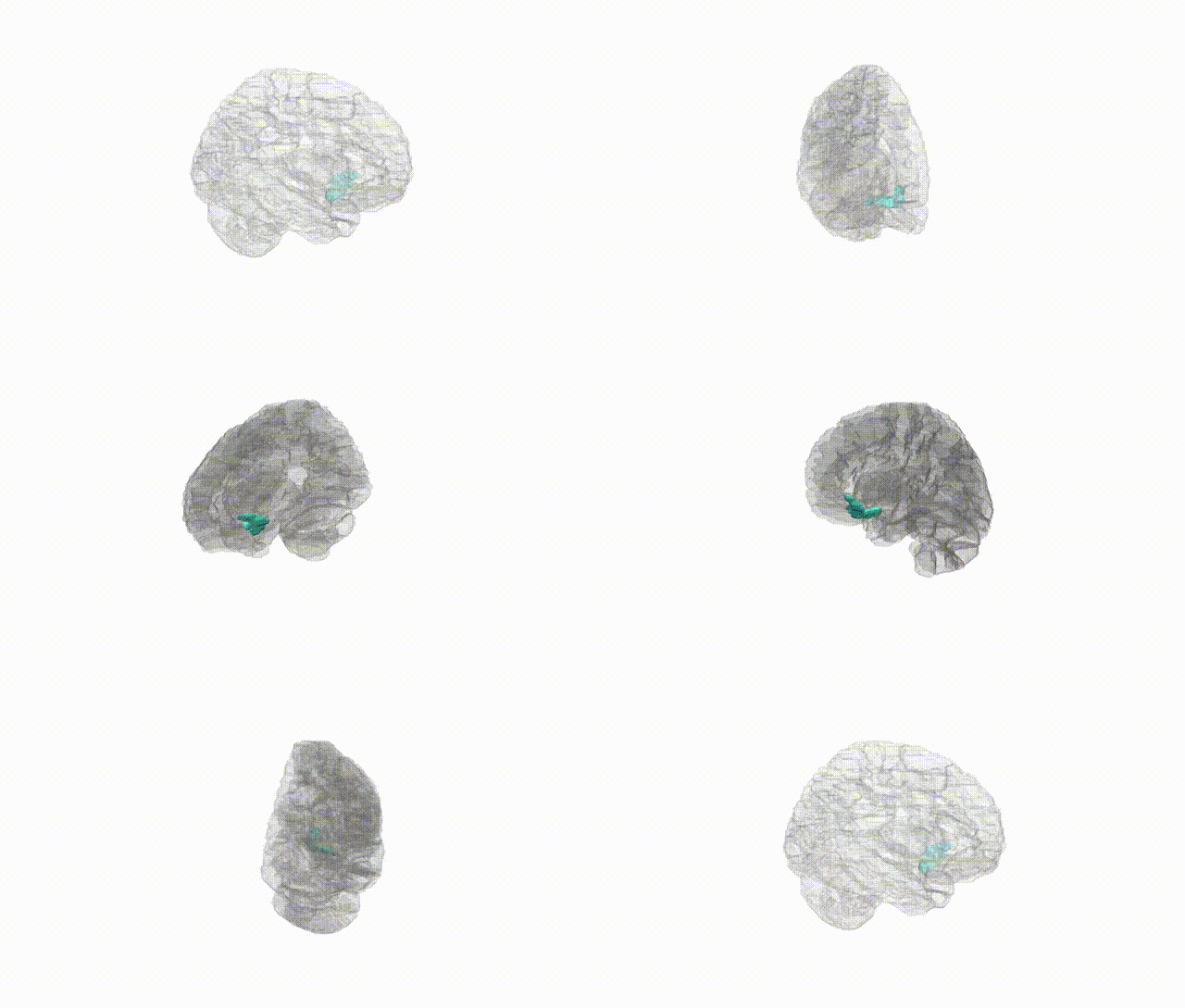
Full Quality Version: Download MP4
Wali Sidiqyar*, Gaurav Rudravaram*, Elyssa M. McMaster, Trent M. Schwartz, Adam M. Saunders, Kurt G. Schilling, Bennett A. Landman "Introducing SPINS: A Shared Public Visualization Library of Neuroanatomical Structures." Medical Imaging with Deep Learning- short paper
This resource is licensed under CC0 1.0 Universal (Public Domain).