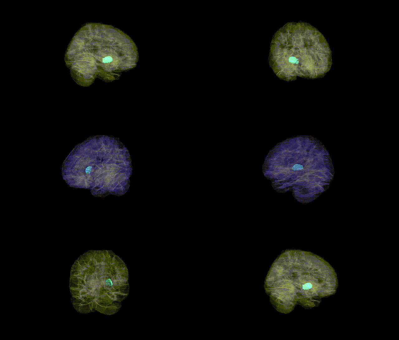
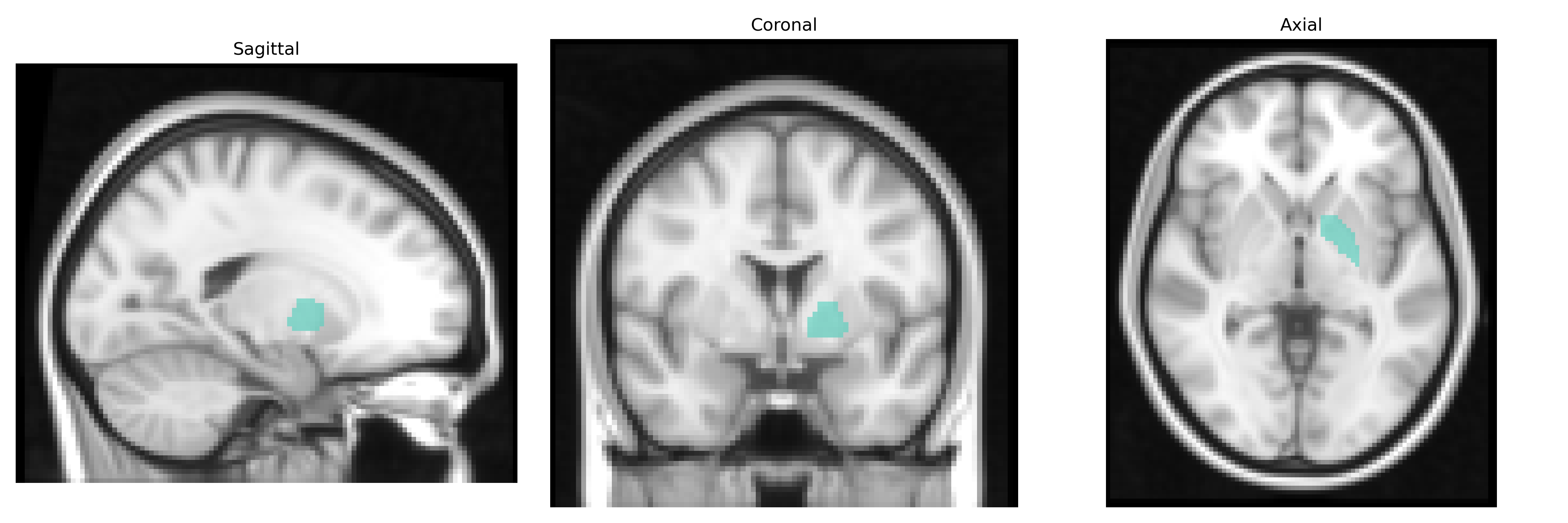
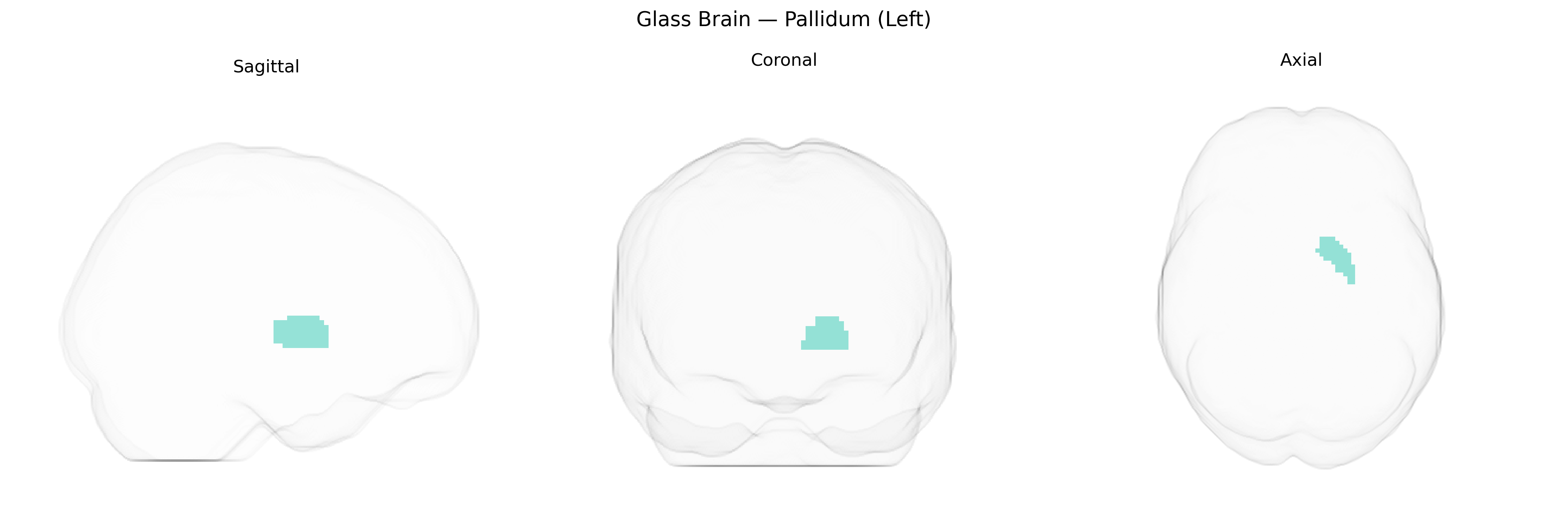
The left pallidum, corresponding to the left globus pallidus within the basal ganglia, is a subcortical gray matter structure located medial to the putamen and lateral to the internal capsule in the deep cerebral hemispheres. It consists of internal (medial) and external (lateral) segments that play a critical role in the regulation of voluntary movement through inhibitory GABAergic projections to thalamic and brainstem motor nuclei. Functionally, the pallidum participates in motor control, muscle tone regulation, and the integration of cortical inputs via striatal pathways, and is implicated in movement disorders such as Parkinson’s disease and dystonia when its circuitry is disrupted. As part of the extrapyramidal system, it contributes to the fine-tuning and smoothing of motor commands generated by cortical motor areas. Globus pallidus
The left pallidum (part of the basal ganglia and often labeled as the left globus pallidus in imaging genetics) has been implicated in multiple genome-wide association studies of subcortical brain volumes, most prominently through large ENIGMA and UK Biobank analyses identifying common variants in or near genes involved in neurodevelopment, synaptic function, and dopamine signaling (e.g., putamen/pallidum–associated loci near DRD2, DCC, and genes regulating axon guidance and neuronal migration). Pallidal volume is moderately heritable, and polygenic influences overlap with risk for Parkinson’s disease, schizophrenia, and major depressive disorder, consistent with the pallidum’s role in motor and motivational circuits; several GWAS have shown that alleles increasing risk for these disorders are also associated with altered pallidal structure or connectivity. Additional associations link left pallidum volume or activity to genetic liability for obsessive–compulsive disorder, Tourette syndrome, and attention-deficit/hyperactivity disorder, reflecting basal ganglia involvement in habit formation and inhibitory control. Imaging–genetics studies further indicate that dopaminergic and glutamatergic pathway variants, as well as genes affecting iron metabolism and myelination, may contribute to individual differences in left pallidum morphology and function, though specific loci remain less consistently replicated than for some other subcortical nuclei.
Overview generated by GPT-4o (2026).
Region ID: 7021
Hemisphere: left
Atlas: AAL2
Full Quality Version: Download MP4
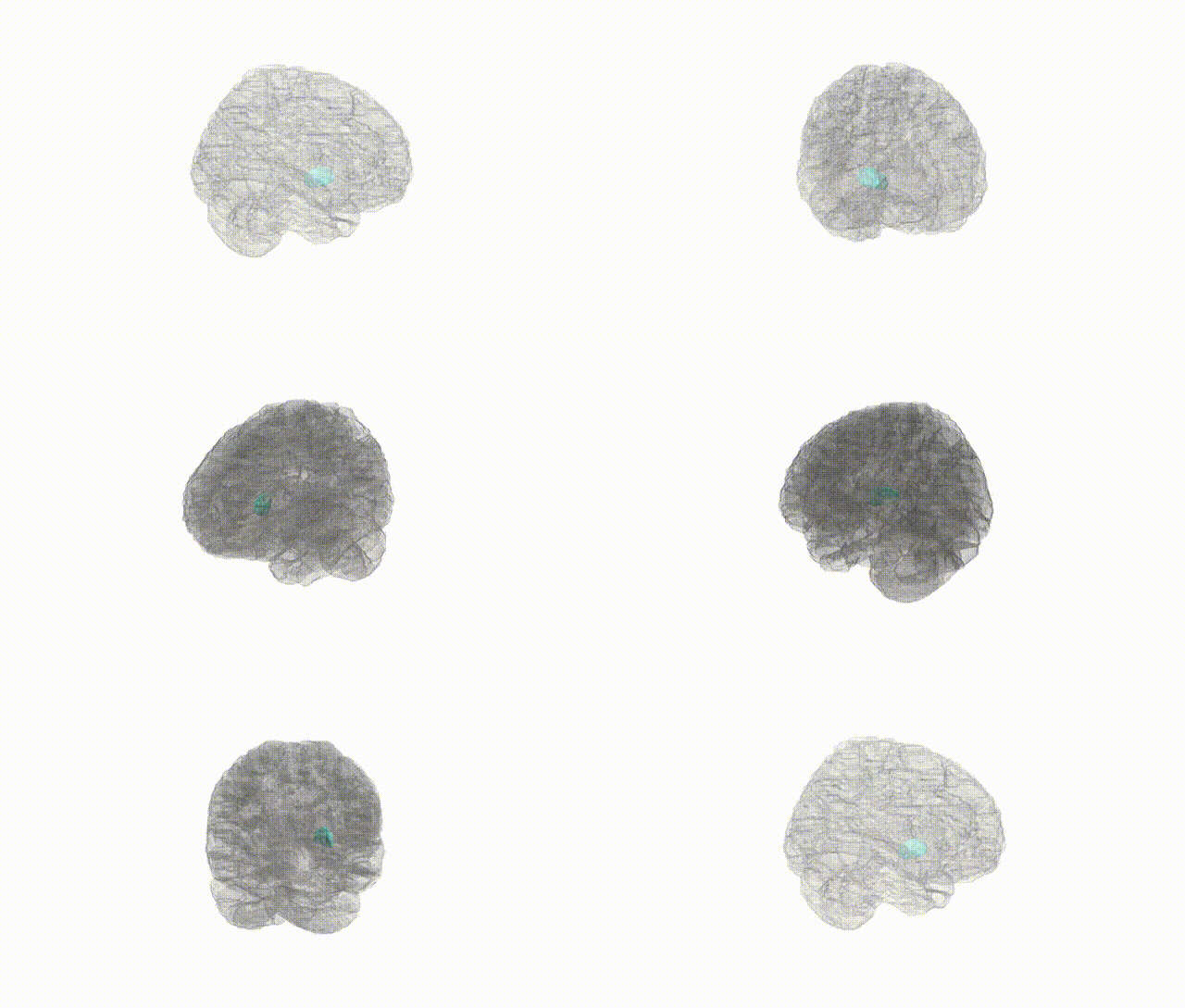
Full Quality Version: Download MP4
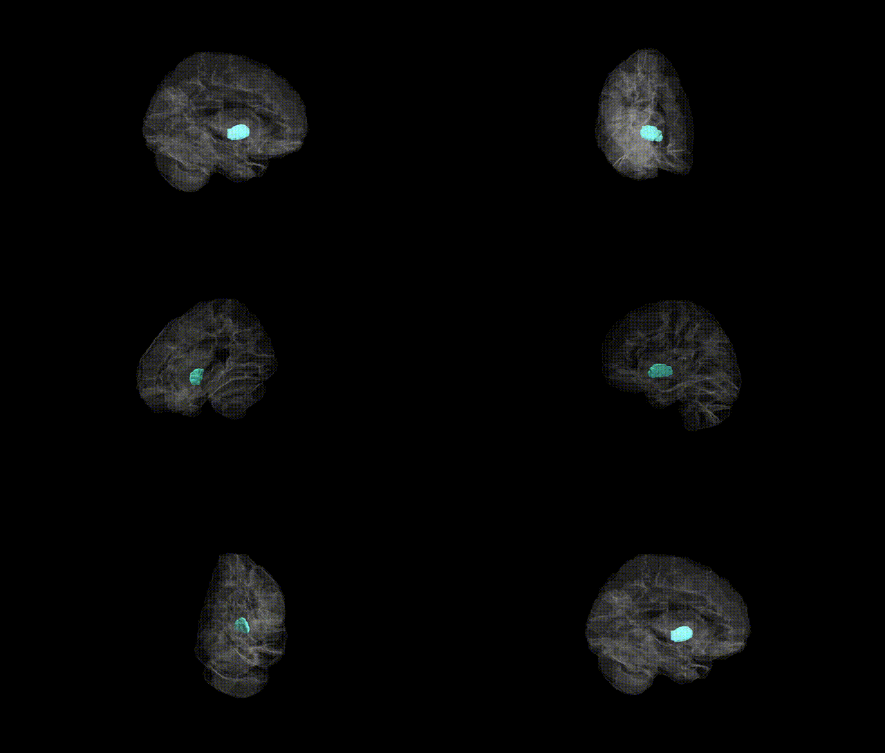
Full Quality Version: Download MP4
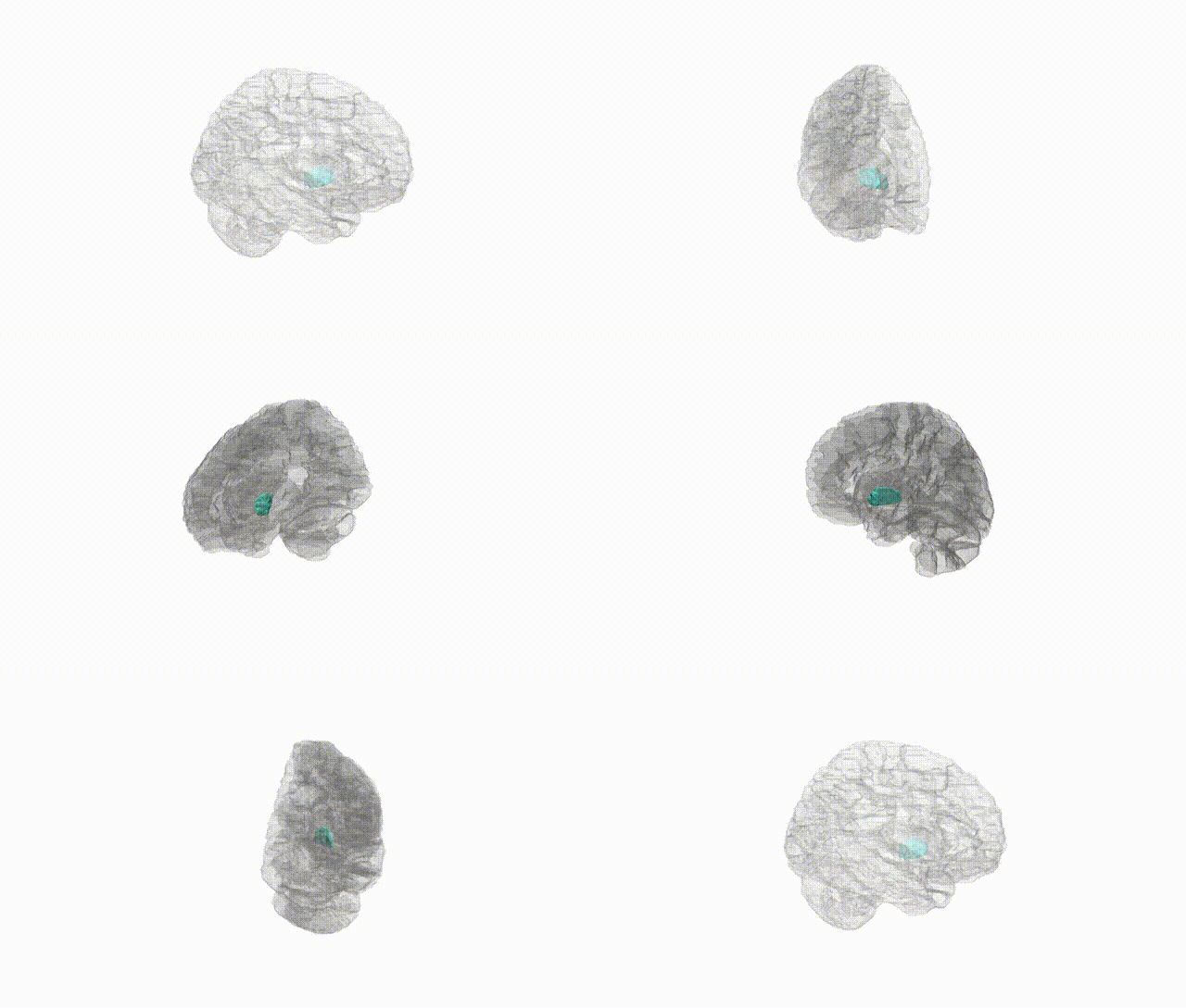
Full Quality Version: Download MP4
Wali Sidiqyar*, Gaurav Rudravaram*, Elyssa M. McMaster, Trent M. Schwartz, Adam M. Saunders, Kurt G. Schilling, Bennett A. Landman "Introducing SPINS: A Shared Public Visualization Library of Neuroanatomical Structures." Medical Imaging with Deep Learning- short paper
This resource is licensed under CC0 1.0 Universal (Public Domain).