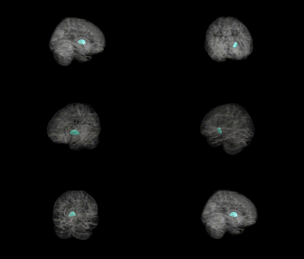
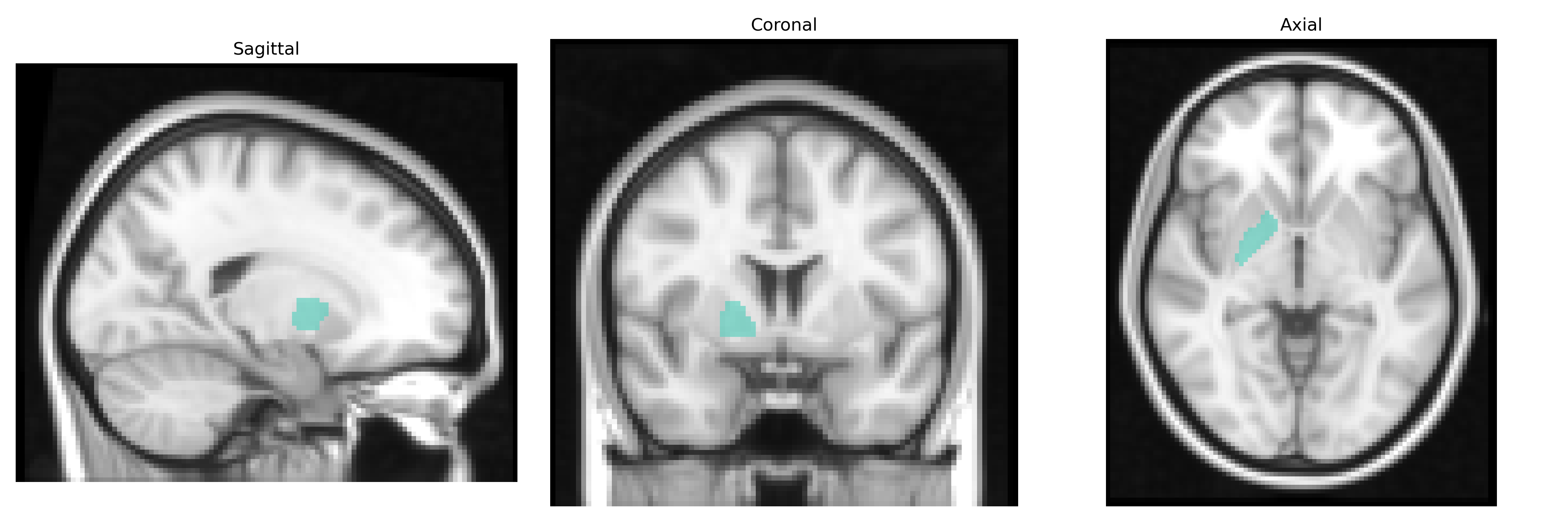
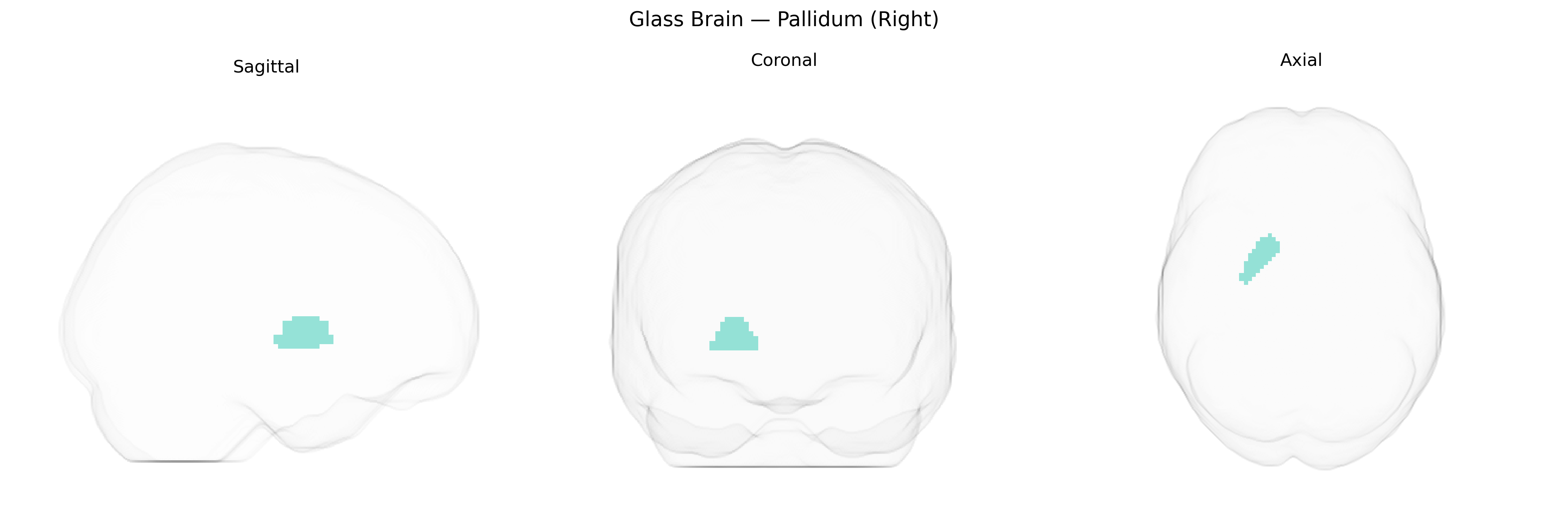
The right pallidum, part of the basal ganglia, is a subcortical gray matter structure located in the medial portion of the temporal lobe’s deep telencephalic region, comprising primarily the globus pallidus. It plays a critical role in the regulation of voluntary movement through its position in the indirect and direct pathways of motor control, integrating inhibitory GABAergic signals and modulating thalamocortical activity. The pallidum participates in motor planning, muscle tone regulation, and the suppression of unwanted movements, and is also implicated in cognitive and limbic circuits affecting motivation and behavioral regulation. Pathophysiological alterations of the pallidum are associated with movement disorders such as Parkinson’s disease and dystonia, as well as certain neuropsychiatric conditions. There is no direct link for “right pallidum”; see the related structure Globus pallidus.
Genetic associations involving the right pallidum (right globus pallidus) from AAL2-based and related neuroimaging-genetics studies suggest that individual differences in its volume and function are moderately heritable and linked to multiple common variants implicated in basal ganglia circuitry, dopaminergic signaling, and neurodevelopment. Large-scale GWAS of subcortical volumes (e.g., ENIGMA, UK Biobank) have identified loci near or within genes such as DRD2, MAOB, BCL2L1, and others that influence pallidal volume bilaterally, with some effects showing lateralization, including on the right side. These variants often overlap with genes implicated in Parkinson’s disease, schizophrenia, major depressive disorder, and obsessive-compulsive disorder, in which structural and functional abnormalities of the right pallidum have been reported. Polygenic risk scores for schizophrenia, bipolar disorder, ADHD, and depression have shown associations with altered right pallidal volume or activity, reflecting shared genetic architectures between basal ganglia morphology and psychiatric/neurological risk. Additional GWAS and candidate-gene studies link right pallidum structure and connectivity to traits such as cognitive performance, motor control, reward processing, and impulsivity, often via genes involved in synaptic plasticity, axonal guidance, and glutamatergic and GABAergic signaling, underscoring the region’s role as a genetically influenced hub in cortico-striato-pallido-thalamo-cortical loops.
Overview generated by GPT-4o (2026).
Region ID: 7022
Hemisphere: right
Atlas: AAL2
Full Quality Version: Download MP4
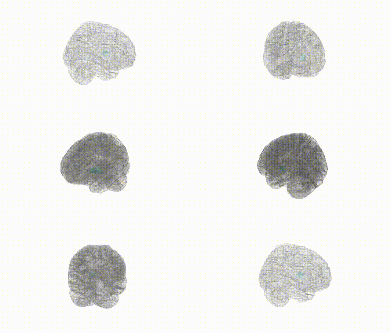
Full Quality Version: Download MP4
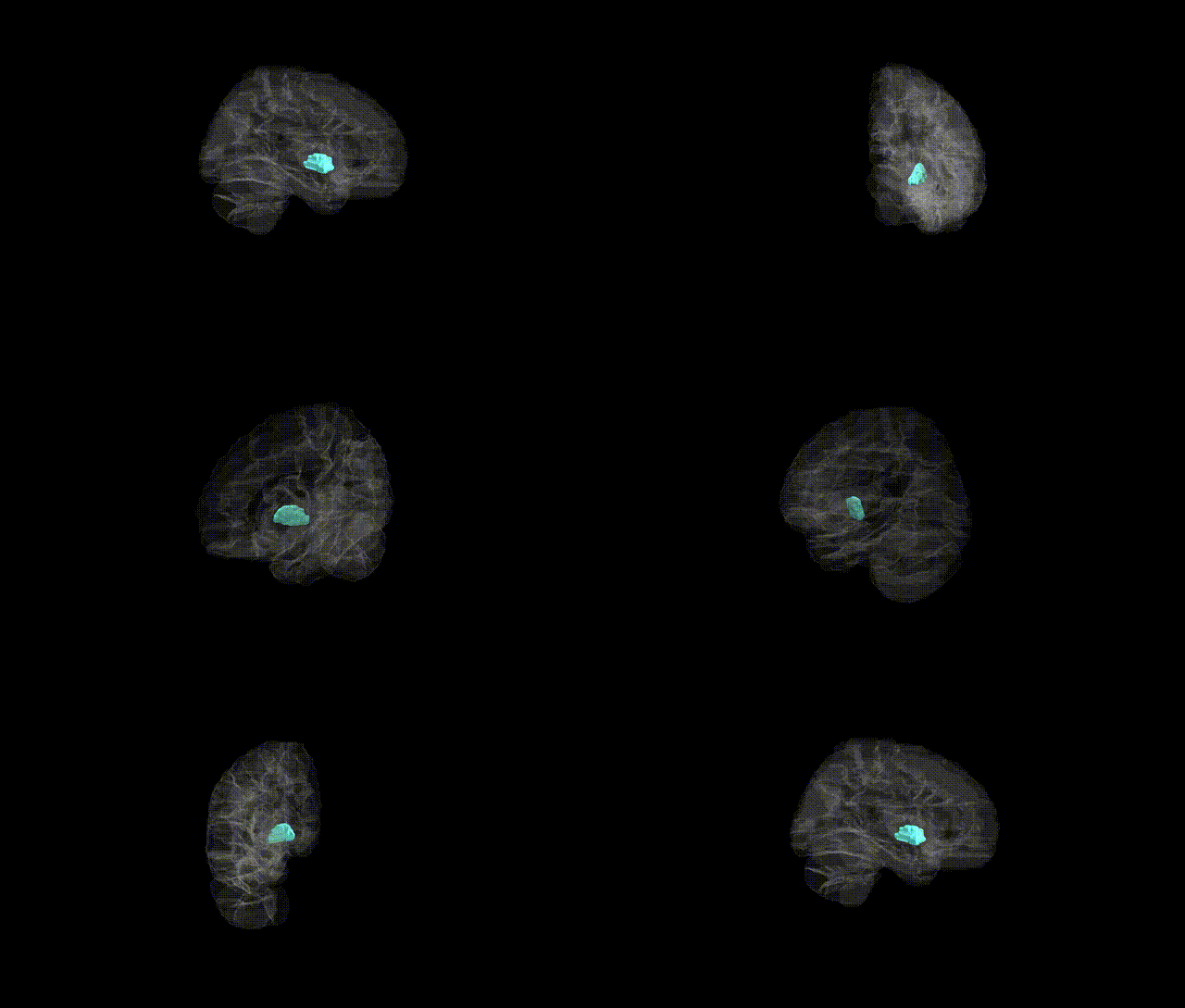
Full Quality Version: Download MP4
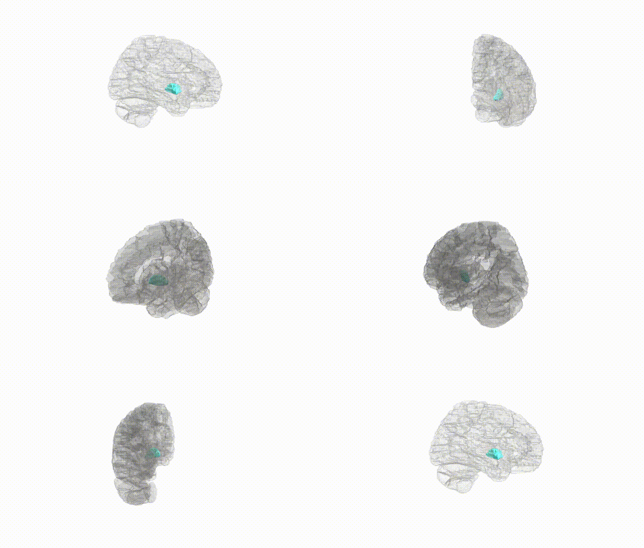
Full Quality Version: Download MP4
Wali Sidiqyar*, Gaurav Rudravaram*, Elyssa M. McMaster, Trent M. Schwartz, Adam M. Saunders, Kurt G. Schilling, Bennett A. Landman "Introducing SPINS: A Shared Public Visualization Library of Neuroanatomical Structures." Medical Imaging with Deep Learning- short paper
This resource is licensed under CC0 1.0 Universal (Public Domain).