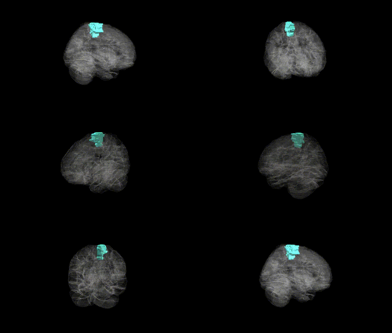
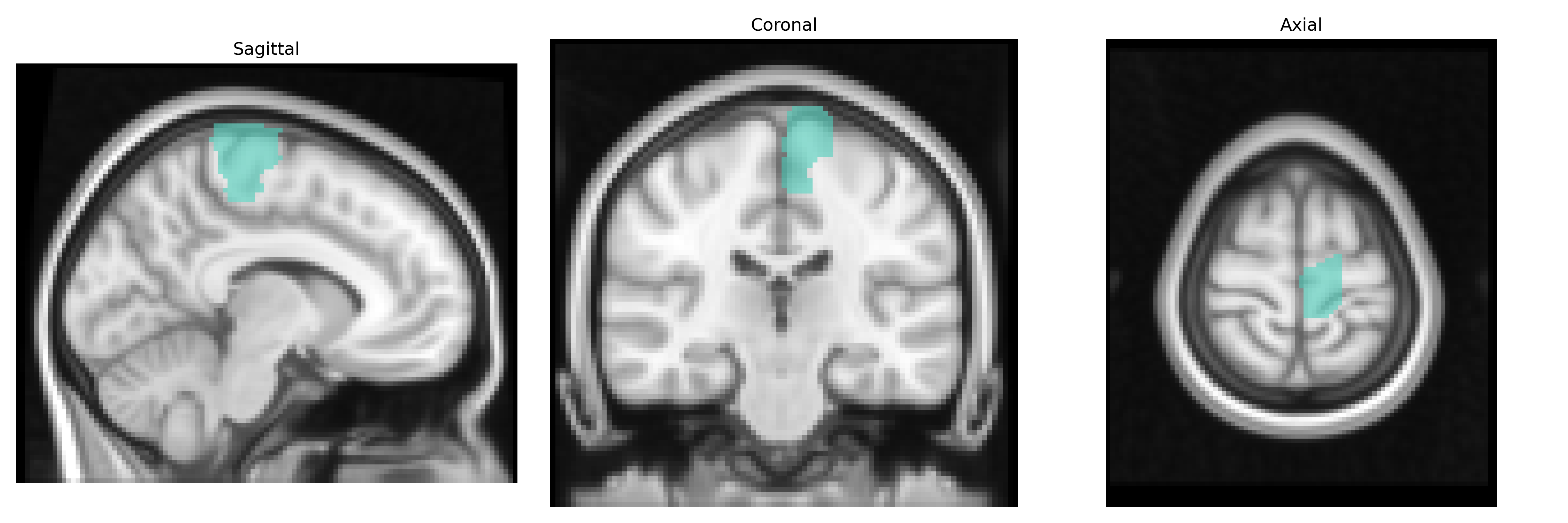
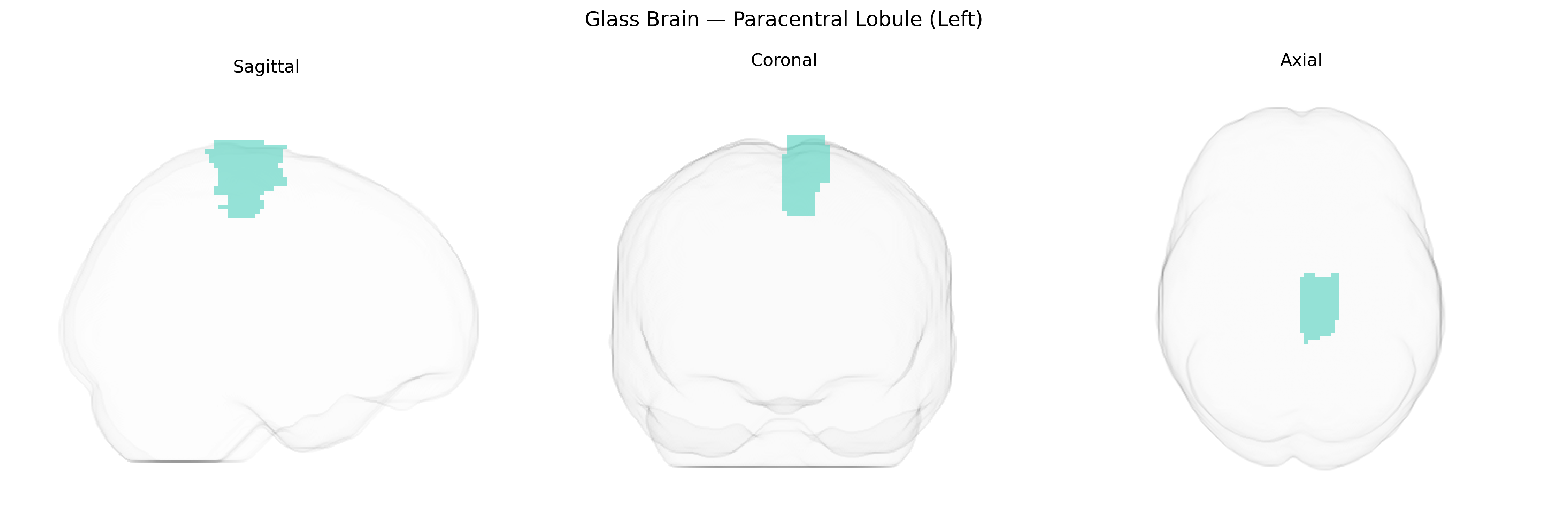
The left paracentral lobule is a medial cortical region located on the dorsal surface of the cerebral hemisphere, bridging the posterior portion of the superior frontal gyrus and the anterior segment of the precuneus around the central sulcus. It encompasses parts of the primary motor and primary somatosensory cortices corresponding predominantly to the lower limb and perineal representations, and is critically involved in voluntary motor control, somatosensory processing, and sphincter regulation for the contralateral leg and pelvic floor. Vascular supply arises mainly from branches of the anterior cerebral artery, and its white matter connections integrate with motor, premotor, and somatosensory networks. In the AAL2 atlas, the left paracentral lobule is defined as a distinct anatomical region based on gyral and sulcal landmarks within the medial parietal–frontal transition zone. There is no dedicated Wikipedia article for the left paracentral lobule; a closely related structure is the Paracentral lobule.
The left paracentral lobule, encompassing medial portions of the primary motor and somatosensory cortex for lower extremities, has been implicated in several genetic and GWAS-based associations, though typically as part of broader sensorimotor or cortical-structure analyses rather than as an isolated AAL2-defined region. Large neuroimaging GWAS consortia such as ENIGMA and UK Biobank have identified multiple common variants influencing regional cortical thickness, surface area, and gyrification in medial sensorimotor regions overlapping the paracentral lobule, with loci often near genes involved in neurodevelopment, synaptic function, and axon guidance (e.g., variants in or near genes like MAPT, FOXP1, and CADM2 in motor-related and cortical-structure GWAS). Structural and functional alterations in paracentral areas have shown genetic correlations with polygenic risk scores for motor neuron disease and multiple sclerosis, consistent with the region’s role in motor and sensory processing of the lower limbs. In Parkinson’s disease and hereditary spastic paraplegia, imaging-genetic studies report genotype-dependent differences in activation or connectivity involving the paracentral lobule during motor tasks. Moreover, GWAS of traits such as gait speed, balance, and physical activity sometimes report associated variants whose imaging follow-up studies highlight sensorimotor cortex involvement, including the paracentral lobule, suggesting a partial mediating role of this region in genetically influenced motor and somatosensory phenotypes. Overall, while few studies focus exclusively on the “left paracentral lobule” as labeled in AAL2, convergent genetic and imaging evidence links its structure and function to inherited variation in motor control, lower-limb sensorimotor function, and neurodegenerative or demyelinating disorders affecting these systems.
Overview generated by GPT-4o (2026).
Region ID: 6401
Hemisphere: left
Atlas: AAL2
Full Quality Version: Download MP4
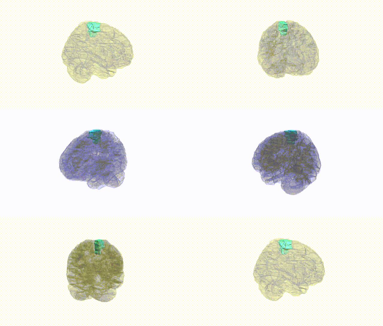
Full Quality Version: Download MP4
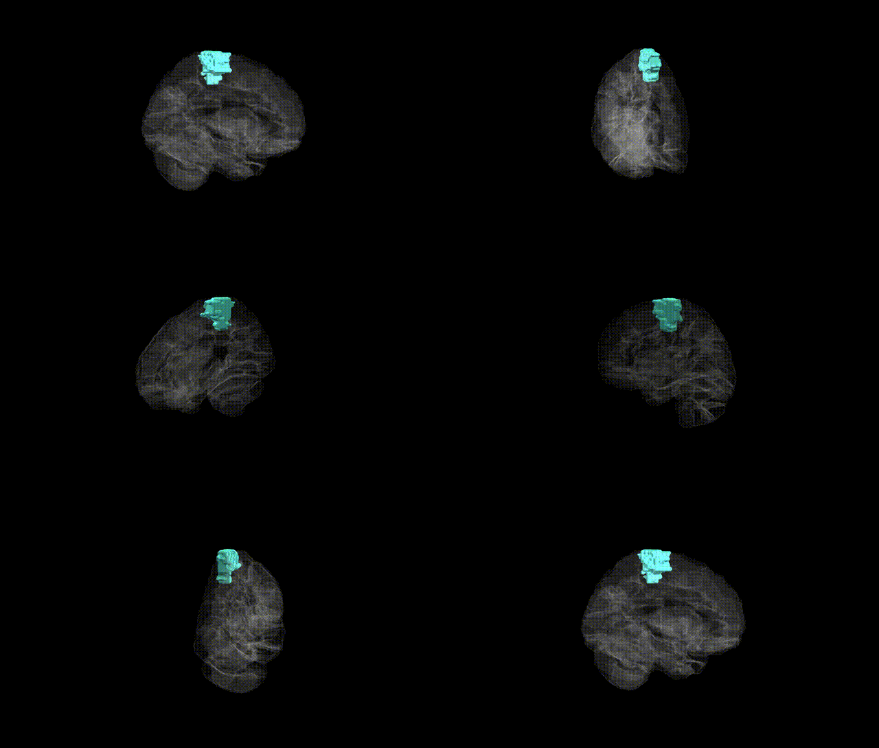
Full Quality Version: Download MP4
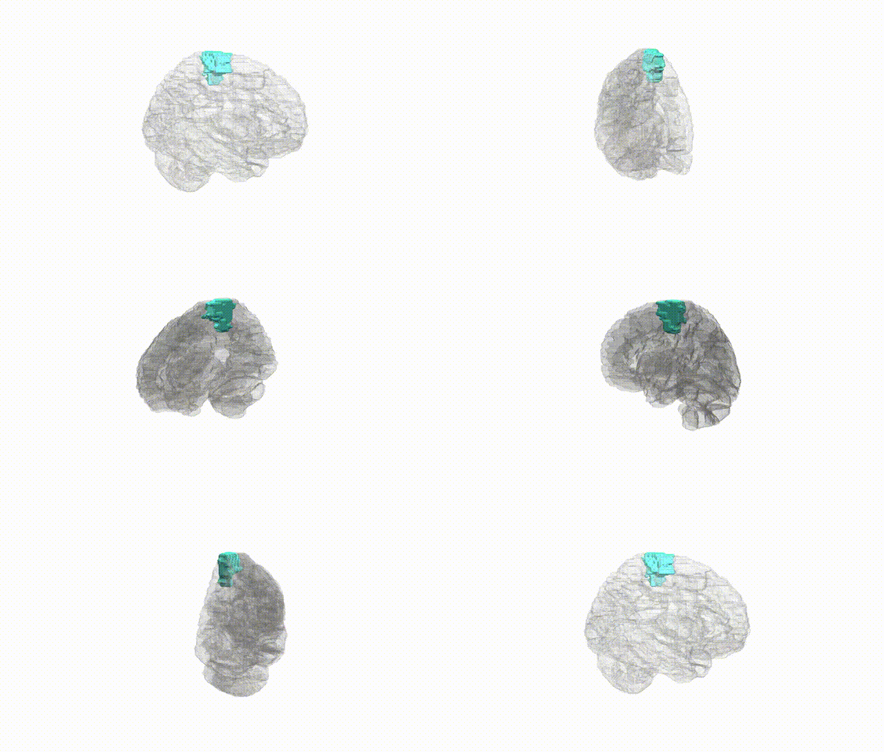
Full Quality Version: Download MP4
Wali Sidiqyar*, Gaurav Rudravaram*, Elyssa M. McMaster, Trent M. Schwartz, Adam M. Saunders, Kurt G. Schilling, Bennett A. Landman "Introducing SPINS: A Shared Public Visualization Library of Neuroanatomical Structures." Medical Imaging with Deep Learning- short paper
This resource is licensed under CC0 1.0 Universal (Public Domain).