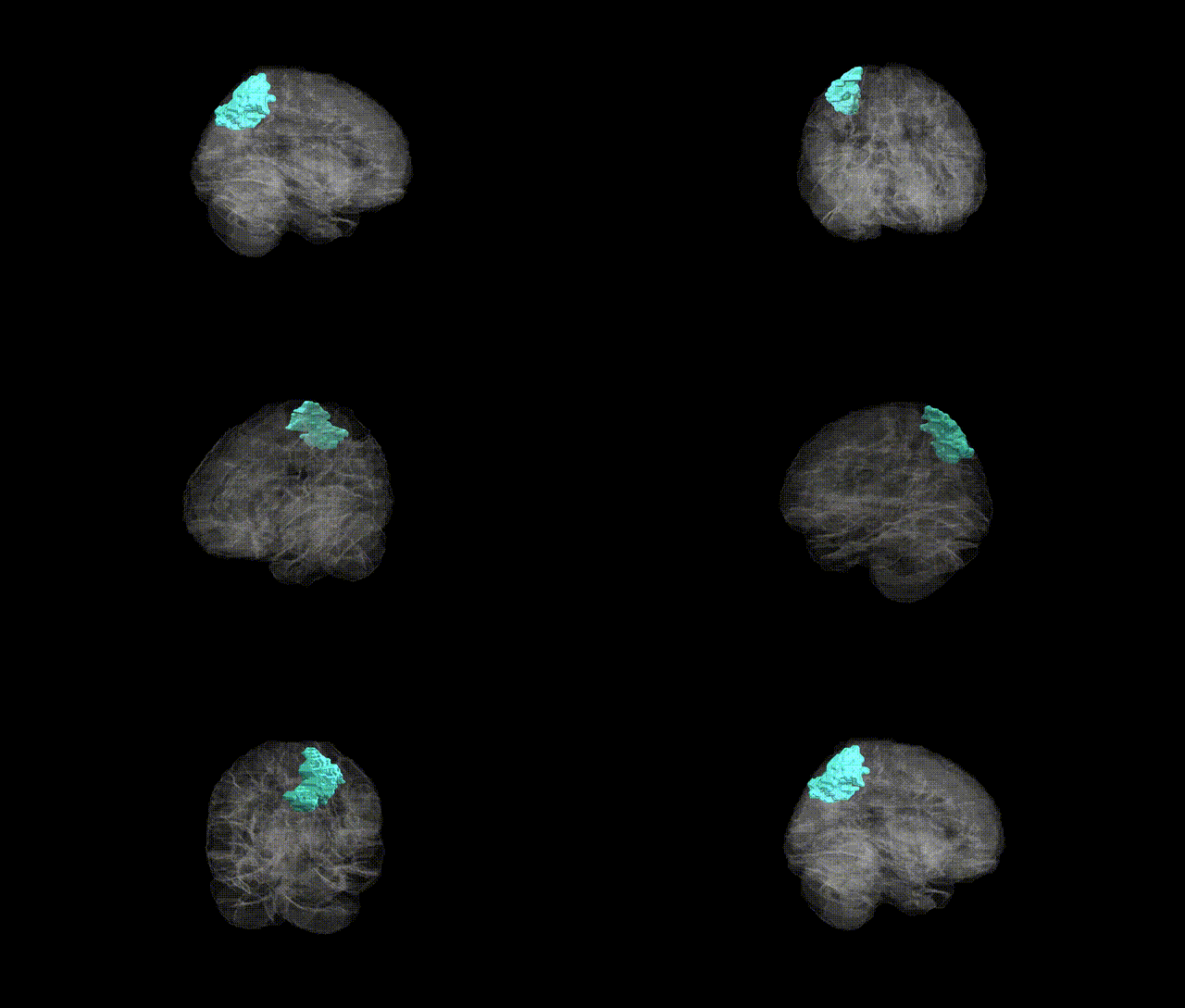
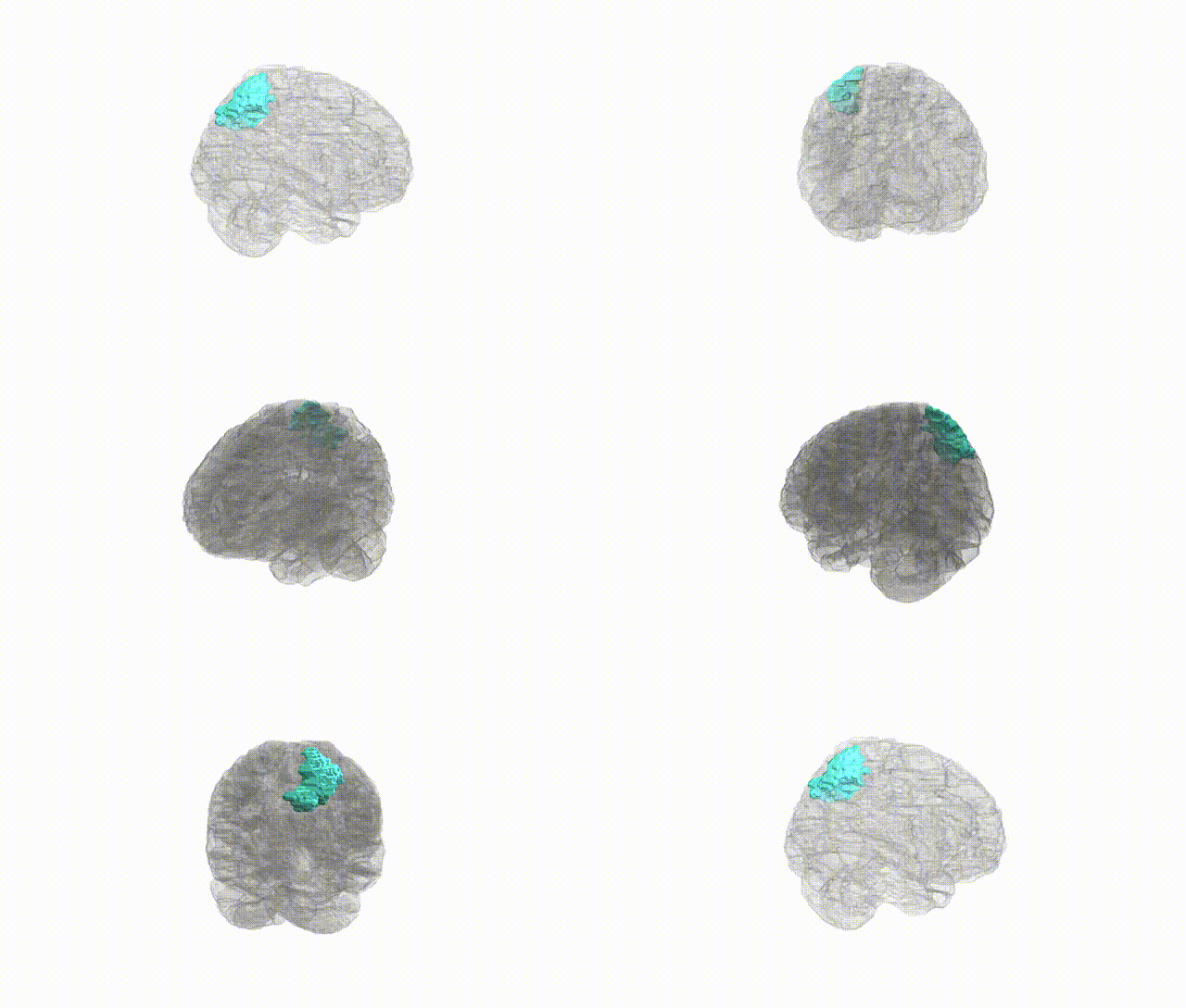
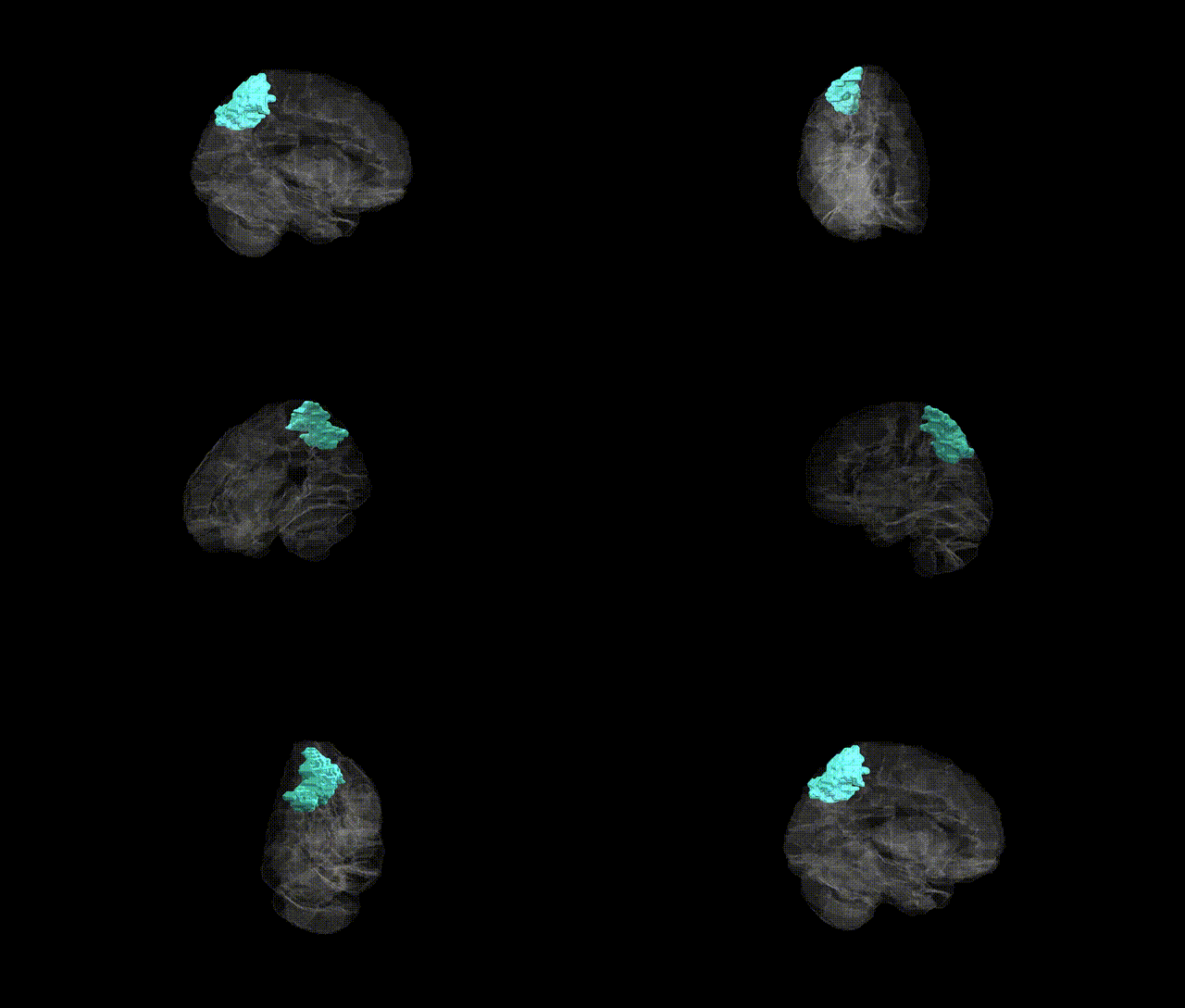
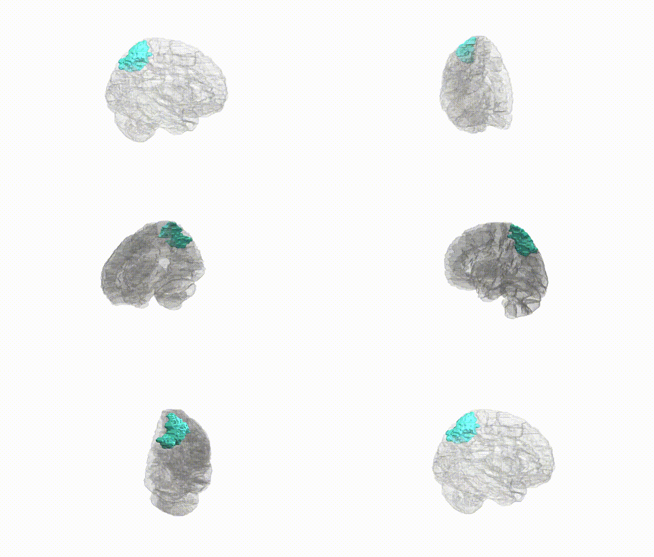
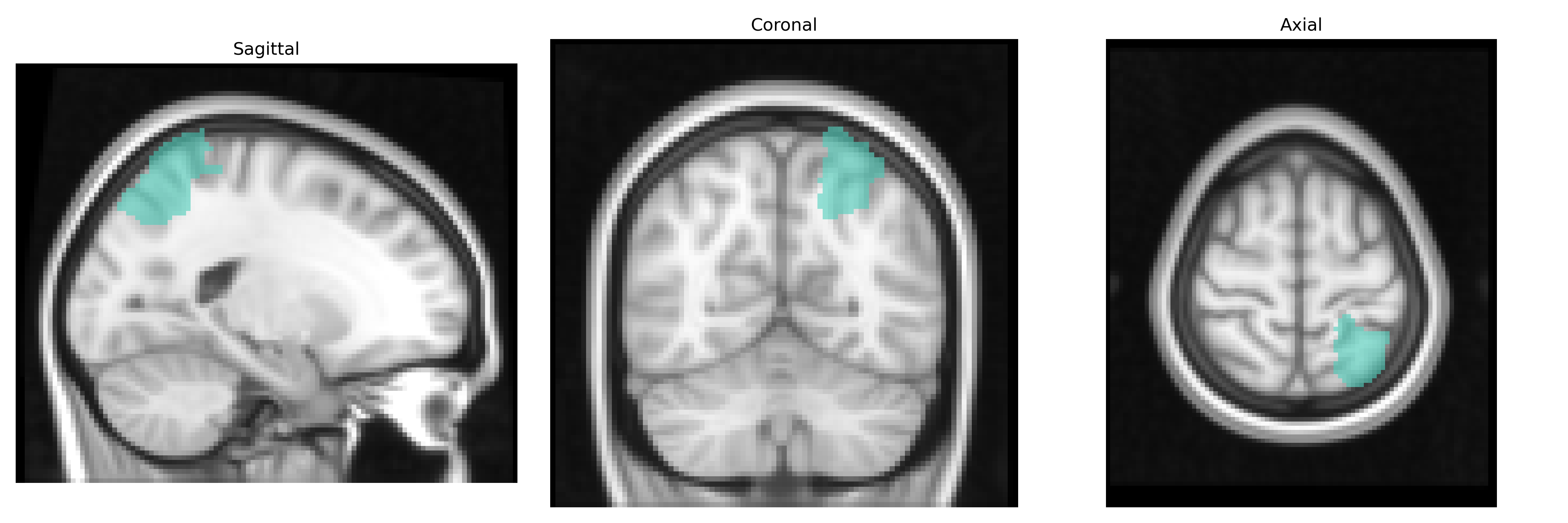
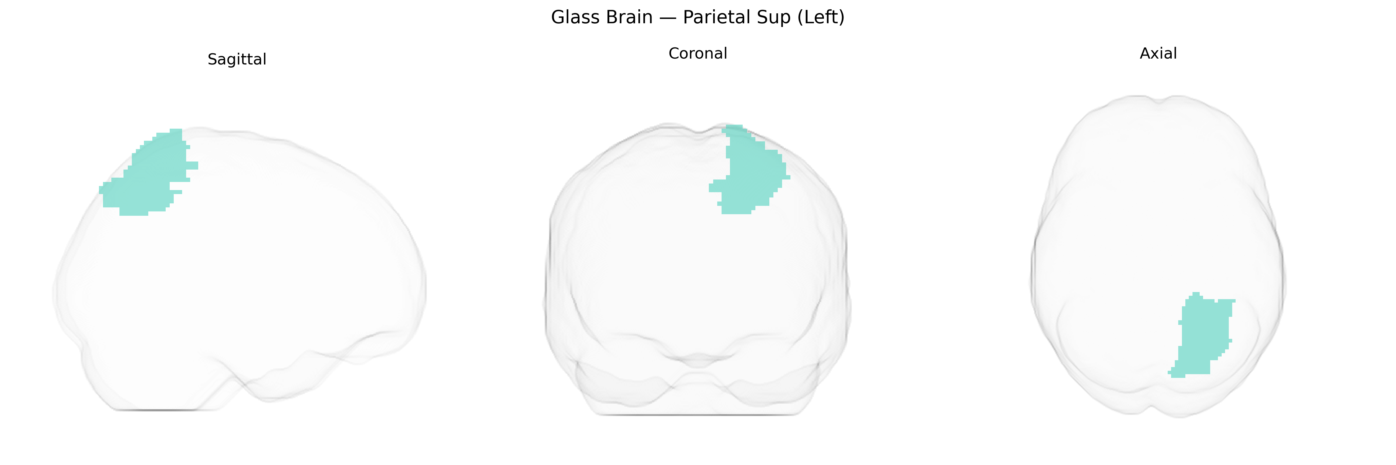
The left Superior Parietal lobule (Parietal Sup Left in the AAL2 atlas) is a dorsal parietal cortical region located posterior to the postcentral gyrus and superior to the intraparietal sulcus, forming part of the posterior parietal cortex. Cytoarchitectonically, it corresponds largely to Brodmann area 7, with contributions from adjacent parietal areas, and is heavily interconnected with premotor, visual, somatosensory, and inferior parietal regions. Functionally, it plays key roles in visuospatial integration, sensorimotor transformation, body-centered coordinate mapping, and the guidance of attention and limb movements in space, including reaching and object manipulation. It also contributes to higher-order spatial cognition and aspects of working memory related to spatial information. A closely related structure is the Superior parietal lobule: Superior parietal lobule.
The left superior parietal lobule (Parietal Sup L in the AAL2 atlas) has been implicated in several genetic imaging and GWAS studies, largely via cortical thickness, surface area, and functional connectivity measures rather than direct “region-specific” gene effects. Large consortia such as ENIGMA and UK Biobank have identified polygenic influences on superior parietal morphology, with common variants in or near genes involved in synaptic function, neurodevelopment, and cell adhesion (e.g., MAPT region, BDNF, GRIN2B, CNTNAP2, and various Wnt pathway genes) contributing modest but significant effects on parietal cortical measures. Parietal regions, including the left superior parietal lobule, show heritable differences in structure and connectivity that have been associated with cognitive traits such as general intelligence, working memory, visuospatial attention, and mathematical ability, with GWAS of these traits often highlighting genes involved in neuronal growth and plasticity that correlate with parietal metrics. In neuropsychiatric and neurodevelopmental disorders, genetic risk for schizophrenia, autism spectrum disorder, and ADHD has been associated with altered parietal morphology and connectivity, and polygenic risk scores for these conditions predict structural and functional variation in superior parietal areas. Similarly, Alzheimer’s disease and frontotemporal dementia genetic risk (including APOE and MAPT haplotypes) has been linked to accelerated cortical thinning and hypometabolism in parietal regions, though these associations are typically distributed across wider parietal and posterior cortical networks rather than confined to the left superior parietal lobule specifically. Overall, the genetic architecture influencing left Parietal Sup reflects highly polygenic, pleiotropic effects that overlap with broader parietal and frontoparietal networks supporting attention, executive function, and visuospatial processing.
Overview generated by GPT-4o (2026).
Region ID: 6101
Hemisphere: left
Atlas: AAL2
Full Quality Version: Download MP4
Full Quality Version: Download MP4
Full Quality Version: Download MP4
Full Quality Version: Download MP4
Wali Sidiqyar*, Gaurav Rudravaram*, Elyssa M. McMaster, Trent M. Schwartz, Adam M. Saunders, Kurt G. Schilling, Bennett A. Landman "Introducing SPINS: A Shared Public Visualization Library of Neuroanatomical Structures." Medical Imaging with Deep Learning- short paper
This resource is licensed under CC0 1.0 Universal (Public Domain).