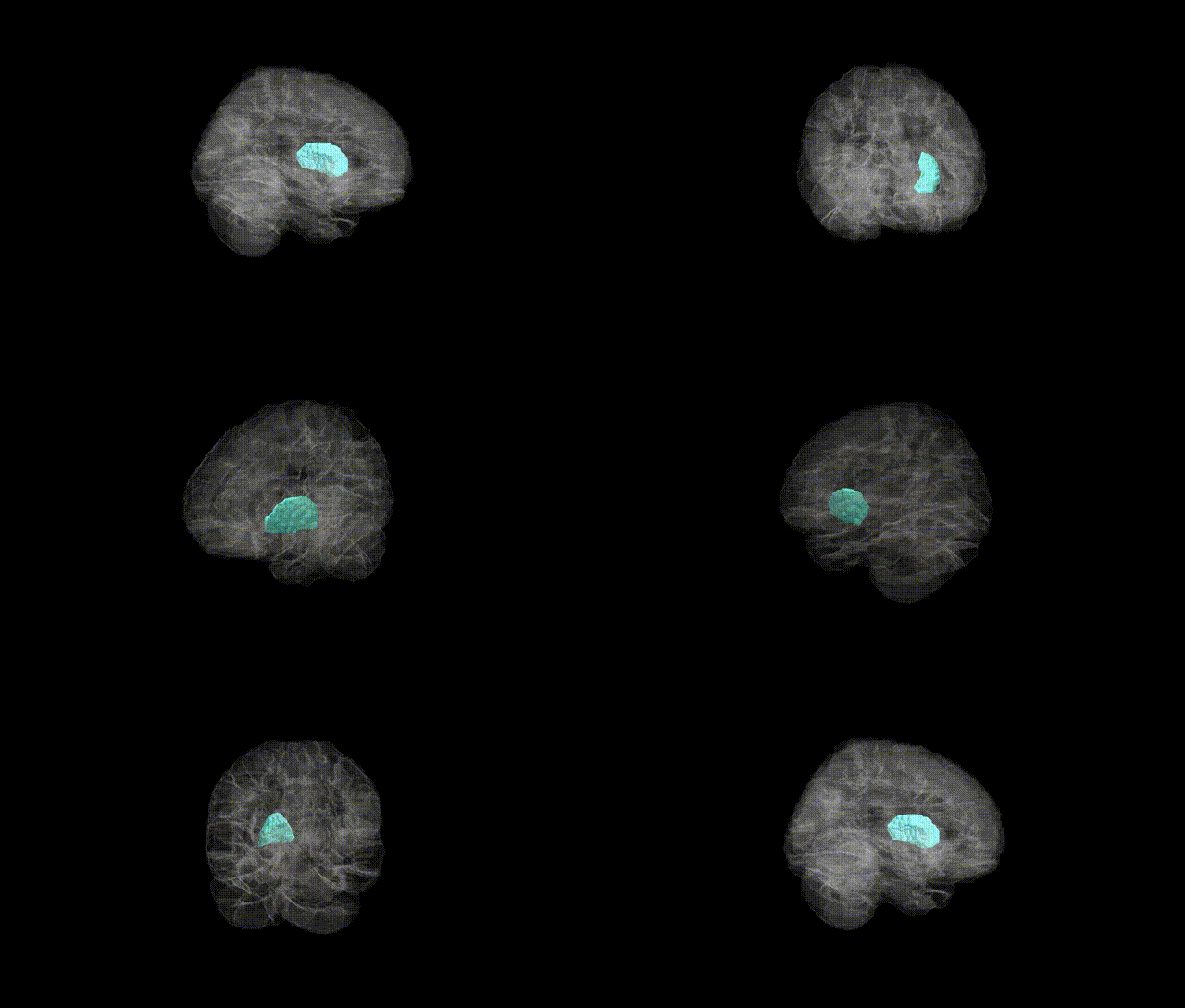
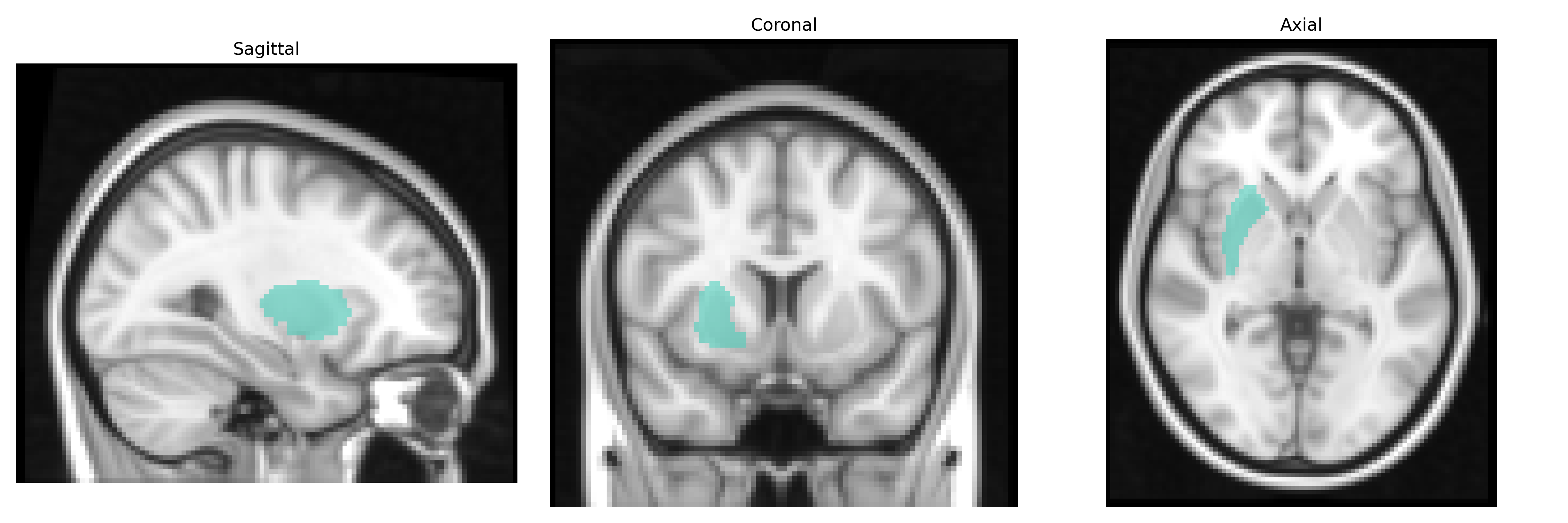
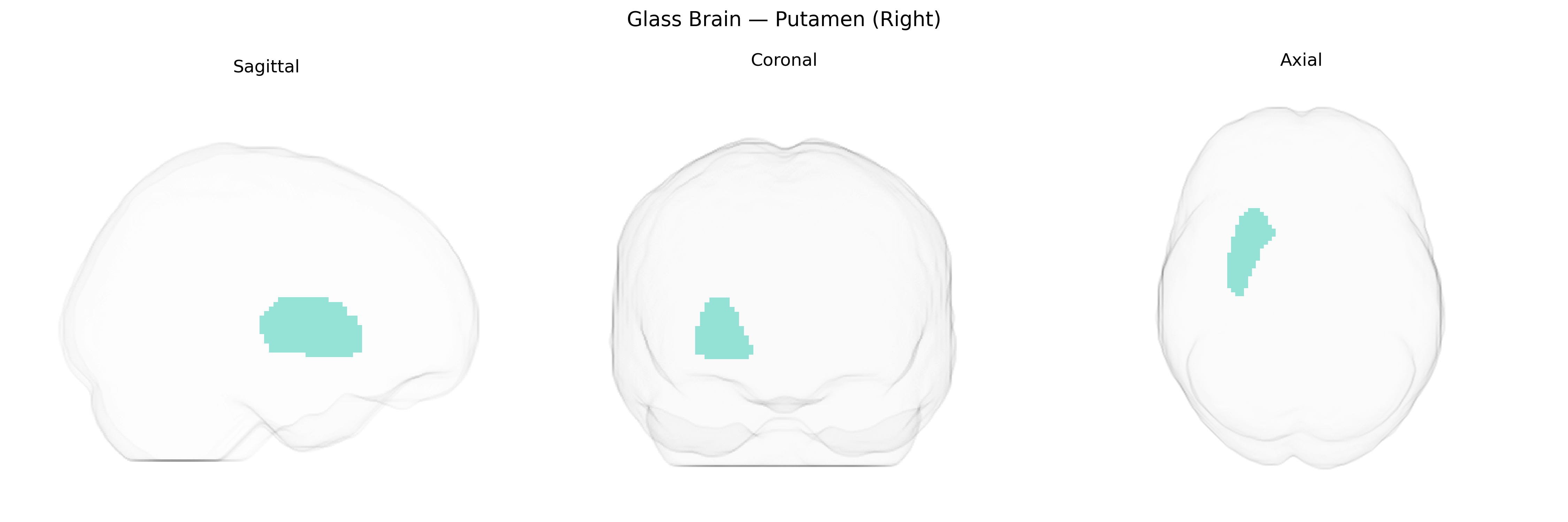
The right Putamen is a subcortical gray matter structure within the basal ganglia, lateral to the globus pallidus and deep to the insular cortex, and is part of the striatum along with the caudate nucleus. It receives dense glutamatergic input from sensorimotor and associative cortical areas and dopaminergic projections from the substantia nigra, integrating motor, cognitive, and limbic information. Functionally, the right Putamen is critically involved in motor planning, execution, and habit formation, as well as in procedural learning and reinforcement-based behavior. It participates in cortico-striato-thalamo-cortical loops that modulate voluntary movement, and its dysfunction is implicated in movement disorders such as Parkinson’s disease, dystonia, and Huntington’s disease, as well as in certain neuropsychiatric conditions. Putamen
The right putamen, as defined in the AAL2 atlas, has been implicated in multiple genetic and GWAS findings related to neuropsychiatric traits and brain structure. Large-scale imaging genetics consortia (e.g., ENIGMA, UK Biobank) have identified common variants in and near genes such as KLB, DCC, DRD2, PDE10A, BDNF, and several loci on chromosomes 11 and 14 that are associated with putamen volume and microstructure. DRD2 and PDE10A, key components of striatal dopaminergic signaling, have been repeatedly linked to putaminal morphology and function, supporting their role in reward processing and motor control. Polygenic risk scores for schizophrenia, bipolar disorder, major depressive disorder, and ADHD show associations with altered putamen volume or connectivity, and GWAS of Parkinson’s disease and dystonia highlight dopaminergic and synaptic genes whose effects converge on basal ganglia circuits including the putamen. Additional GWAS of traits such as general cognitive ability, substance use (alcohol, nicotine), and risk-taking behavior report overlapping loci that influence both these behaviors and putamen structure or activity, suggesting that genetic variation in neurodevelopmental, synaptic, and neurotransmitter pathways contributes to individual differences in right putamen anatomy and its vulnerability to psychiatric and movement disorders.
Overview generated by GPT-4o (2026).
Region ID: 7012
Hemisphere: right
Atlas: AAL2
Full Quality Version: Download MP4
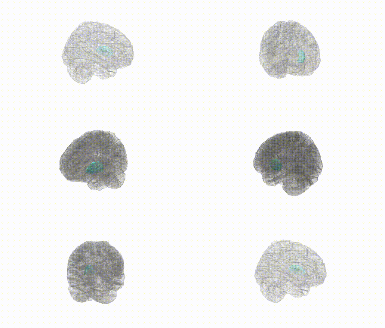
Full Quality Version: Download MP4
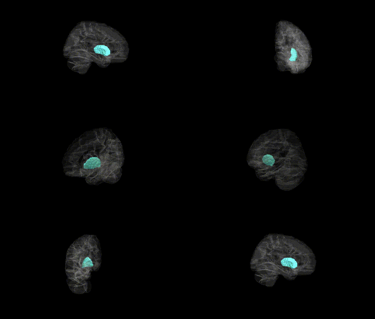
Full Quality Version: Download MP4
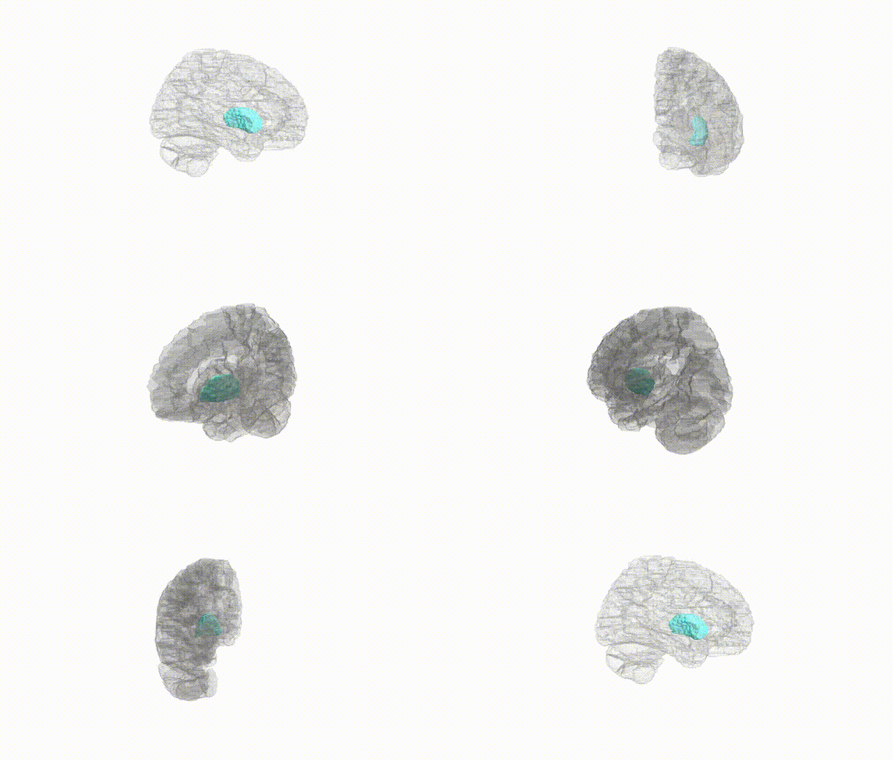
Full Quality Version: Download MP4
Wali Sidiqyar*, Gaurav Rudravaram*, Elyssa M. McMaster, Trent M. Schwartz, Adam M. Saunders, Kurt G. Schilling, Bennett A. Landman "Introducing SPINS: A Shared Public Visualization Library of Neuroanatomical Structures." Medical Imaging with Deep Learning- short paper
This resource is licensed under CC0 1.0 Universal (Public Domain).