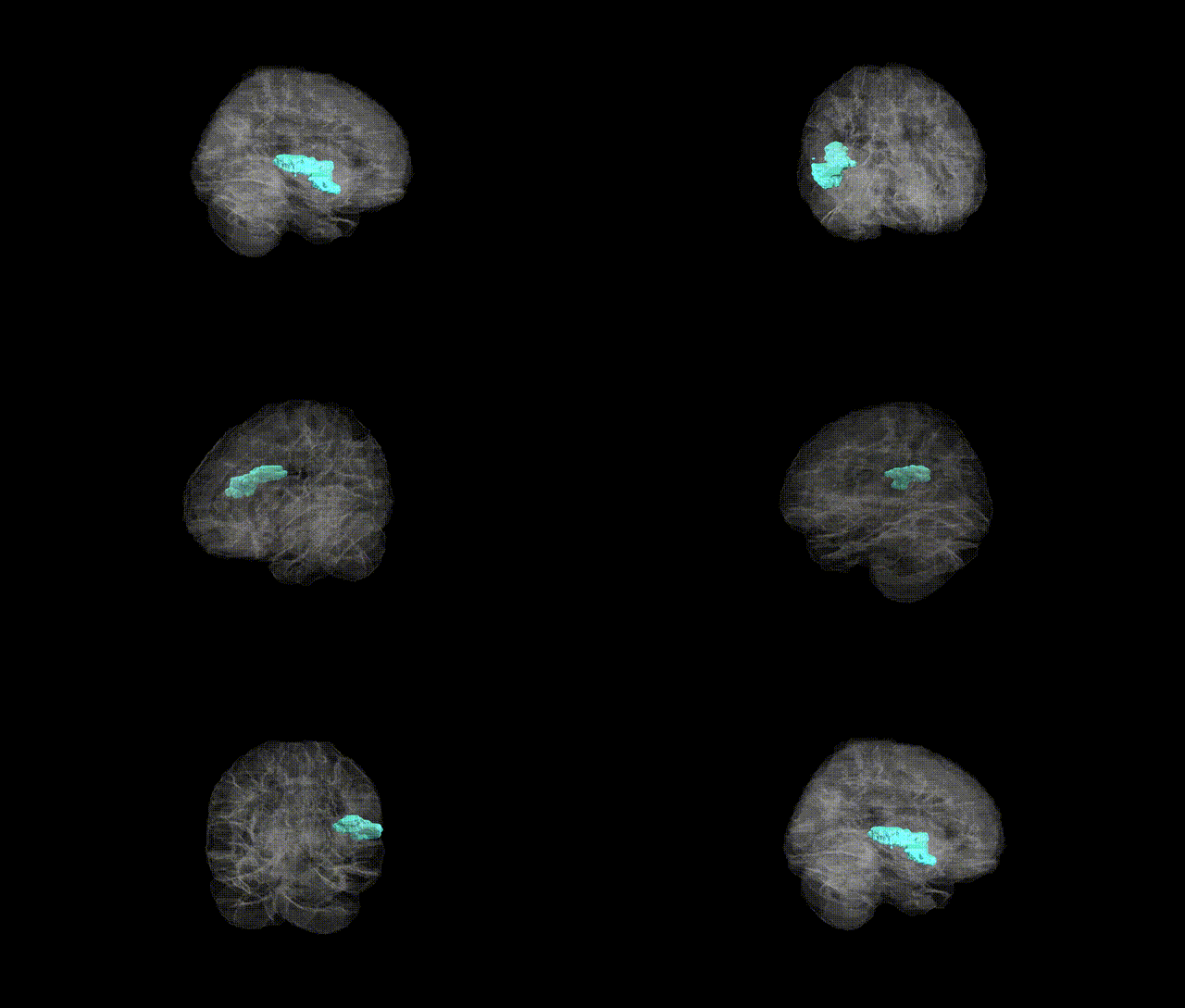
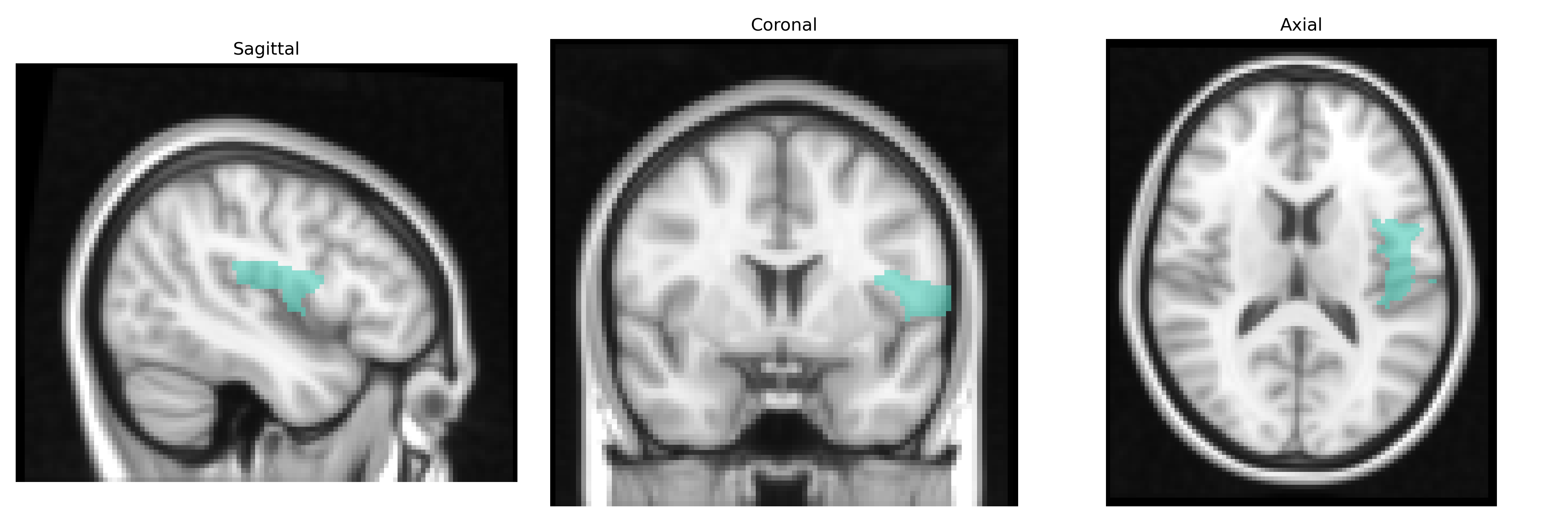
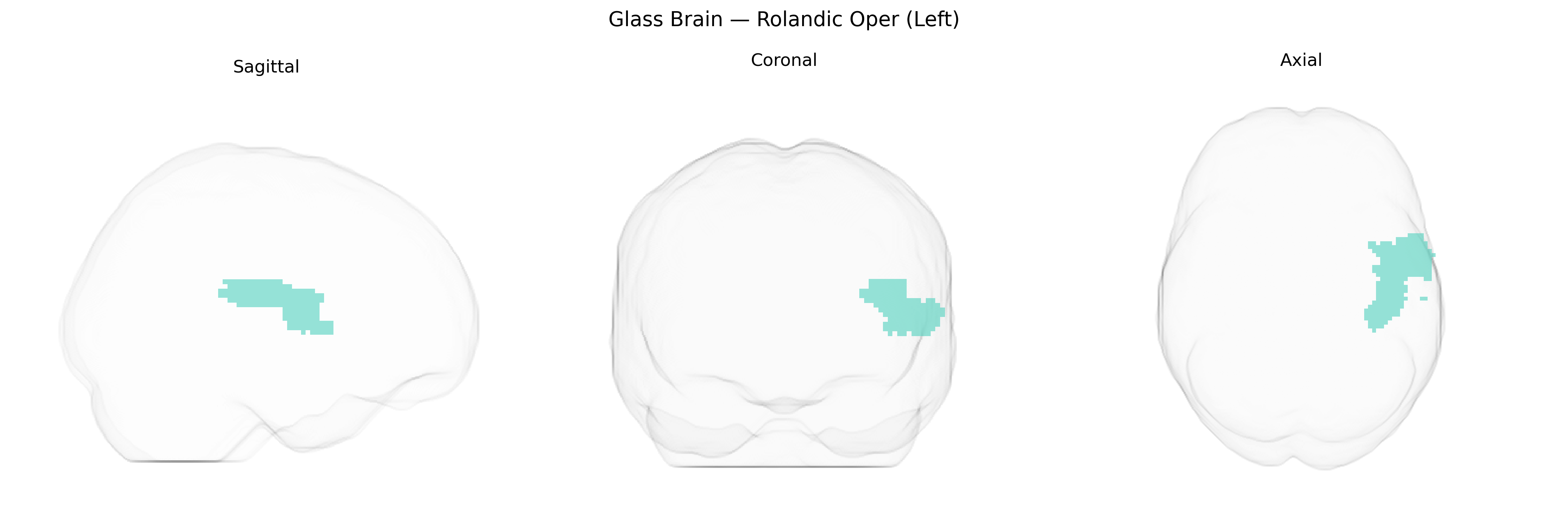
The left Rolandic operculum (Rolandic Oper (Left)) in the AAL2 atlas corresponds to the opercular portion of the inferior frontal and parietal regions overlying the insula along the central (Rolandic) sulcus. It participates in sensorimotor integration, particularly for oral–facial, laryngeal, and pharyngeal musculature, and is frequently implicated in speech production, articulation, and orofacial motor control, as well as in somatosensory processing related to the tongue, jaw, and throat. This region forms part of the perisylvian network involved in language and praxis and is structurally contiguous with the frontal and parietal opercula that cover the insula. There is no direct link for “Rolandic operculum,” but it is part of the broader Operculum (brain).
The left Rolandic operculum, a perisylvian region implicated in sensorimotor integration for speech and orofacial functions, has been indirectly associated with several genetic influences through imaging genetics and GWAS of brain structure and neuropsychiatric traits, although few studies target it as a primary ROI. Large-scale brain MRI GWAS (e.g., ENIGMA and UK Biobank) have identified variants in genes such as HMGA2, IGF1, and multiple loci near FOXP2, DCDC2, and CNTNAP2 that affect cortical thickness and surface area in nearby perisylvian and opercular areas, some of which map onto the Rolandic operculum in AAL2-based parcellations. This region’s structural and functional characteristics have been linked genetically to speech and language traits, developmental language disorder, and reading disability, with risk variants in FOXP2 and DCDC2 showing associations with altered perisylvian morphology. GWAS of stuttering and childhood speech sound disorders, while still underpowered, suggest polygenic overlap with language and motor-planning genes that influence opercular and adjacent inferior frontal and insular cortices. Moreover, multi-locus polygenic risk scores for schizophrenia, autism spectrum disorder, and ADHD have been correlated with altered thickness, gyrification, or connectivity in the Rolandic operculum and contiguous frontal opercular regions, implicating synaptic and neurodevelopmental genes such as CACNA1C, GRIN2A, and NRXN1. Finally, studies of taste and somatosensory perception, as well as chronic pain and migraine GWAS, have reported associations between variants in ion-channel and neuroinflammatory genes and structural or functional measures in opercular–insular complexes, which in AAL2-based analyses often include the left Rolandic operculum, highlighting this region as a genetically modulated hub for sensorimotor, language, and interoceptive processes rather than a site with single, well-established locus-specific effects.
Overview generated by GPT-4o (2026).
Region ID: 2331
Hemisphere: left
Atlas: AAL2
Full Quality Version: Download MP4
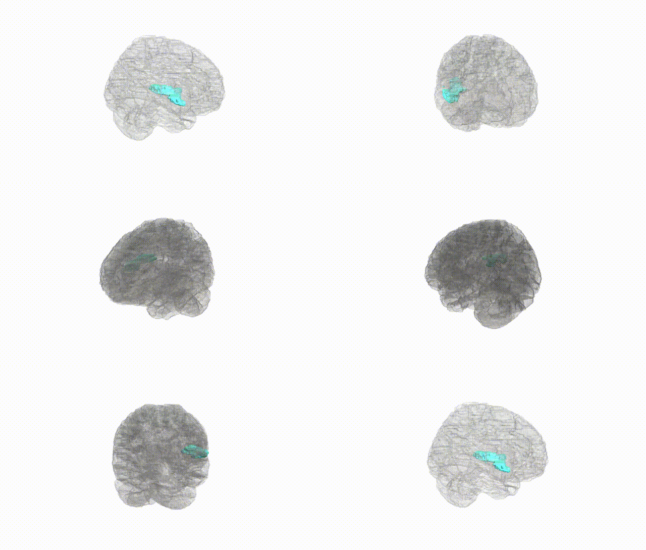
Full Quality Version: Download MP4
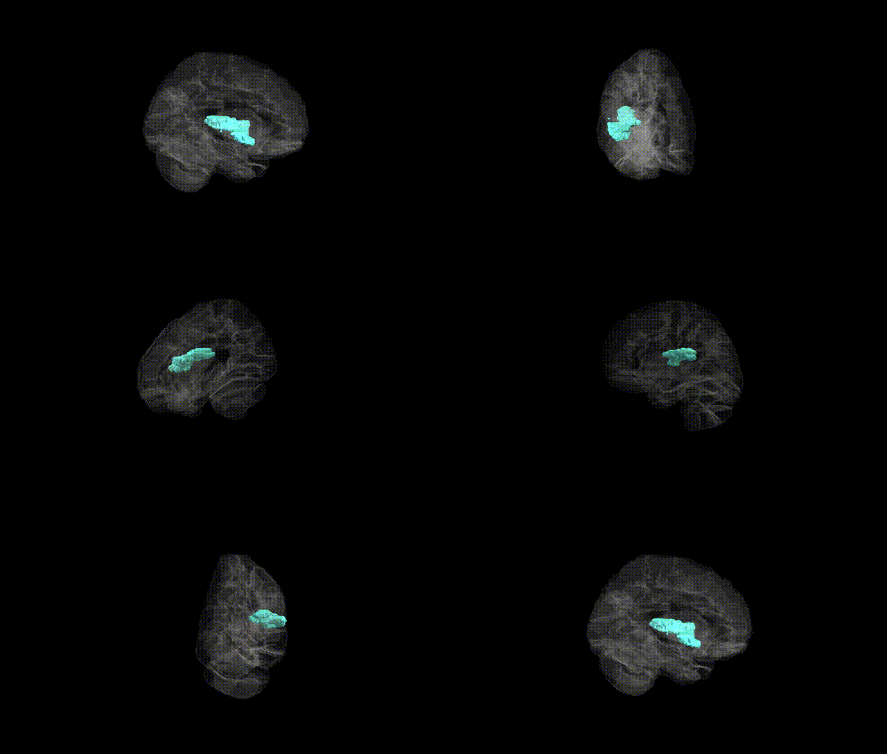
Full Quality Version: Download MP4
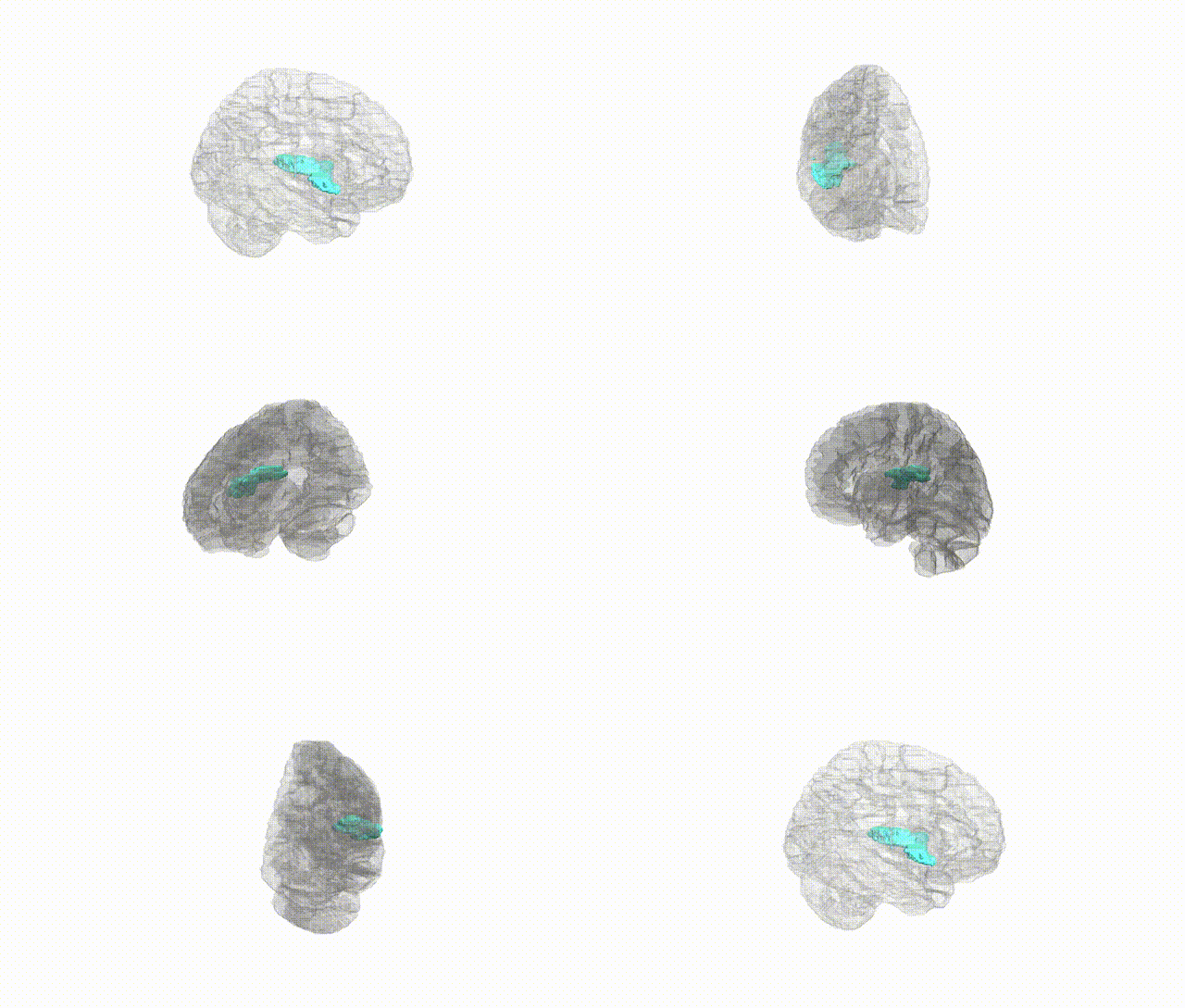
Full Quality Version: Download MP4
Wali Sidiqyar*, Gaurav Rudravaram*, Elyssa M. McMaster, Trent M. Schwartz, Adam M. Saunders, Kurt G. Schilling, Bennett A. Landman "Introducing SPINS: A Shared Public Visualization Library of Neuroanatomical Structures." Medical Imaging with Deep Learning- short paper
This resource is licensed under CC0 1.0 Universal (Public Domain).