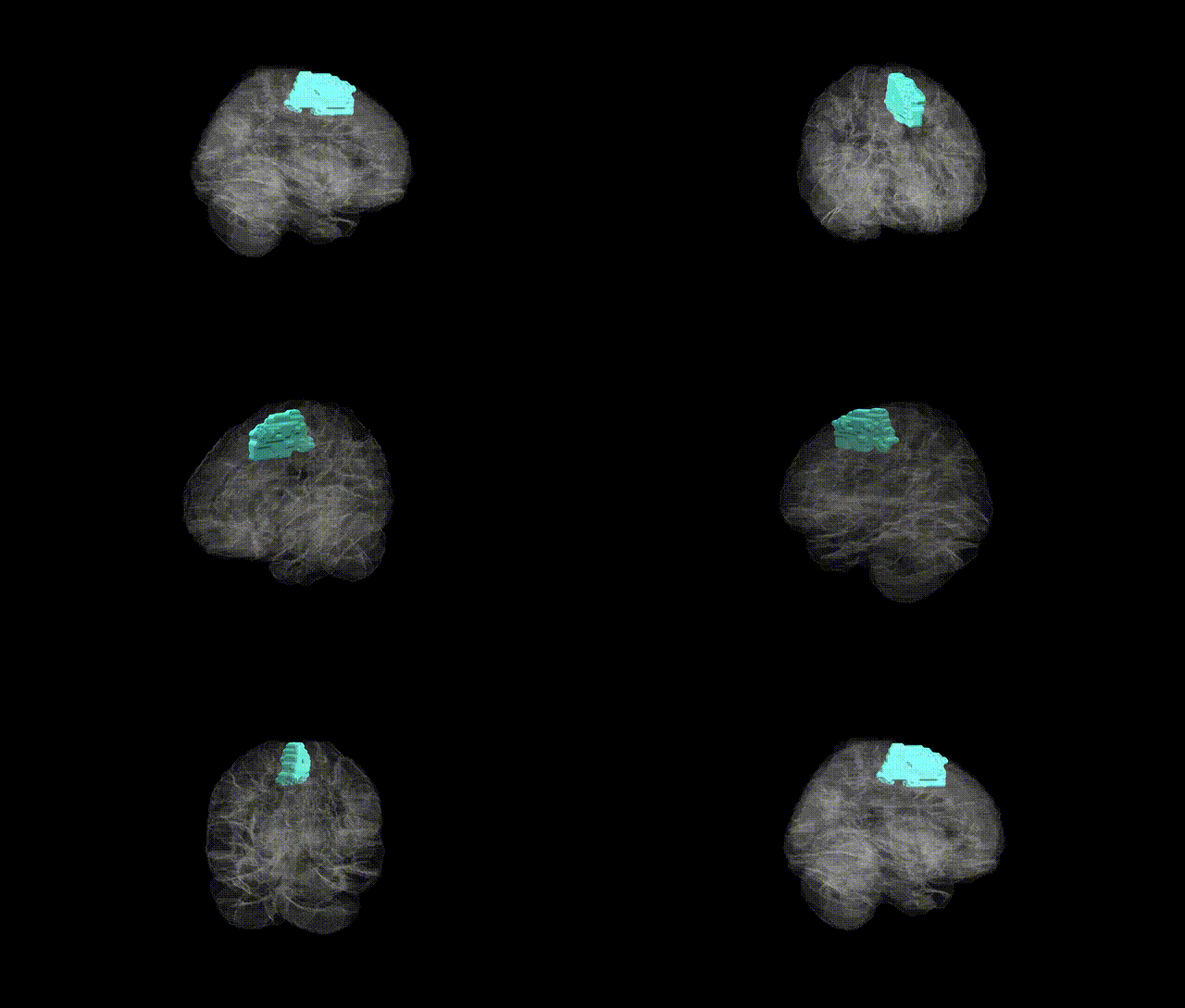
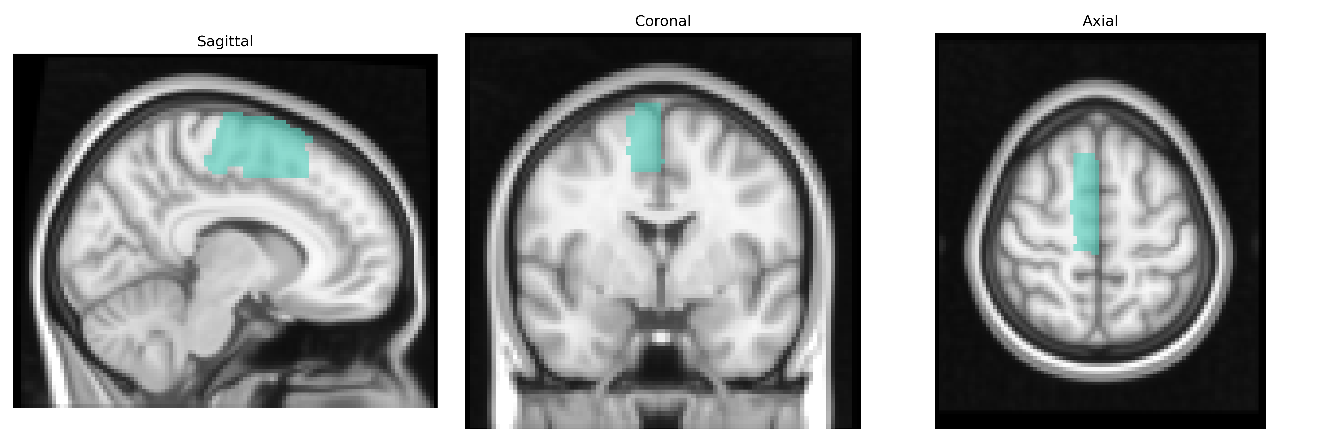
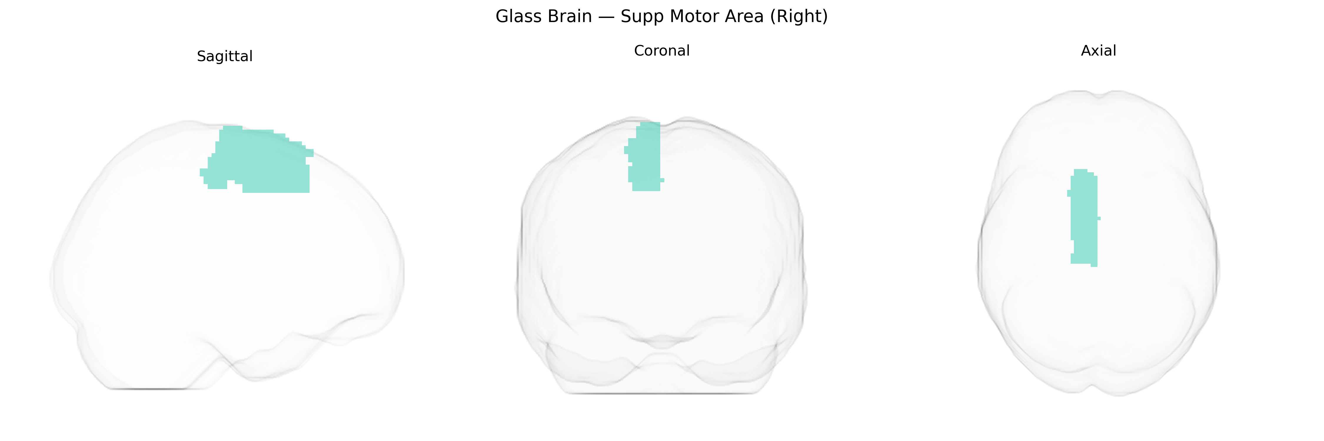
The Right Supplementary Motor Area (SMA) is a medial frontal cortical region located on the dorsomedial surface of the right hemisphere, anterior to the primary motor cortex and within the superior frontal gyrus. It is involved in higher-order aspects of motor control, including planning, initiation, sequencing, and coordination of internally generated movements, as well as bimanual and complex motor actions. The SMA also contributes to motor imagery, temporal organization of movement, and the integration of motor programs with cognitive processes such as decision-making and attention. Functionally, it forms part of the motor network interconnected with premotor cortex, primary motor cortex, basal ganglia, and cerebellum. There is no direct Wikipedia article specific to the “Right Supplementary Motor Area”; a related entry is the bilateral supplementary motor area: Supplementary motor area.
The right supplementary motor area (SMA), as defined in the AAL2 atlas, has been indirectly implicated in several genetically influenced traits and disorders through imaging genetics and GWAS-based imaging studies, though few findings target this subregion in isolation; common themes involve motor control, response inhibition, and higher-order cognitive functions. Variants in genes affecting glutamatergic and GABAergic signaling (such as GRIN2B, GAD1, and SLC6A1) and synaptic plasticity (for example BDNF Val66Met) have been associated with altered SMA activation or structure during motor sequence learning, response inhibition, and working memory tasks. Large-scale imaging GWAS have linked polygenic risk scores for schizophrenia, ADHD, autism spectrum disorder, and major depressive disorder to altered cortical thickness or functional connectivity in medial premotor regions encompassing the SMA, while specific schizophrenia- and ADHD-associated loci (e.g., in CACNA1C, DRD2, and other dopaminergic or calcium-channel genes) show convergent effects on fronto-striatal and SMA networks involved in cognitive control. Right SMA involvement has also been reported in genetic studies of Tourette syndrome and dystonia, in which variants in genes governing basal ganglia–cortical circuitry (such as those in dopaminergic pathways and TOR1A in dystonia) correspond to abnormal activation or connectivity of SMA during motor tasks. Additionally, GWAS of traits like impulsivity, risk-taking, and reaction time implicate polygenic architectures that modulate activity and connectivity of the right SMA within broader inhibitory control networks, although individual loci are rarely mapped exclusively to this region due to the distributed nature of genetic effects on brain structure and function.
Overview generated by GPT-4o (2026).
Region ID: 2402
Hemisphere: right
Atlas: AAL2
Full Quality Version: Download MP4
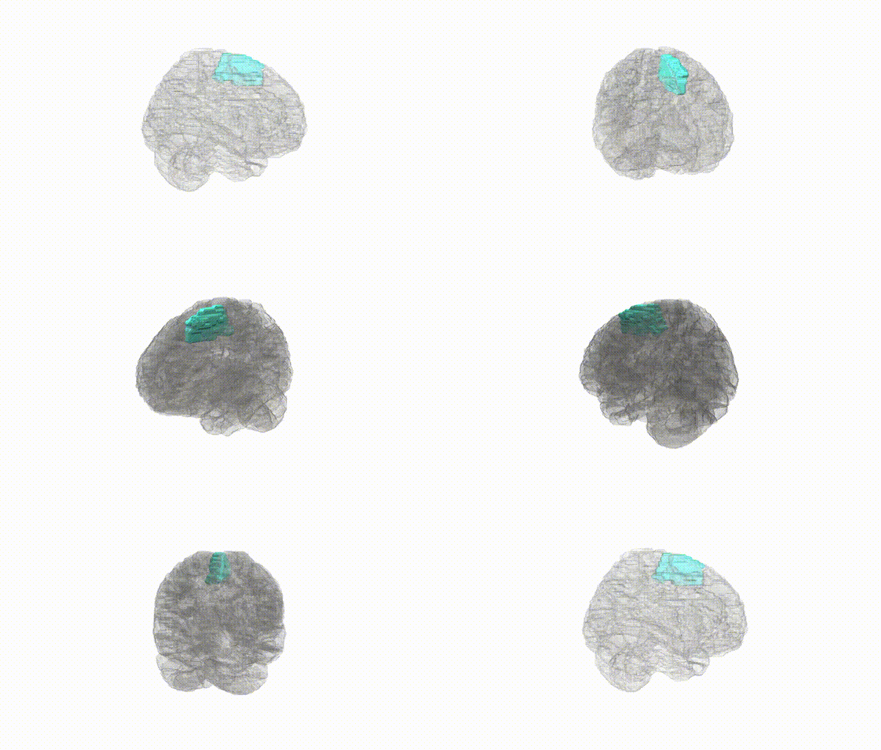
Full Quality Version: Download MP4
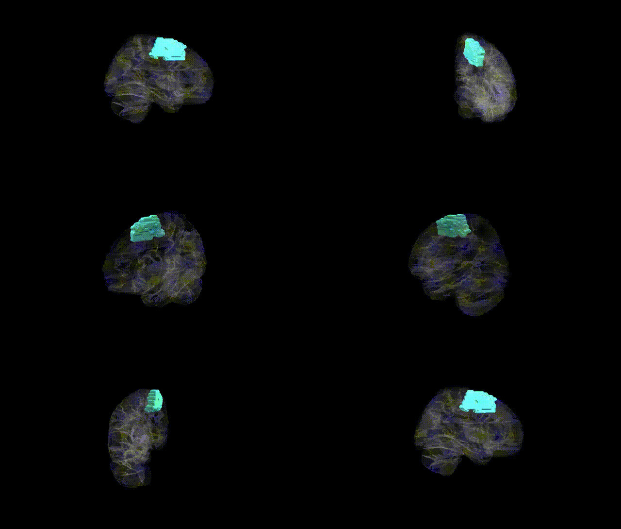
Full Quality Version: Download MP4
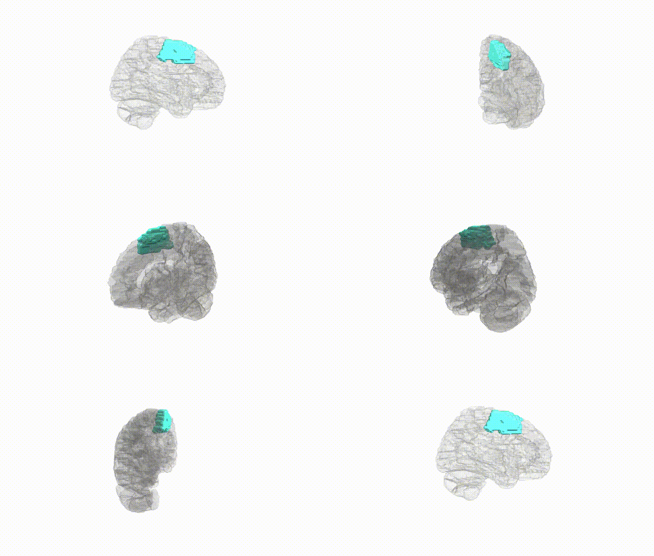
Full Quality Version: Download MP4
Wali Sidiqyar*, Gaurav Rudravaram*, Elyssa M. McMaster, Trent M. Schwartz, Adam M. Saunders, Kurt G. Schilling, Bennett A. Landman "Introducing SPINS: A Shared Public Visualization Library of Neuroanatomical Structures." Medical Imaging with Deep Learning- short paper
This resource is licensed under CC0 1.0 Universal (Public Domain).