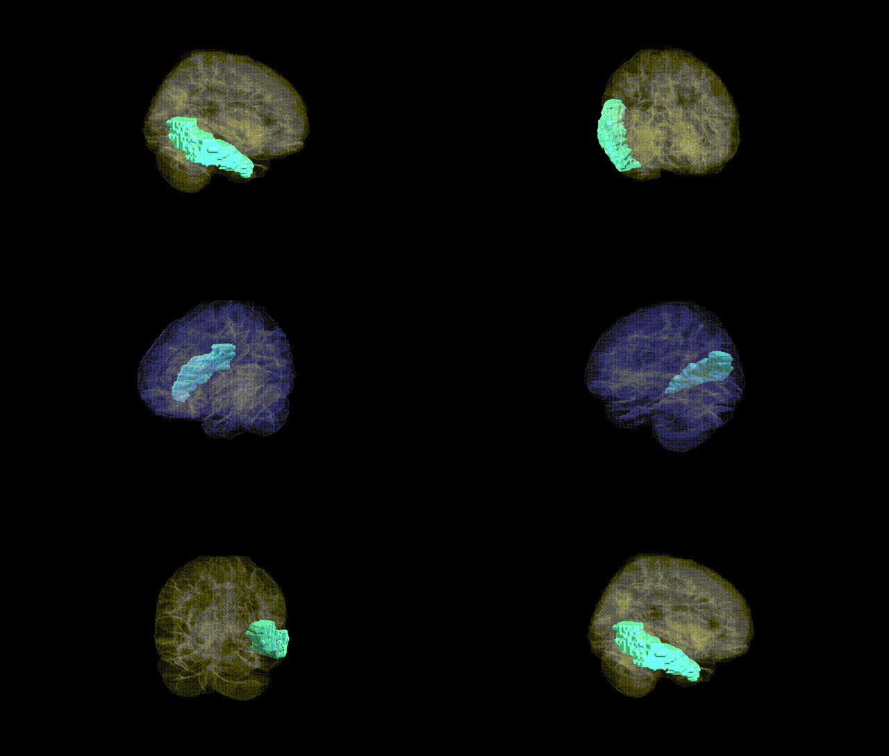
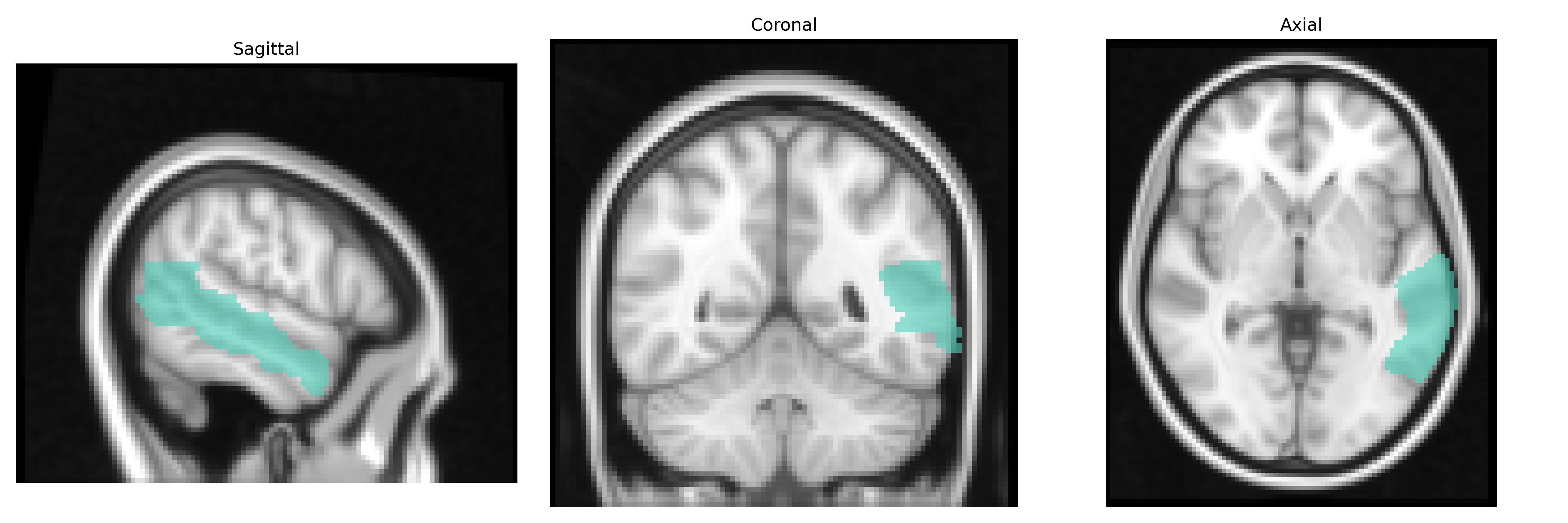
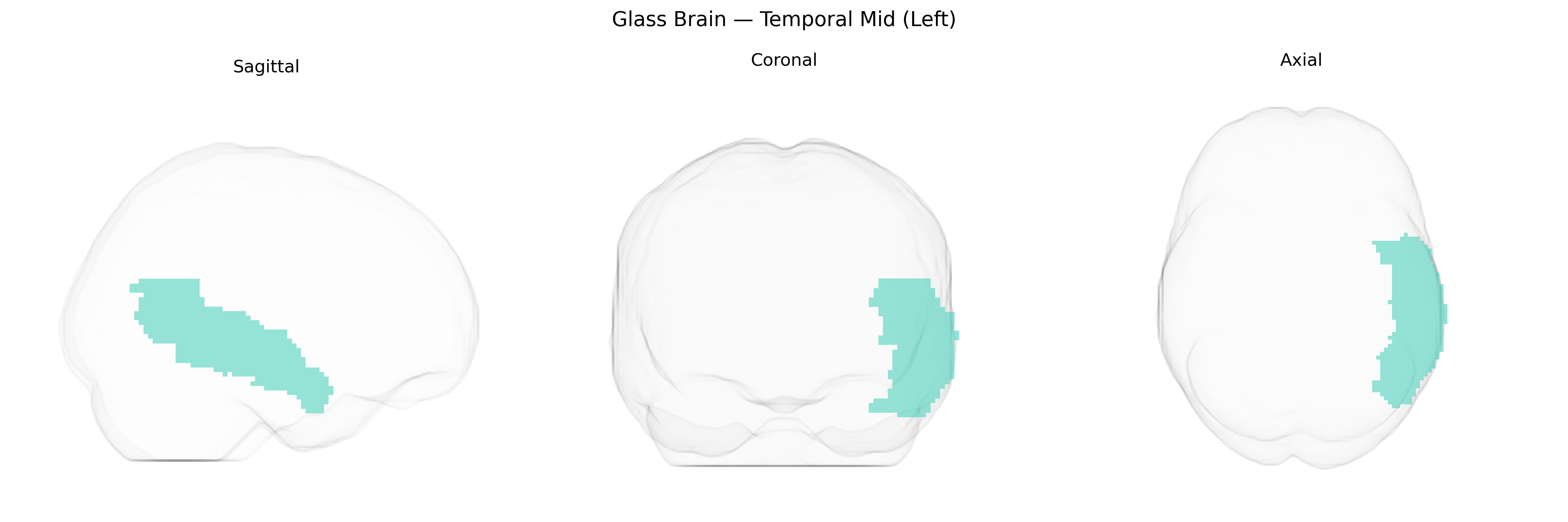
The left Temporal Mid (Left) region in the AAL2 atlas corresponds to the middle temporal gyrus of the left cerebral hemisphere, a lateral temporal lobe structure implicated in semantic processing, language comprehension, and integration of auditory and visual information. Cytoarchitectonically, it forms part of association cortex receiving multimodal input from auditory, visual, and limbic areas, and it contributes to higher-order functions such as lexical-semantic access, narrative comprehension, and aspects of social cognition including interpretation of biological motion and facial expressions. Functionally, this region is frequently engaged in tasks involving word meaning, sentence processing, and conceptual knowledge retrieval, and is interconnected with inferior frontal, angular, and superior temporal regions in language and semantic networks. There is no direct link for “Temporal Mid (Left)” as an AAL2 label; a closely related structure is the Middle temporal gyrus.
The left middle temporal gyrus (Temporal Mid Left in the AAL2 atlas), a core component of the language and semantic network, has been repeatedly implicated in genetic and GWAS-based studies of neuropsychiatric and cognitive traits, though typically as part of broader temporal or cortical measures rather than a uniquely isolated locus. Imaging–genetics and ENIGMA consortia analyses have linked common variants in genes such as HMGA2, IGF1, and those near the 12q14 and 3q28 regions to temporal lobe and cortical surface area or thickness that prominently include the middle temporal gyrus, while polygenic scores for educational attainment and general cognitive ability correlate with structural and functional differences in this region. GWAS of schizophrenia, bipolar disorder, and major depressive disorder implicate temporal lobe–enriched risk loci (e.g., CACNA1C, GRIN2A, complement genes such as C4) whose expression and imaging correlates include altered left middle temporal volume or activity, especially in language and auditory hallucination circuits in schizophrenia. In autism spectrum disorder and specific language impairment, convergent genetic findings in synaptic and chromatin-remodeling genes (e.g., SHANK3, CNTNAP2, FOXP2-related pathways) have been associated with atypical development and connectivity of the left middle temporal cortex within perisylvian language networks. Large-scale GWAS of Alzheimer’s disease and related dementias (e.g., APOE, CLU, PICALM, BIN1) also show downstream effects on temporal lobe atrophy patterns that prominently involve the left middle temporal gyrus, which is critical for semantic memory; similarly, GWAS of temporal lobe epilepsy identify risk loci influencing temporal structures, although region-specific genetic effects for the left middle temporal gyrus remain less precisely mapped.
Overview generated by GPT-4o (2026).
Region ID: 8201
Hemisphere: left
Atlas: AAL2
Full Quality Version: Download MP4
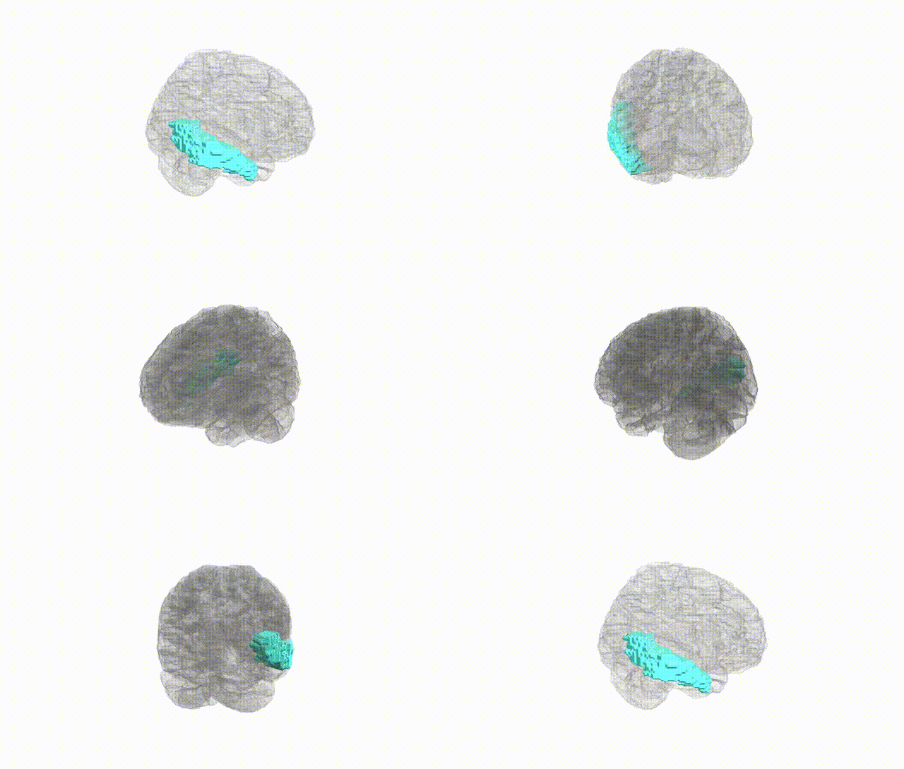
Full Quality Version: Download MP4
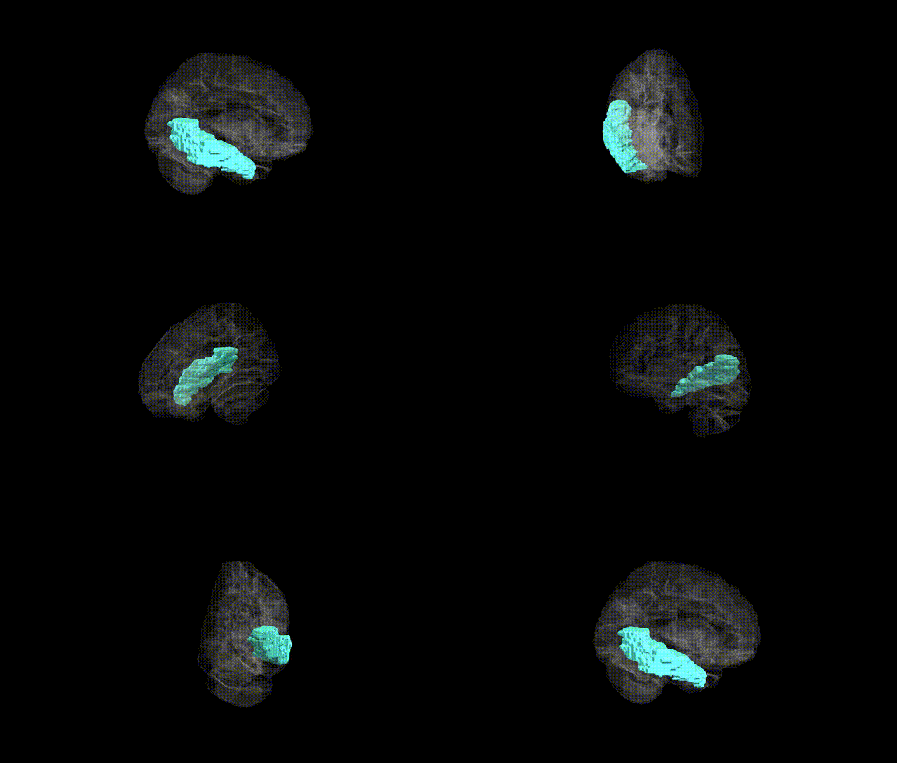
Full Quality Version: Download MP4
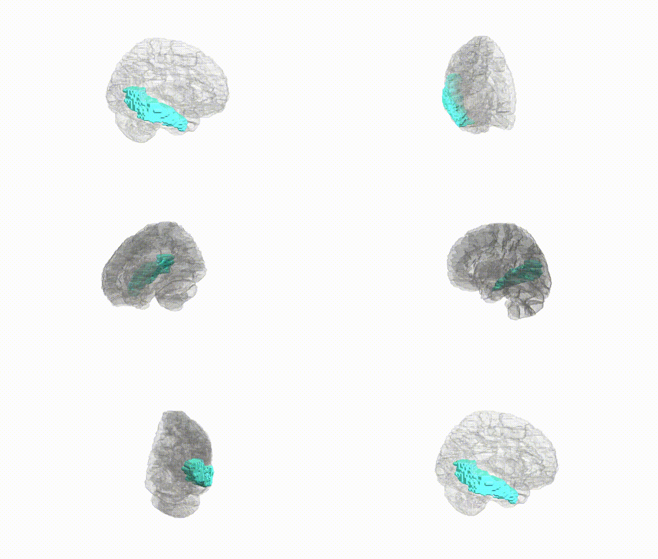
Full Quality Version: Download MP4
Wali Sidiqyar*, Gaurav Rudravaram*, Elyssa M. McMaster, Trent M. Schwartz, Adam M. Saunders, Kurt G. Schilling, Bennett A. Landman "Introducing SPINS: A Shared Public Visualization Library of Neuroanatomical Structures." Medical Imaging with Deep Learning- short paper
This resource is licensed under CC0 1.0 Universal (Public Domain).