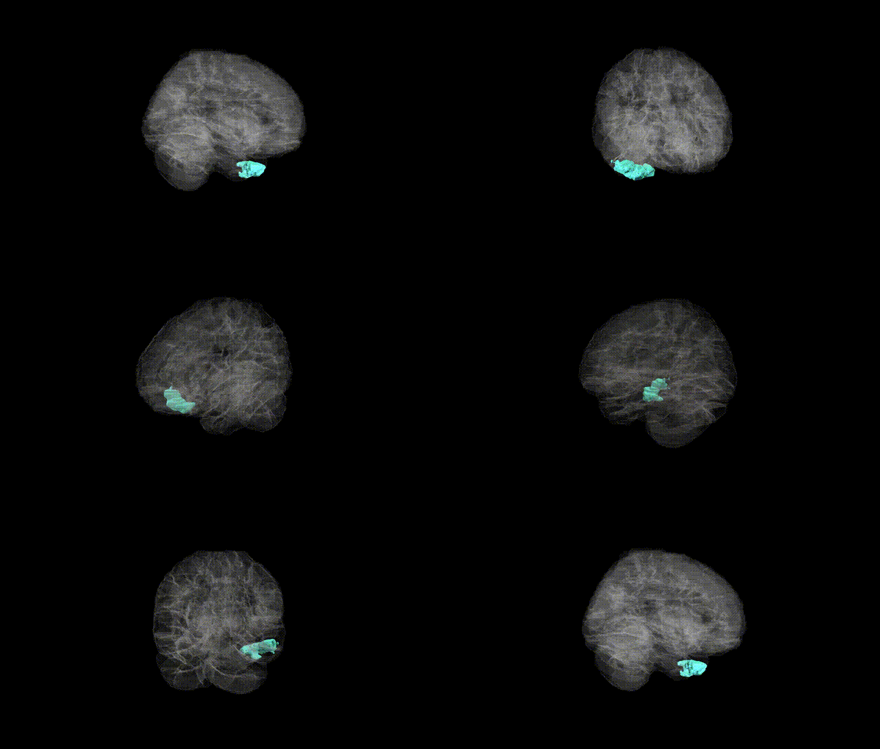
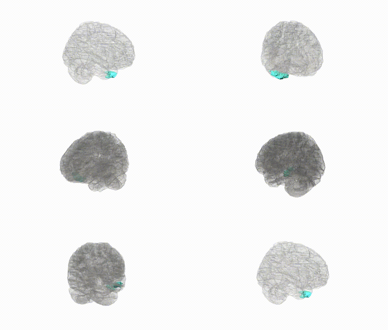
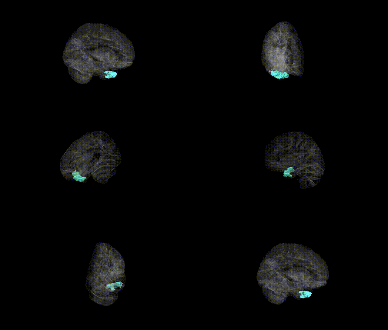
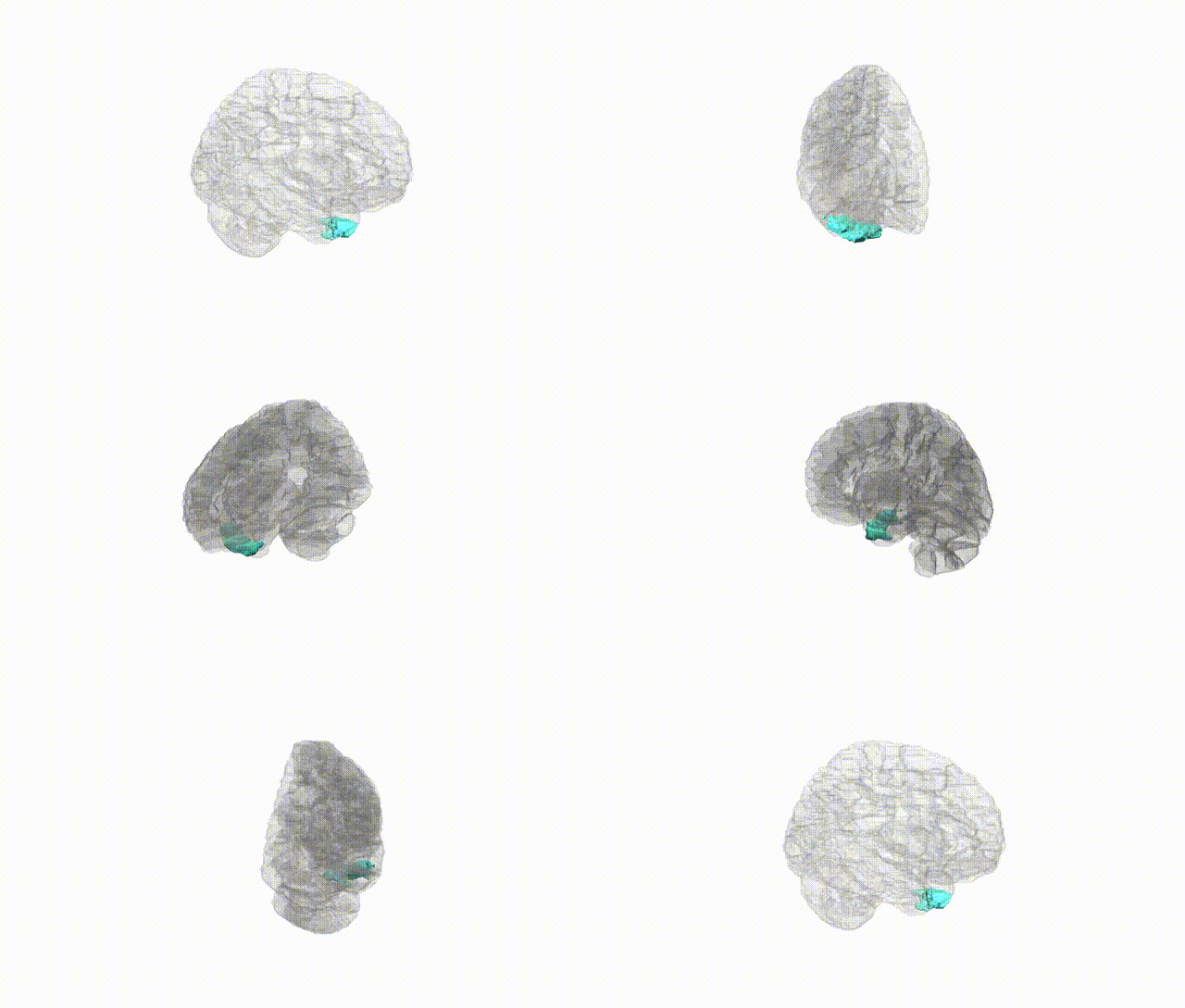
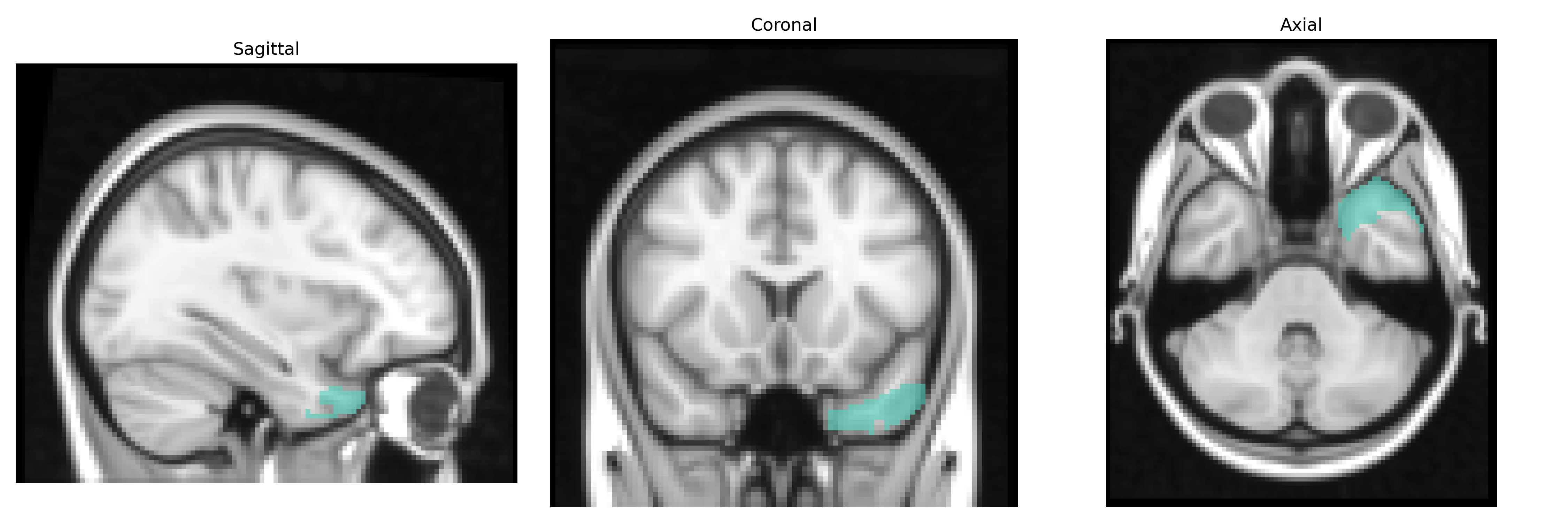
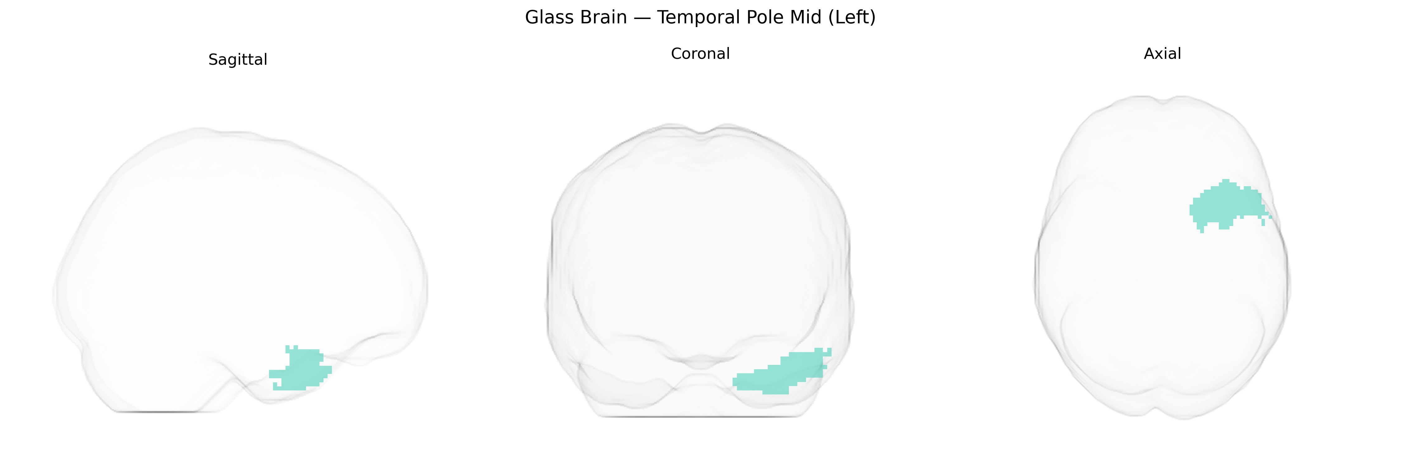
The left Temporal Pole Mid (Left), as defined in the AAL2 atlas, corresponds to the anterior-most portion of the middle temporal gyrus in the left temporal lobe, forming part of the temporal pole. This region is involved in high-level semantic processing, social and emotional cognition, and integration of multimodal sensory information, including language-related functions such as narrative comprehension and the processing of abstract concepts. Anatomically, it lies anterior to the superior and inferior temporal gyri, bordered posteriorly by more caudal segments of the middle temporal gyrus, and is heavily interconnected with limbic and prefrontal cortices, including the amygdala, orbitofrontal cortex, and medial prefrontal areas. Functionally, it contributes to the representation of semantic memory, social knowledge, and conceptual associations, and is implicated in disorders affecting semantic cognition, such as semantic variant primary progressive aphasia. There is no direct Wikipedia article for “Temporal Pole Mid”; a closely related structure is the Temporal pole.
The left temporal pole (AAL2: Temporal_Pole_Mid_L), a key hub for semantic memory, social cognition, and emotional processing, shows consistent heritability in imaging genetics studies and appears in multiple GWAS of brain structure and connectivity. Large-scale MRI GWAS (e.g., ENIGMA, UK Biobank) have identified associations between variants near genes involved in neurodevelopment, synaptic function, and axon guidance (such as MAPT, DCC, and related loci) and temporal pole cortical thickness or surface area, although findings are often reported for broader temporal or anterior temporal regions rather than the specific AAL2 label. Genetically influenced variation in temporal pole structure and connectivity has been implicated in risk for temporal lobe epilepsy, Alzheimer’s disease, and frontotemporal dementia, with MAPT, GRN, and C9orf72 showing convergent evidence in disorders that prominently affect anterior temporal structures. Polygenic risk for schizophrenia, bipolar disorder, major depression, and autism spectrum disorder has also been linked to altered volume or cortical thickness in anterior temporal regions, supporting a shared genetic architecture between psychiatric risk and temporal pole morphology. GWAS of social behavior, language-related traits, and emotional processing occasionally report imaging endophenotypes involving the anterior temporal cortex, suggesting that common variants shaping these behavioral phenotypes partly act through development and maintenance of the temporal pole; however, most associations remain modest, are not specific to the left temporal pole mid region, and often require replication or meta-analytic confirmation.
Overview generated by GPT-4o (2026).
Region ID: 8211
Hemisphere: left
Atlas: AAL2
Full Quality Version: Download MP4
Full Quality Version: Download MP4
Full Quality Version: Download MP4
Full Quality Version: Download MP4
Wali Sidiqyar*, Gaurav Rudravaram*, Elyssa M. McMaster, Trent M. Schwartz, Adam M. Saunders, Kurt G. Schilling, Bennett A. Landman "Introducing SPINS: A Shared Public Visualization Library of Neuroanatomical Structures." Medical Imaging with Deep Learning- short paper
This resource is licensed under CC0 1.0 Universal (Public Domain).