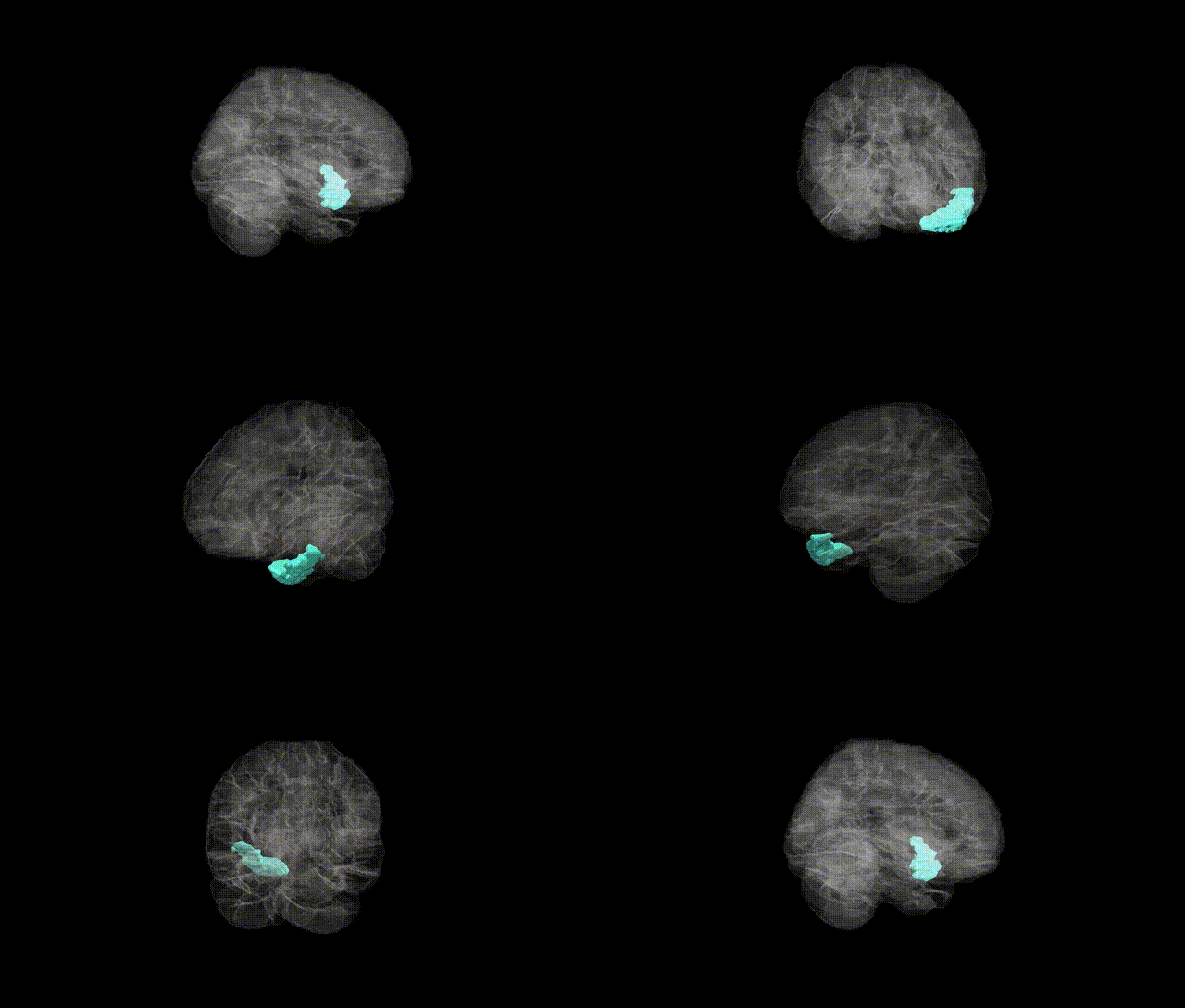
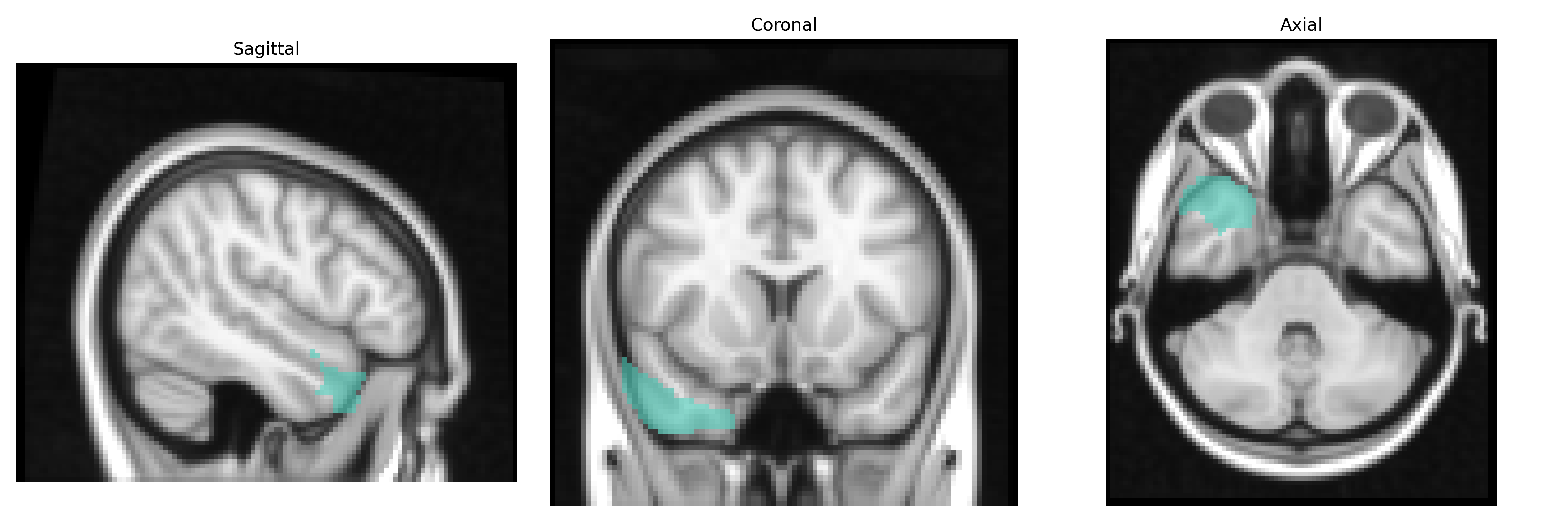
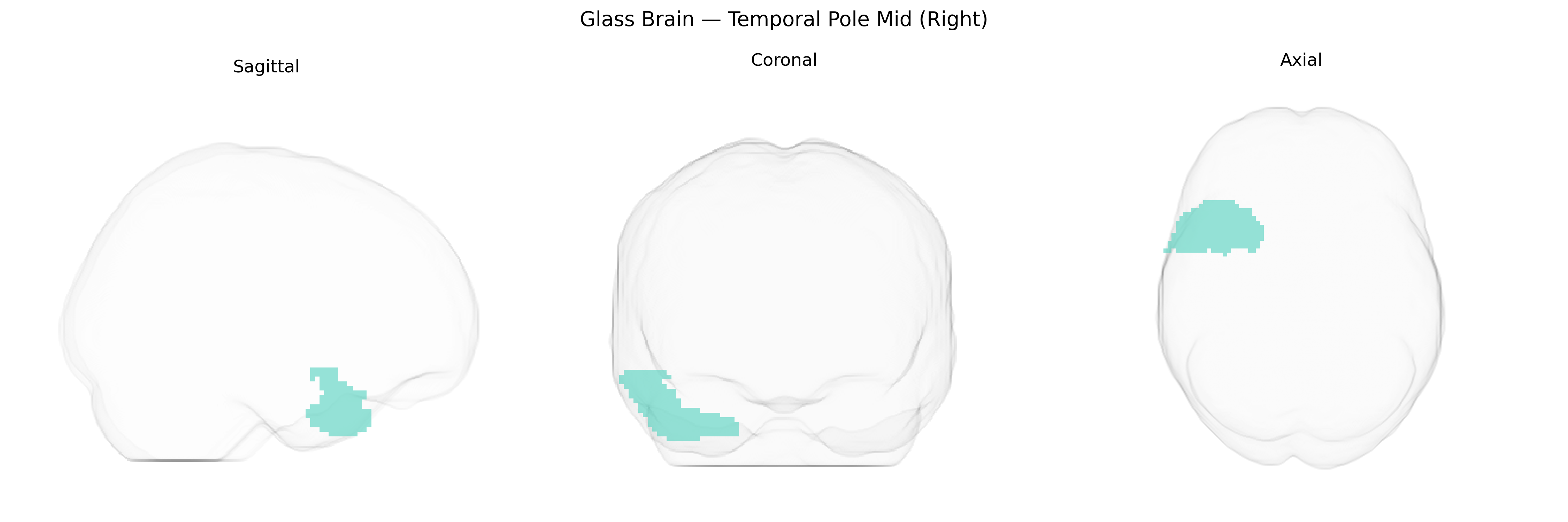
The right Temporal Pole Mid (Right), as defined in the AAL2 atlas, corresponds to the anterior portion of the middle temporal gyrus within the temporal pole of the right cerebral hemisphere. This region is part of the paralimbic cortex and is structurally connected with the amygdala, orbitofrontal cortex, and other temporal lobe regions. Functionally, it is implicated in high-level social and emotional processing, semantic memory, and multimodal integration of sensory inputs, contributing to the representation of complex concepts and socially relevant information. In clinical and neuroimaging studies, the temporal pole has been associated with semantic dementia, social cognition deficits, and aspects of emotional evaluation. There is no direct Wikipedia article for “Temporal Pole Mid (Right),” but it is encompassed within the broader Temporal pole.
The right temporal pole (mid portion) in the AAL2 atlas is implicated by genetic studies largely through imaging genetics and GWAS of brain structure and connectivity rather than region-specific gene mapping. Twin and SNP-based heritability analyses show that temporal pole cortical thickness and surface area are substantially heritable, with polygenic influences overlapping those for general cognitive ability, educational attainment, and psychiatric risk. Large-scale neuroimaging GWAS (e.g., ENIGMA and UK Biobank-based studies) have identified multiple loci (including variants near genes such as KIAA0586, FOXO3, and others related to neurodevelopment, synaptic function, and cell adhesion) associated with temporal lobe cortical measures that encompass or closely approximate the temporal pole, though findings are typically reported at a lobar or parcellation level rather than specifically “right temporal pole mid.” Genetically influenced variation in this region’s volume or thickness has been linked to Alzheimer’s disease risk, frontotemporal dementia, and temporal-variant frontotemporal lobar degeneration, consistent with the temporal pole’s known role in semantic memory and social-emotional processing, and PRS for these neurodegenerative disorders shows associations with temporal lobe atrophy patterns that include the pole. Additionally, schizophrenia, major depression, and autism spectrum disorder GWAS and polygenic risk imaging studies report that higher psychiatric polygenic risk correlates with altered temporal pole structure or connectivity, suggesting shared genetic architectures affecting anterior temporal networks, although no single gene is uniquely or specifically tied to the right temporal pole mid region in current AAL2-based analyses.
Overview generated by GPT-4o (2026).
Region ID: 8212
Hemisphere: right
Atlas: AAL2
Full Quality Version: Download MP4
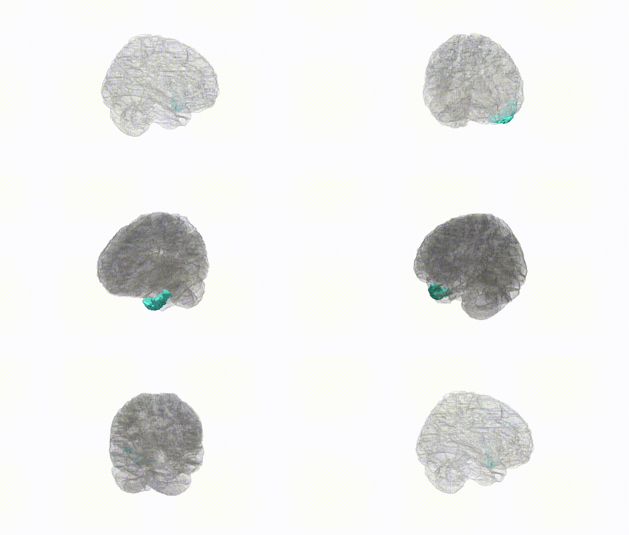
Full Quality Version: Download MP4
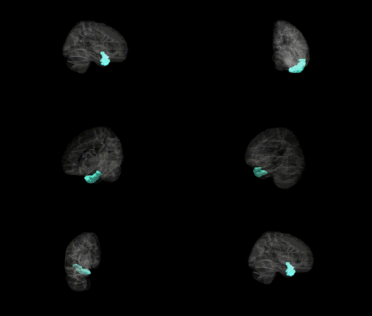
Full Quality Version: Download MP4
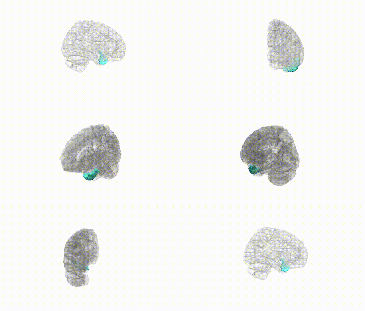
Full Quality Version: Download MP4
Wali Sidiqyar*, Gaurav Rudravaram*, Elyssa M. McMaster, Trent M. Schwartz, Adam M. Saunders, Kurt G. Schilling, Bennett A. Landman "Introducing SPINS: A Shared Public Visualization Library of Neuroanatomical Structures." Medical Imaging with Deep Learning- short paper
This resource is licensed under CC0 1.0 Universal (Public Domain).