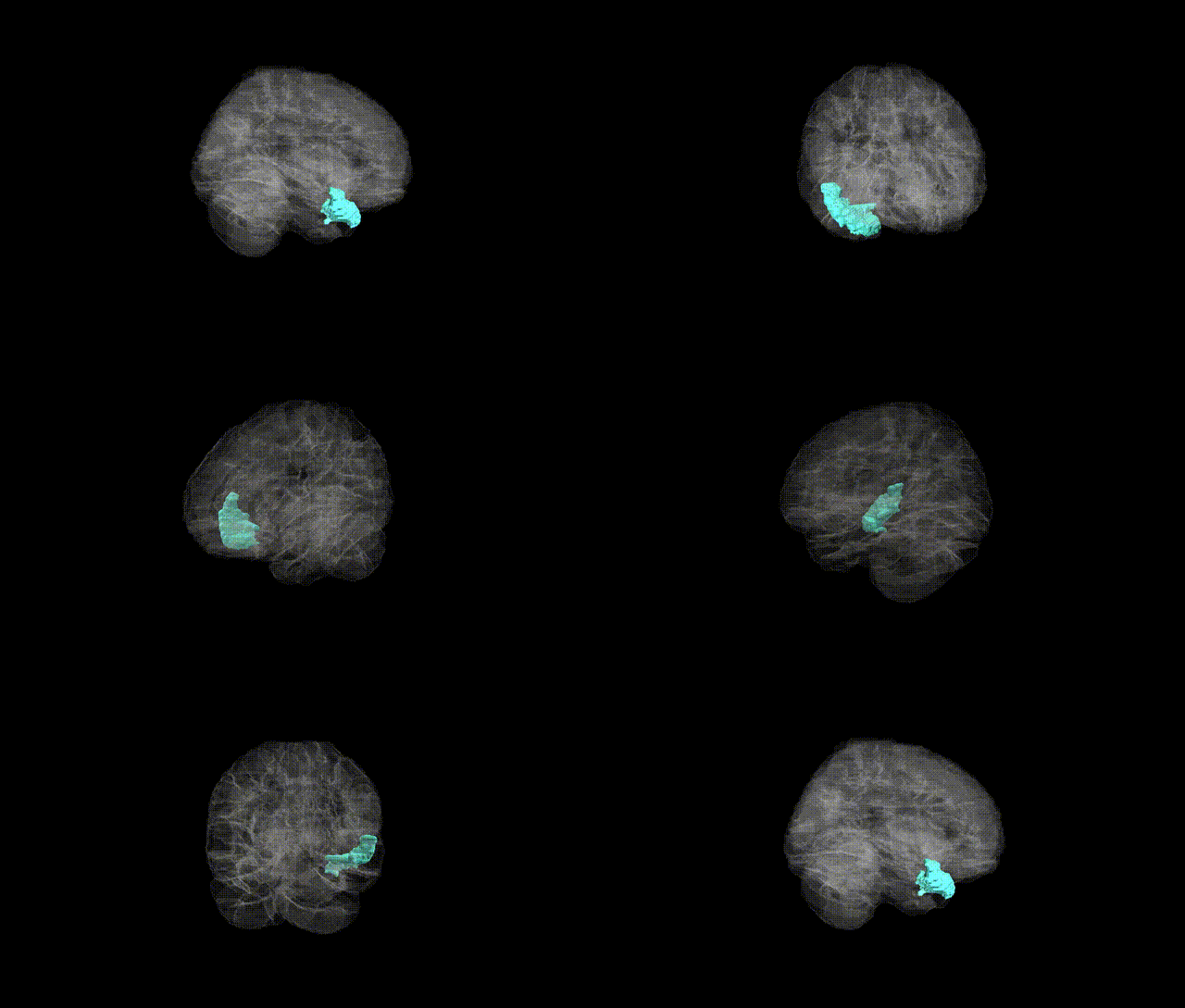
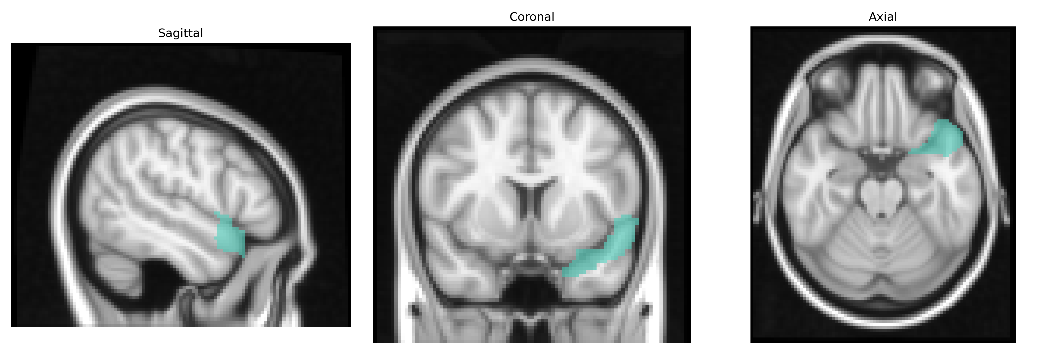
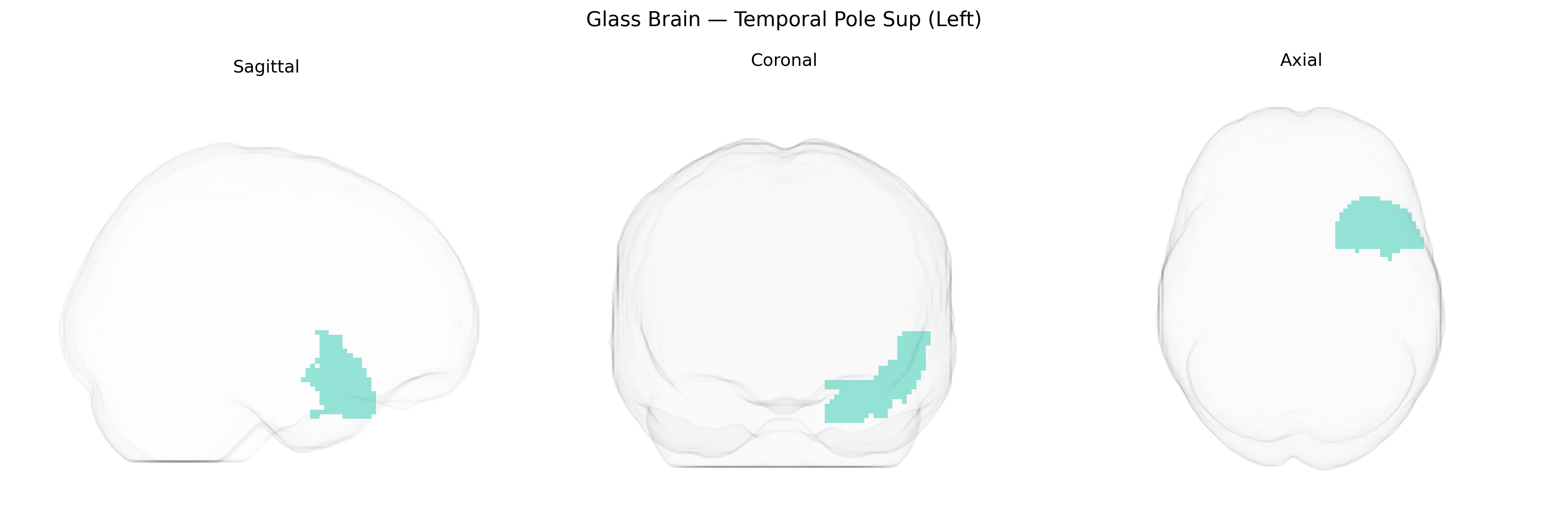
The left Temporal Pole Sup (Left) in the AAL2 atlas corresponds to the anterior portion of the superior temporal gyrus within the temporal pole, a region situated at the most rostral end of the temporal lobe. This area is heavily interconnected with limbic and paralimbic structures, including the amygdala, orbitofrontal cortex, and medial temporal regions, supporting roles in semantic memory, social and emotional processing, and higher-order auditory and language functions. Cytoarchitectonically, it is part of the paralimbic cortex, with transitional features between neocortex and allocortex, and receives multimodal sensory input that is integrated to support person-related knowledge, conceptual processing, and aspects of theory of mind. There is no direct link for “Temporal Pole Sup (Left)”; a related structure is the temporal pole of the temporal lobe: Temporal pole.
The left superior temporal pole (Temporal Pole Sup Left in the AAL2 atlas), a hub for social-emotional processing and semantic memory, has been implicated in several genetic and GWAS-based findings, though often under broader temporal lobe or superior temporal gyrus designations rather than this subregion specifically. Large-scale imaging–genetics consortia (e.g., ENIGMA, UK Biobank) have identified associations between temporal pole volume or thickness and variants in genes involved in neurodevelopment, synaptic function, and axonal guidance (such as MIR137, MAPT, and genes near 17q21.31), as well as polygenic scores for educational attainment and cognitive ability. GWAS of cortical surface measures show that temporal pole structure is moderately heritable and influenced by distributed common variants rather than single large-effect loci. Clinically, genetic risk for temporal lobe epilepsy often involves mesial temporal structures but can encompass anterior temporal regions, including the temporal pole, with rare mutations in genes like LGI1 and DEPDC5 contributing to focal epilepsies affecting this area. In neuropsychiatric disorders, schizophrenia and autism spectrum disorder polygenic risk scores have been associated with alterations in superior temporal and temporal pole morphology and connectivity, while frontotemporal dementia caused by mutations in MAPT, GRN, and C9orf72 frequently shows pronounced anterior temporal (including temporal pole) atrophy. Overall, existing genetic associations for the left superior temporal pole are largely inferred from broader temporal lobe imaging–genetic and disease studies rather than region-specific GWAS focused solely on this AAL2-defined parcel.
Overview generated by GPT-4o (2026).
Region ID: 8121
Hemisphere: left
Atlas: AAL2
Full Quality Version: Download MP4
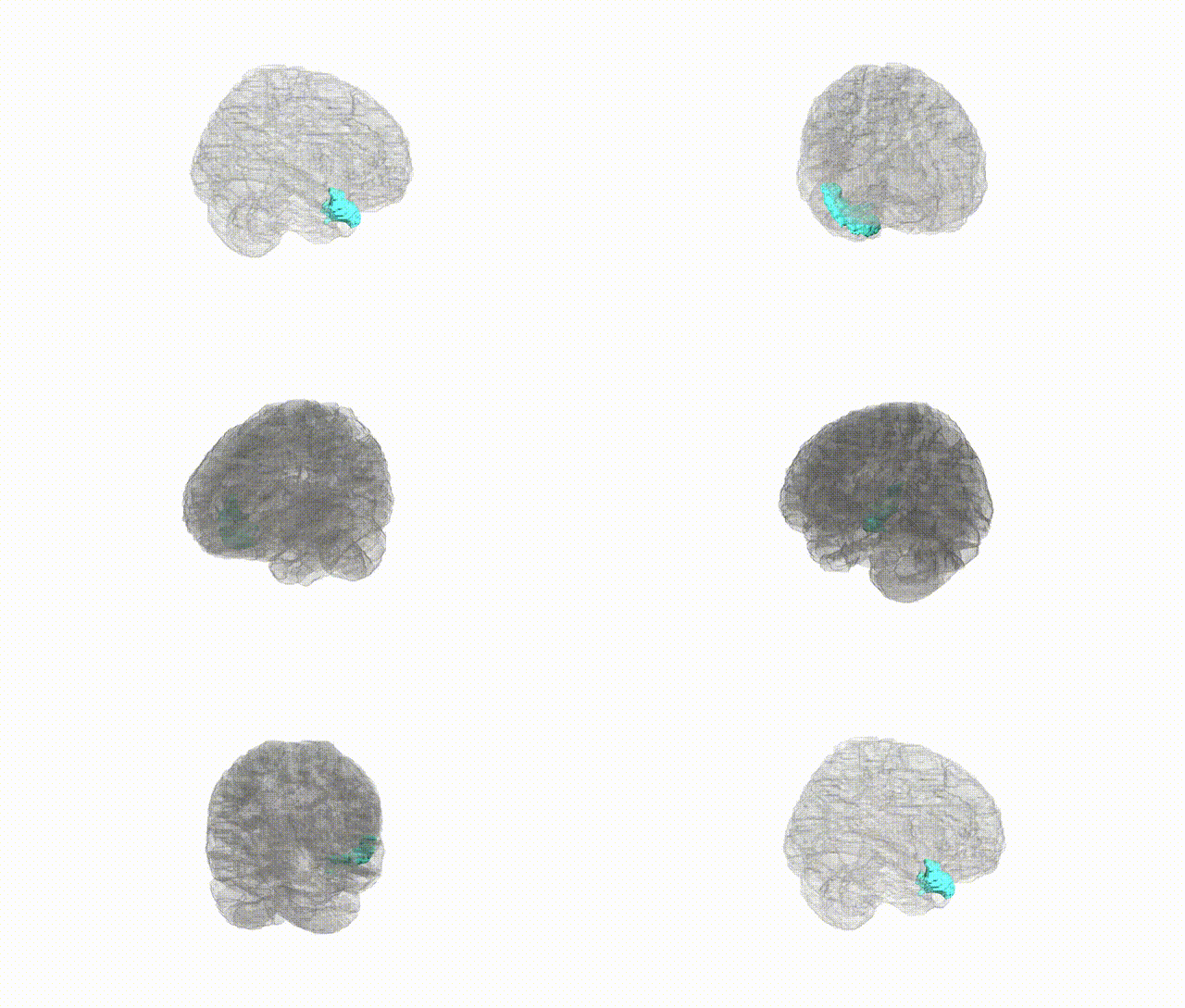
Full Quality Version: Download MP4
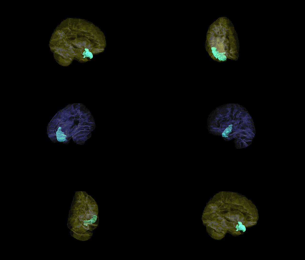
Full Quality Version: Download MP4
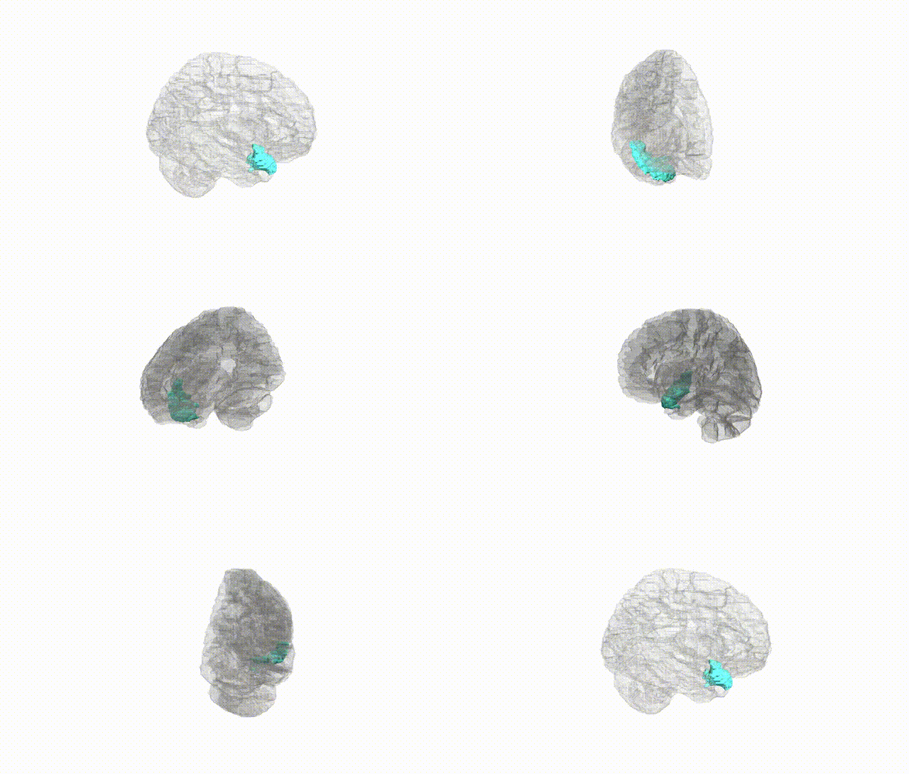
Full Quality Version: Download MP4
Wali Sidiqyar*, Gaurav Rudravaram*, Elyssa M. McMaster, Trent M. Schwartz, Adam M. Saunders, Kurt G. Schilling, Bennett A. Landman "Introducing SPINS: A Shared Public Visualization Library of Neuroanatomical Structures." Medical Imaging with Deep Learning- short paper
This resource is licensed under CC0 1.0 Universal (Public Domain).