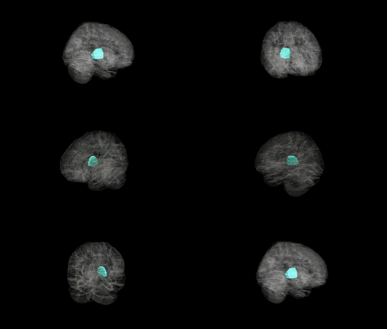
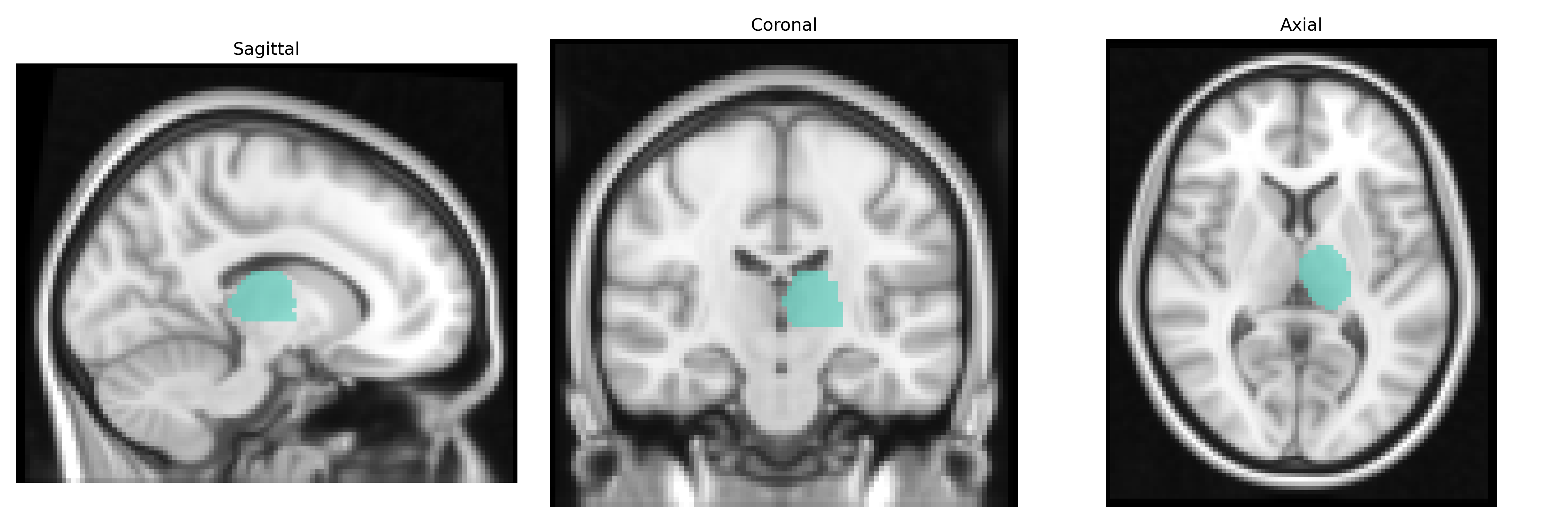
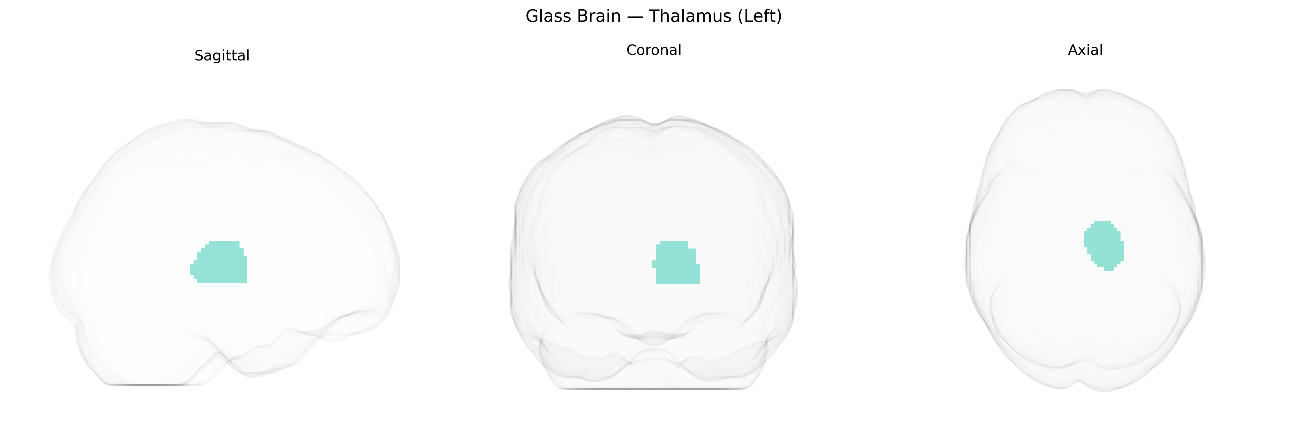
The left thalamus is a paired, ovoid diencephalic structure composed of multiple nuclei that serve as a major relay and integration center for sensory, motor, and associative information projecting to the cerebral cortex. It is situated on the left side of the third ventricle, superior to the brainstem and medial to the internal capsule, and receives inputs from ascending sensory pathways (excluding most olfactory signals), cerebellar and basal ganglia circuits, and various limbic and cortical regions. Functionally, the left thalamus is involved in modulation of somatosensory processing, motor planning and execution, attention, arousal, and aspects of cognition and language that may be lateralized to the dominant hemisphere. Its nuclei maintain topographically organized connections with cortical regions, forming thalamocortical loops essential for normal consciousness, perception, and behavioral responsiveness. There is no direct Wikipedia article for “Left Thalamus”; see the related structure Thalamus.
The left thalamus, as defined in the AAL2 atlas, has been implicated in multiple genetic and GWAS findings linking its volume, connectivity, and function to specific loci and neuropsychiatric traits. Large-scale imaging genetics consortia (e.g., ENIGMA, UK Biobank analyses) have identified common variants near genes such as MAPT, BCL2L1, SMARCA4, and in thalamic-development pathways that influence bilateral and left thalamic volume, with several hits reaching genome-wide significance. Polygenic risk scores for schizophrenia, bipolar disorder, and major depressive disorder show associations with reduced thalamic volume or altered thalamocortical connectivity, and specific schizophrenia risk loci (e.g., in CACNA1C, GRIN2A) have been linked indirectly through their effects on thalamic structure and function. Thalamic alterations related to APOE and other Alzheimer’s disease risk variants have also been reported, particularly involving atrophy and disrupted networks including the left thalamus. Additional associations include variants influencing sleep and circadian traits (e.g., in MEIS1 and other restless legs syndrome genes) and pain processing, consistent with the thalamus’s role in sensory relay, while broader brain-structure GWAS indicate that many thalamic genetic effects are shared across hemispheres but can show lateralized strength, including in the left thalamus.
Overview generated by GPT-4o (2026).
Region ID: 7101
Hemisphere: left
Atlas: AAL2
Full Quality Version: Download MP4
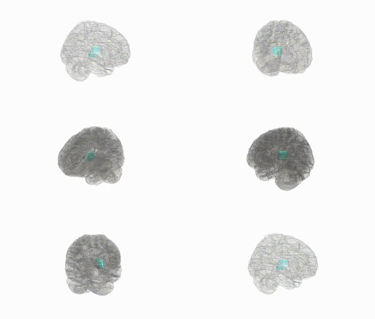
Full Quality Version: Download MP4
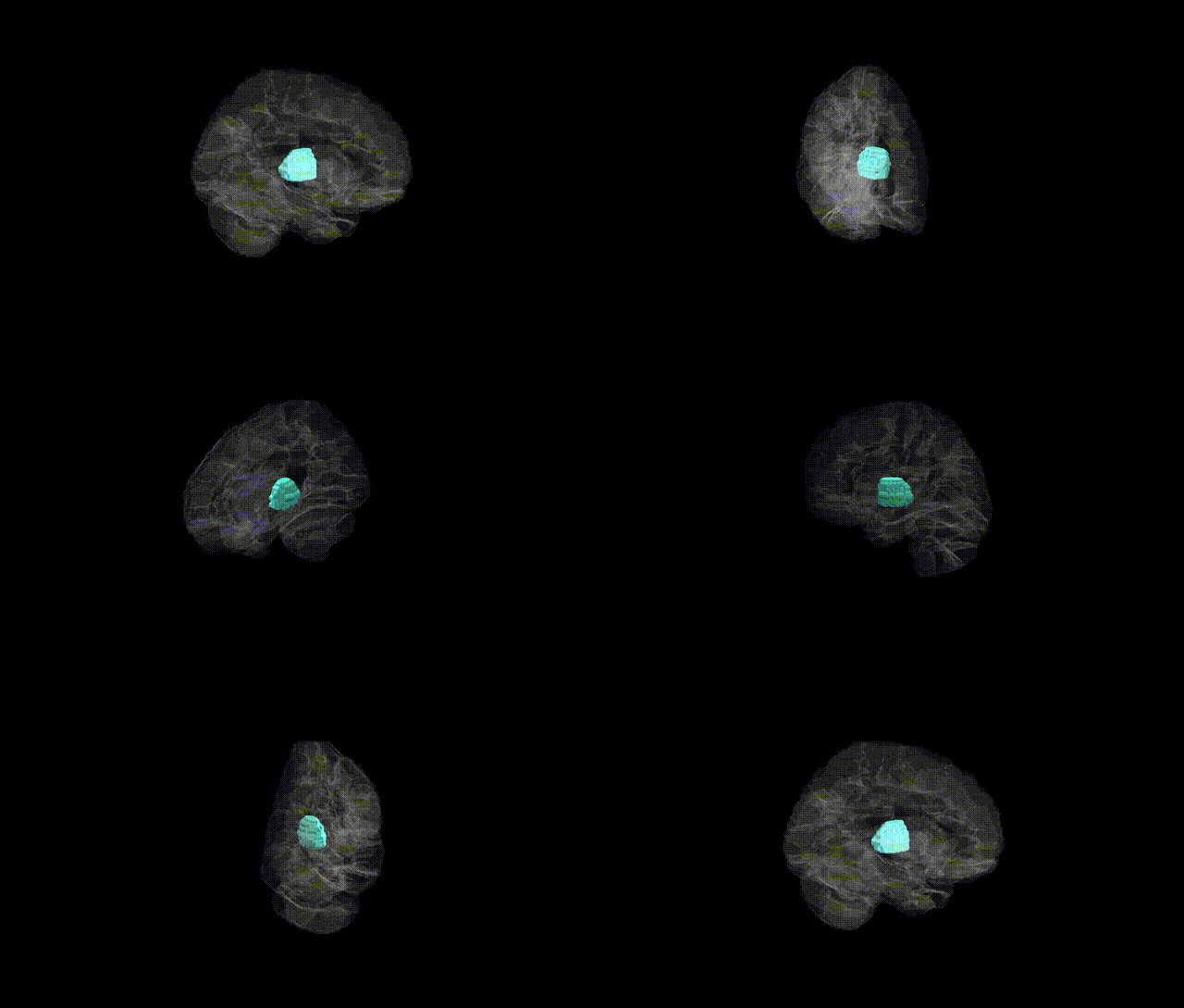
Full Quality Version: Download MP4
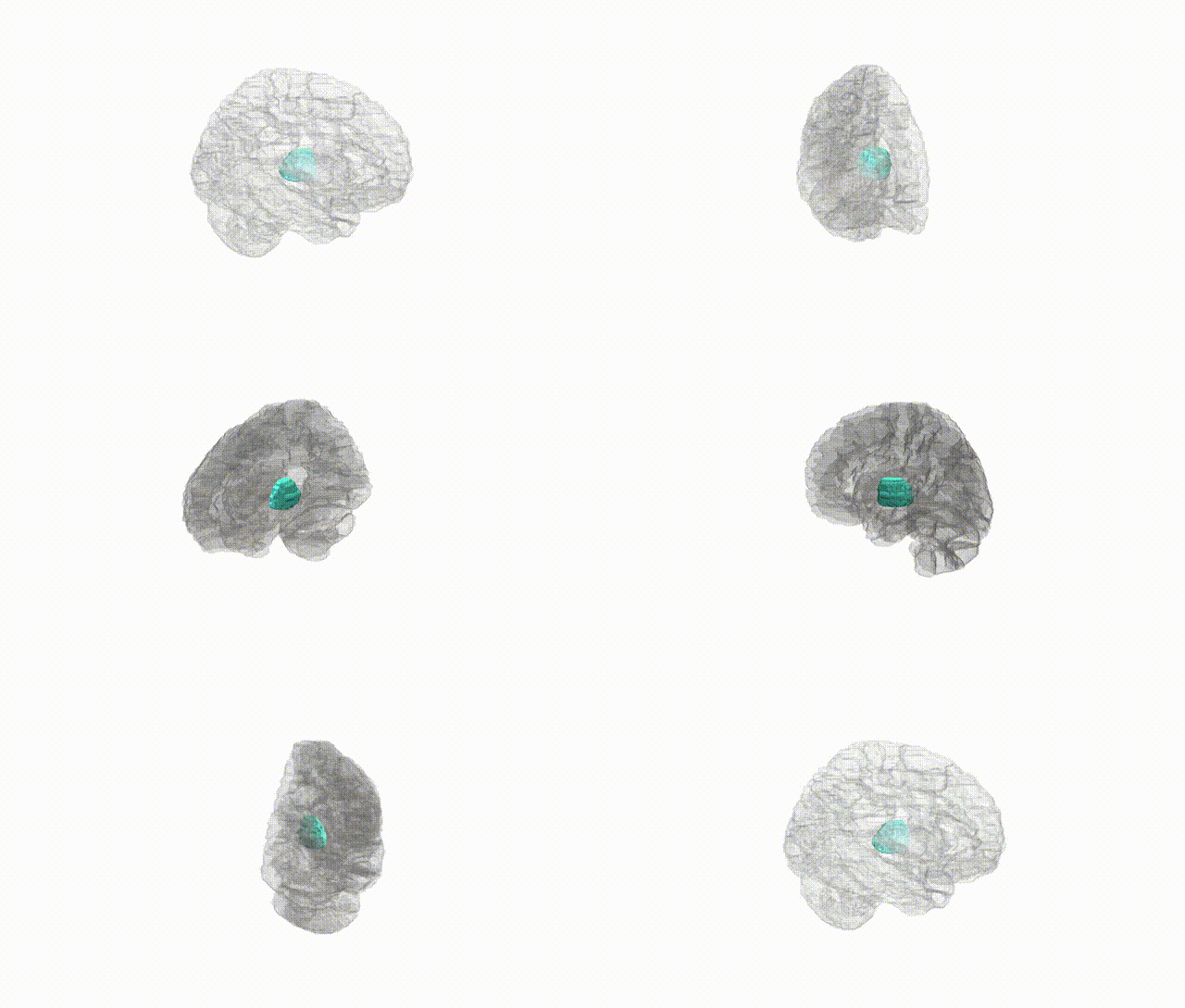
Full Quality Version: Download MP4
Wali Sidiqyar*, Gaurav Rudravaram*, Elyssa M. McMaster, Trent M. Schwartz, Adam M. Saunders, Kurt G. Schilling, Bennett A. Landman "Introducing SPINS: A Shared Public Visualization Library of Neuroanatomical Structures." Medical Imaging with Deep Learning- short paper
This resource is licensed under CC0 1.0 Universal (Public Domain).