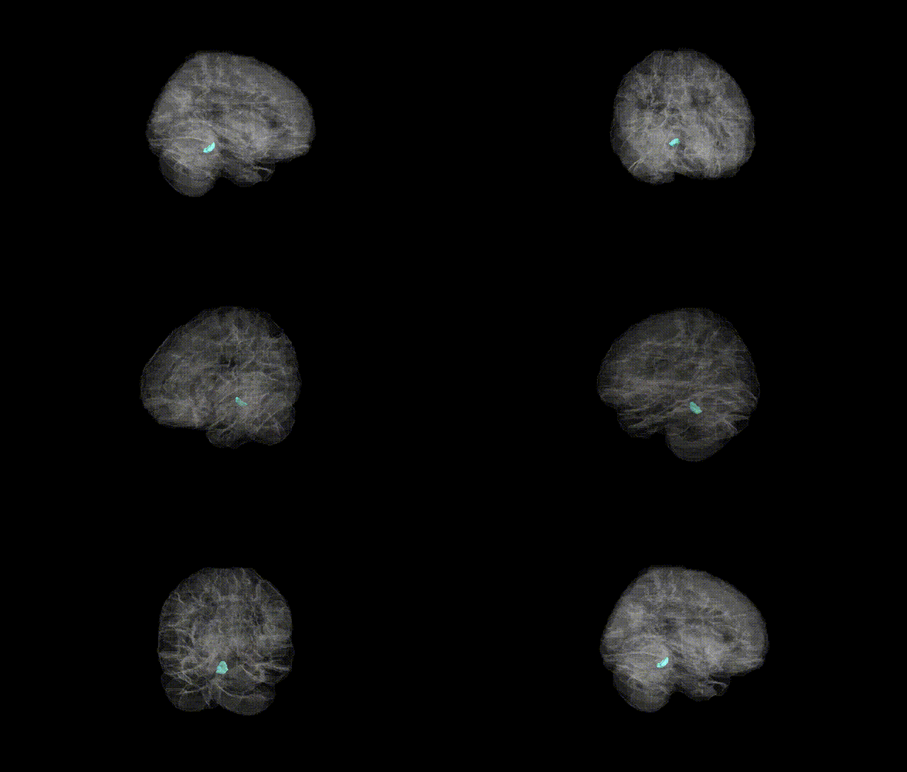
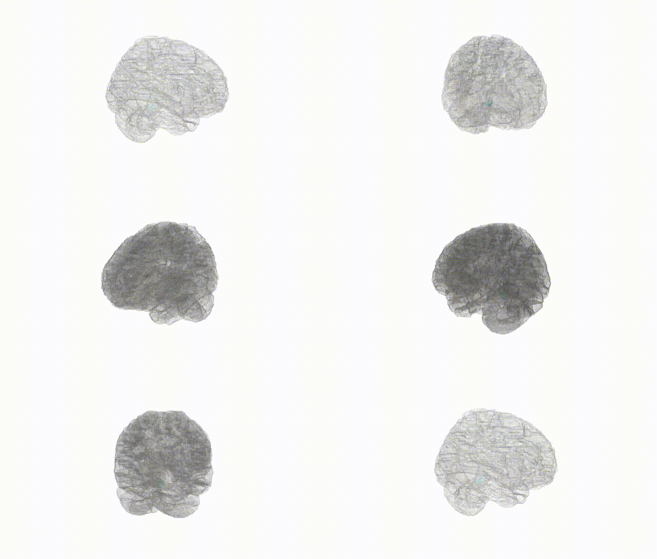
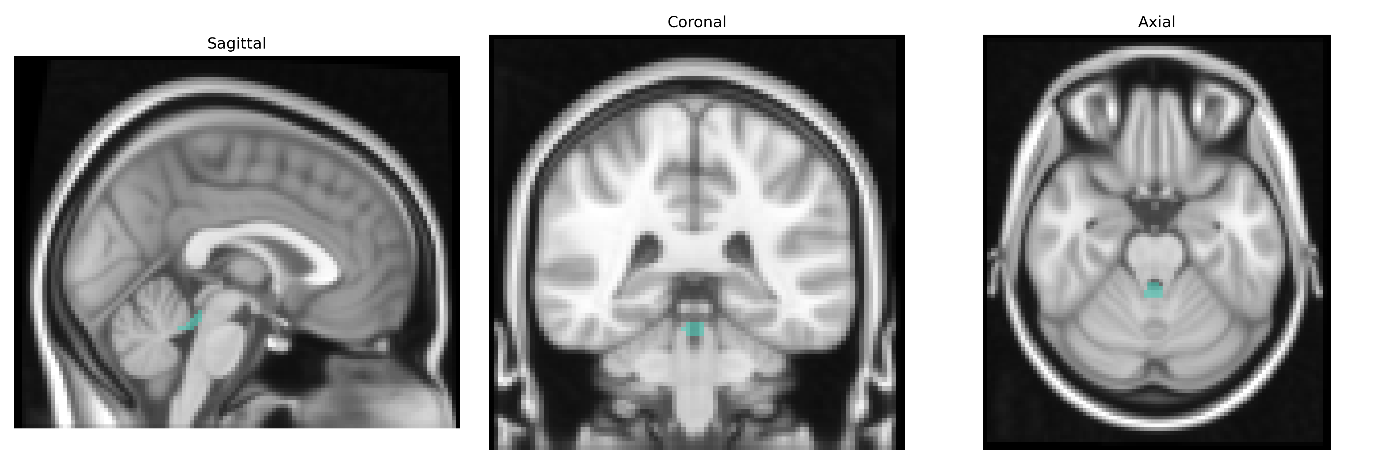
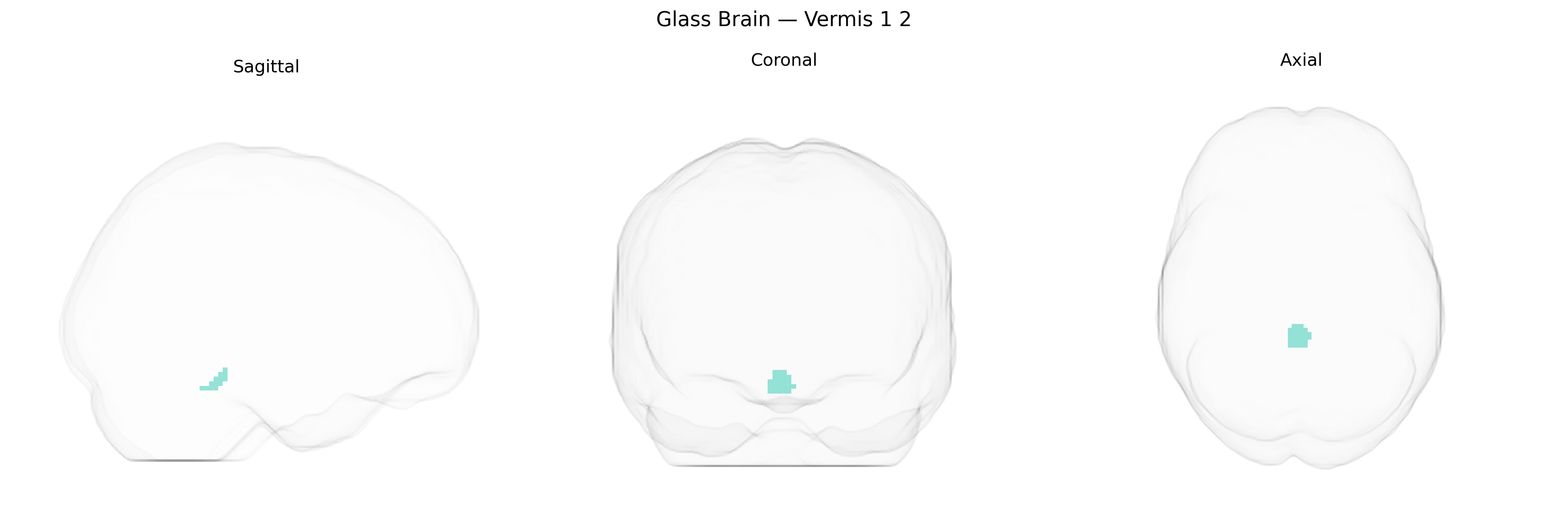
The bilateral Vermis 1–2 region in the AAL2 atlas corresponds to the anterior segments of the cerebellar vermis, located along the midline of the anterior lobe of the cerebellum. This region is composed primarily of lobules I and II and is involved in the coordination of axial and proximal limb movements, as well as basic postural control, through integration of somatosensory and vestibular inputs. Neuronal circuitry in Vermis 1–2 includes Purkinje cells projecting inhibitory outputs to deep cerebellar nuclei (particularly the fastigial nucleus), which in turn modulate brainstem and thalamic motor pathways. Functionally, this region is associated with regulation of muscle tone, gait, and balance, and lesions can lead to truncal ataxia and disturbances of stance and locomotion. There is no direct link for Vermis 1–2; see the related structure Cerebellar vermis.
Genetic associations specifically targeting the bilateral Vermis 1–2 region (anterior cerebellar vermis) in the AAL2 atlas are limited, but converging imaging‑genetics and GWAS evidence implicates this region within broader cerebellar and frontocerebellar circuits. Large brain-structure GWAS (e.g., ENIGMA, UK Biobank) have identified common variants in genes related to neurodevelopment, synaptic plasticity, and axon guidance (such as those in the MAPT, CELSR, and FOXP families) that influence total cerebellar volume and midline vermal structures, though effects are generally distributed rather than Vermis 1–2–specific. Disorders with robust genetic underpinnings that show structural or functional abnormalities involving the anterior vermis include autism spectrum disorder (with risk variants in genes such as CHD8, SHANK3, and SCN2A), attention‑deficit/hyperactivity disorder (implicating dopaminergic and neurodevelopmental genes like DRD4 and SLC6A3 in networks that include the anterior cerebellum), and schizophrenia and bipolar disorder (where polygenic risk scores and loci in genes such as CACNA1C and ZNF804A have been linked to cerebellar and vermal alterations). Additionally, genes associated with alcohol use disorder (e.g., variants in ADH1B, GABRA2) and spinocerebellar ataxias (e.g., CAG expansions in ATXN1, ATXN2, ATXN3) have been tied to degeneration or volume loss in the anterior vermis, often encompassing Vermis 1–2, although most studies report lobular or global vermal changes rather than isolating this subregion.
Overview generated by GPT-4o (2026).
Region ID: 9100
Hemisphere: bilateral
Atlas: AAL2
Full Quality Version: Download MP4
Full Quality Version: Download MP4
Wali Sidiqyar*, Gaurav Rudravaram*, Elyssa M. McMaster, Trent M. Schwartz, Adam M. Saunders, Kurt G. Schilling, Bennett A. Landman "Introducing SPINS: A Shared Public Visualization Library of Neuroanatomical Structures." Medical Imaging with Deep Learning- short paper
This resource is licensed under CC0 1.0 Universal (Public Domain).