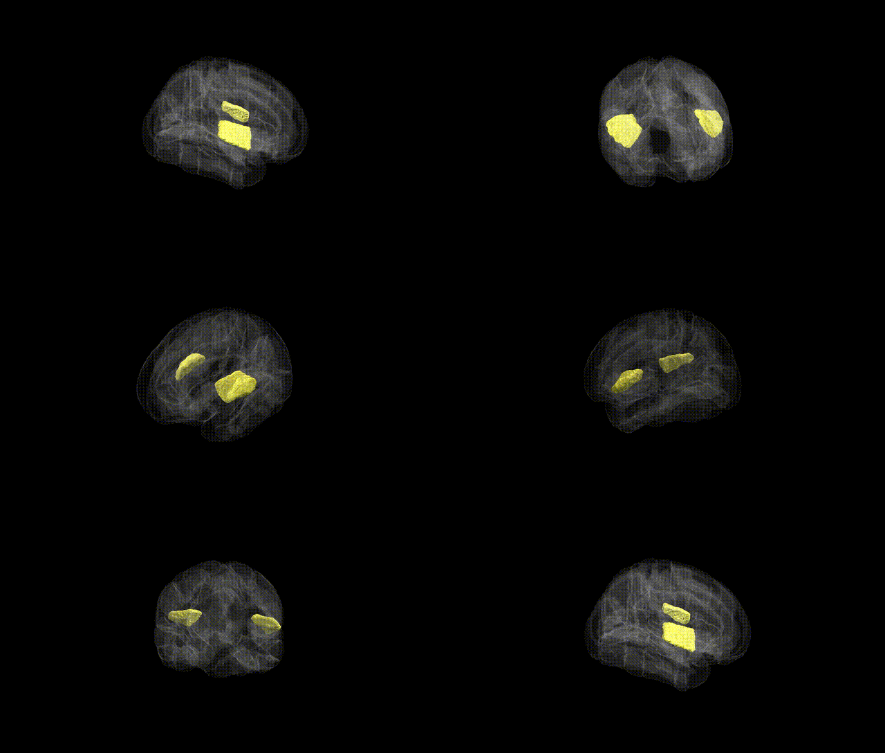
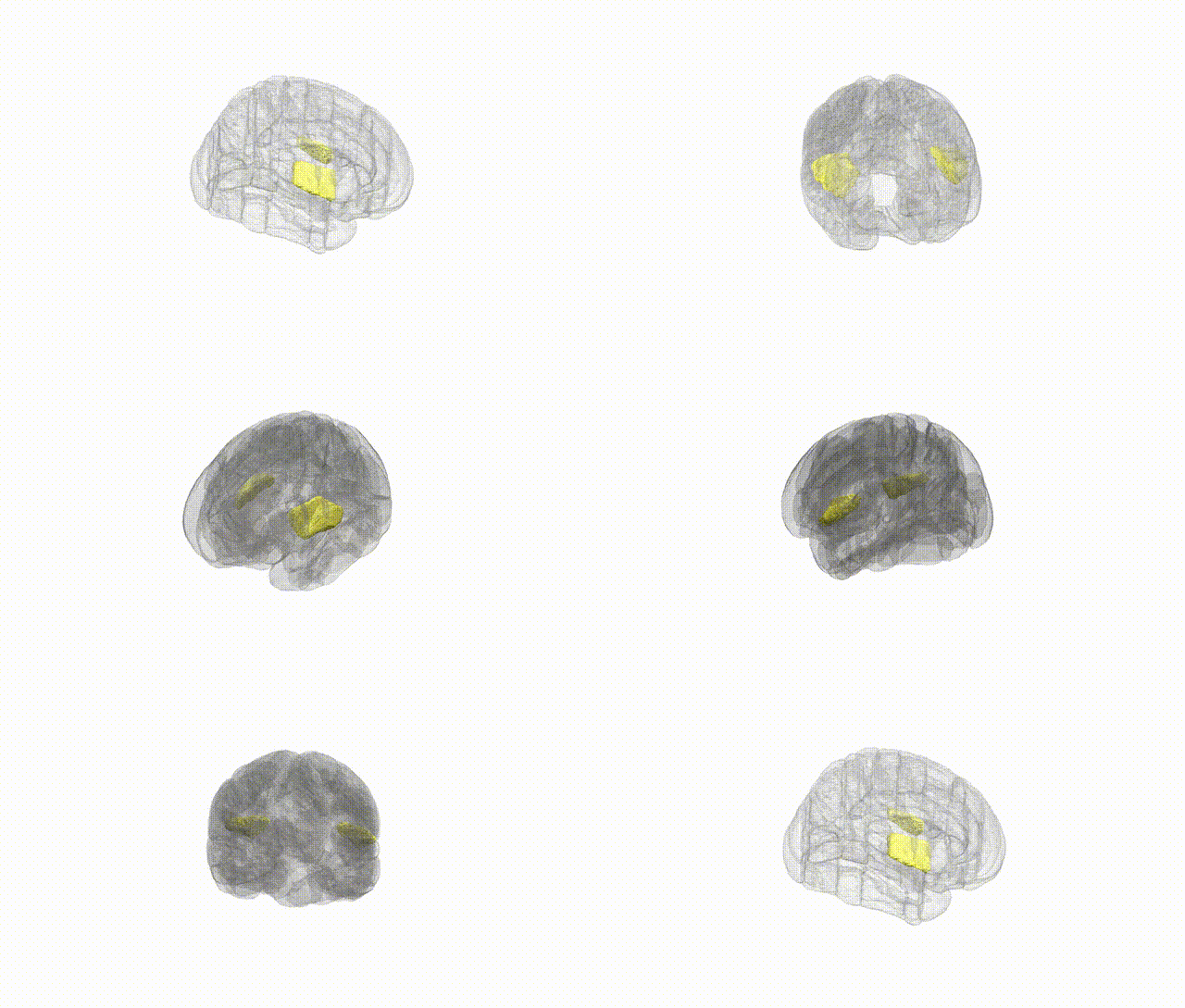
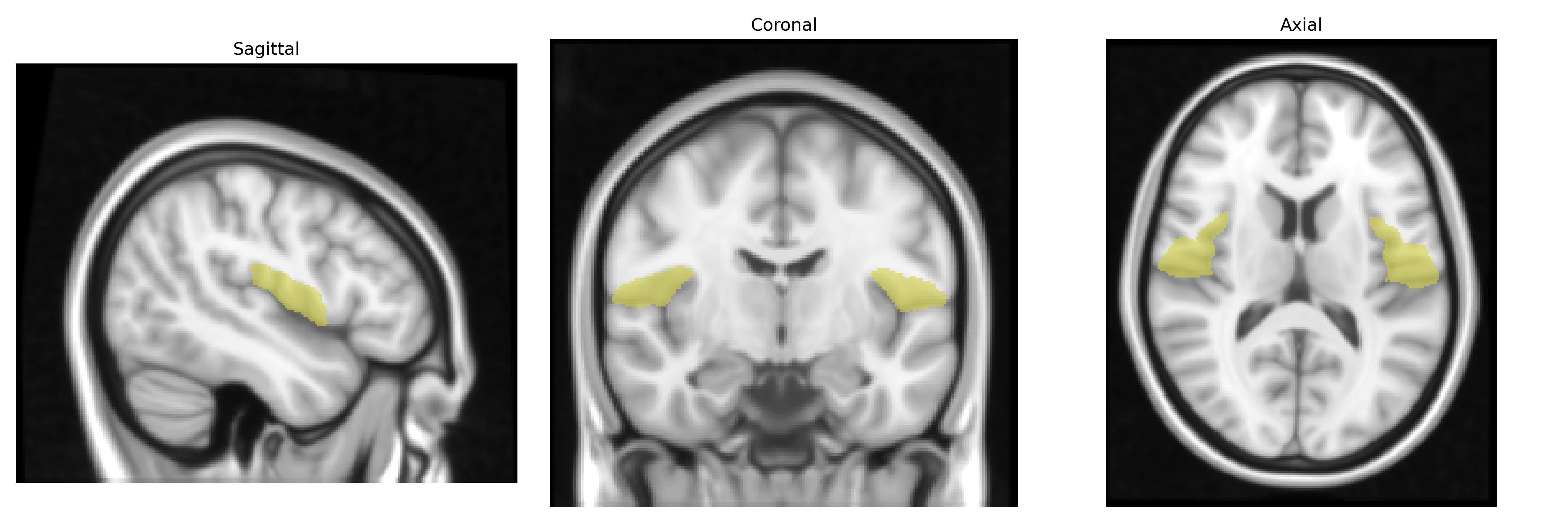
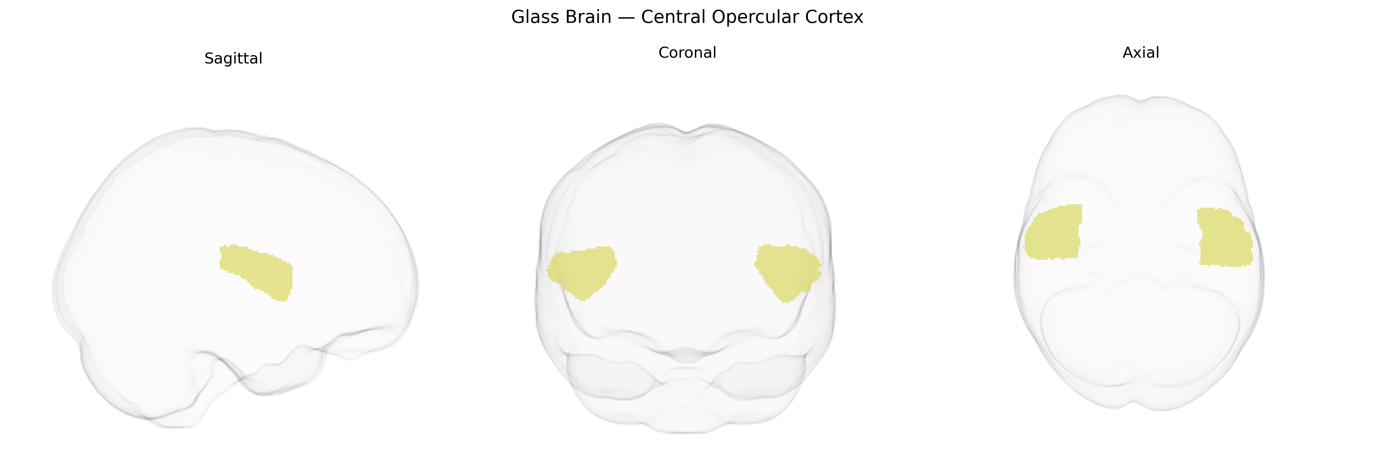
The bilateral Central Opercular Cortex, as defined in the Harvard-Oxford cortical atlas, corresponds to the opercular portion of the parietal and posterior frontal lobes overlying the insula, forming part of the perisylvian region surrounding the lateral sulcus. It participates in sensorimotor processing of the face, tongue, and orofacial musculature, and is implicated in articulation, speech production, and somatosensory integration related to oral and pharyngeal functions. This region forms part of the broader opercular complex, which includes frontal, parietal, and temporal opercula that collectively cover the insular cortex and contribute to multimodal integration, praxis, and language networks. There is no direct link for the Central Opercular Cortex, but it is part of the Operculum (anatomy).
The bilateral Central Opercular Cortex, as defined in the Harvard-Oxford cortical atlas, overlaps functionally and anatomically with perisylvian sensorimotor and language-related regions, and genetic associations typically emerge from large-scale imaging-genetics and neuropsychiatric GWAS rather than region-specific candidate studies. Variants in genes implicated in cortical development, synaptic function, and myelination—such as common polymorphisms near or within genes like FOXP2, CNTNAP2, DCDC2, KIAA0319, and other language/reading-related loci—have been associated with structural and functional alterations in opercular and perisylvian cortices, including changes in cortical thickness, surface area, and activation patterns during speech and phonological tasks. Brain-wide GWAS (e.g., ENIGMA and UK Biobank imaging studies) report that polygenic influences on global cortical morphology, particularly loci enriched for neurodevelopmental pathways, contribute to inter-individual variability in the opercular and adjacent inferior frontal and insular cortices, though specific single-locus effects on the Central Opercular Cortex are typically modest and not always region-selective. Clinically, genetic liability for developmental language disorder, dyslexia, and stuttering, as well as for neuropsychiatric conditions like schizophrenia, autism spectrum disorder, and mood disorders, has been linked to abnormalities in the broader perisylvian and opercular network, suggesting pleiotropic effects of risk variants on this region’s microstructure and connectivity. However, current evidence is largely indirect, arising from associations with network-level or lobar measures rather than the Central Opercular Cortex as an isolated structure, and definitive region-specific genetic signatures remain to be clearly delineated in the literature.
Overview generated by GPT-4o (2026).
Region ID: 42
Hemisphere: bilateral
Atlas: HarvardOxford cort maxprob thr25 1mm
Full Quality Version: Download MP4
Full Quality Version: Download MP4
Wali Sidiqyar*, Gaurav Rudravaram*, Elyssa M. McMaster, Trent M. Schwartz, Adam M. Saunders, Kurt G. Schilling, Bennett A. Landman "Introducing SPINS: A Shared Public Visualization Library of Neuroanatomical Structures." Medical Imaging with Deep Learning- short paper
This resource is licensed under CC0 1.0 Universal (Public Domain).