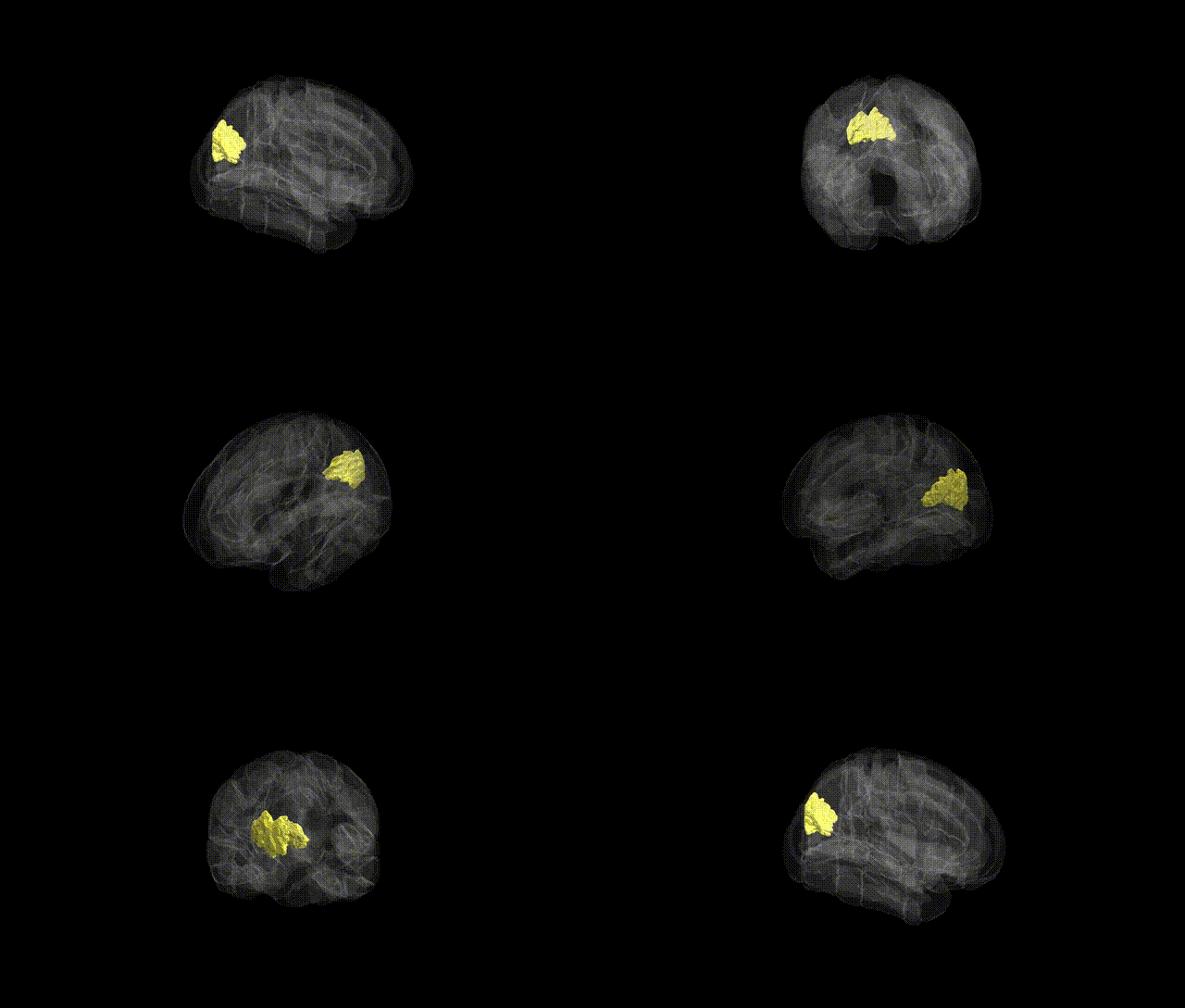
The bilateral cuneal cortex is a medial occipital lobe structure forming part of the primary and associative visual cortices, situated superior to the calcarine sulcus and extending dorsally toward the parieto-occipital sulcus. It corresponds largely to portions of Brodmann areas 17 and 18 and is involved in early-stage processing of visual information, particularly from the lower visual field via retinal inputs relayed through the lateral geniculate nucleus to the striate cortex. Functionally, the cuneal cortex participates in basic feature analysis such as orientation, contrast, and motion, and contributes to visuospatial integration and perception. In the Harvard-Oxford cortical atlas, the bilateral cuneal cortex label delineates this region across both hemispheres based on probabilistic structural boundaries rather than specific cytoarchitectonic subdivisions. There is no direct link for “cuneal cortex”; a closely related and encompassing structure is the Occipital lobe.
The bilateral cuneal cortex, a primary visual and visuospatial processing region in the occipital lobe, has been implicated in several genetic and GWAS-based associations, particularly through imaging genetics and large-scale brain-structure studies. Variants in genes related to neurodevelopment, synaptic function, and myelination (such as those in pathways involving microtubule dynamics, glutamatergic signaling, and neurite outgrowth) have been linked to cuneal cortex thickness, surface area, or activation, with loci on chromosomes including 3, 6, and 17 repeatedly emerging in occipital and visual-cortex–focused imaging GWAS. Cuneal cortex measures have shown heritability and genetic correlations with visual acuity and visual processing traits, as well as with general cognitive ability and educational attainment in multivariate genetic analyses. Psychiatric and neurological GWAS—particularly for schizophrenia, major depressive disorder, and bipolar disorder—have identified polygenic risk loadings associated with structural or functional alterations in the cuneal cortex or adjacent medial occipital regions, suggesting shared genetic architecture between these disorders and visual-network integrity. Additionally, genetic risk for Alzheimer’s disease and small-vessel cerebrovascular disease has been associated with occipital/cuneal cortical thinning and perfusion changes, while variants implicated in migraine and photosensitivity show associations with activity and connectivity in visual cortex networks that include the cuneal cortex. Collectively, available evidence points to the cuneal cortex as a heritable, genetically modulated hub within visual and higher-order networks, with shared polygenic influences spanning sensory processing, cognition, and multiple neuropsychiatric and neurodegenerative disorders, although specific gene–region links remain more diffuse and polygenic than singular.
Overview generated by GPT-4o (2026).
Region ID: 32
Hemisphere: bilateral
Atlas: HarvardOxford cort maxprob thr25 1mm
Full Quality Version: Download MP4
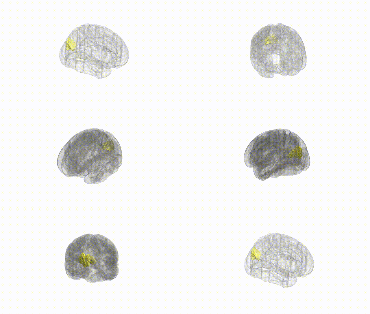
Full Quality Version: Download MP4
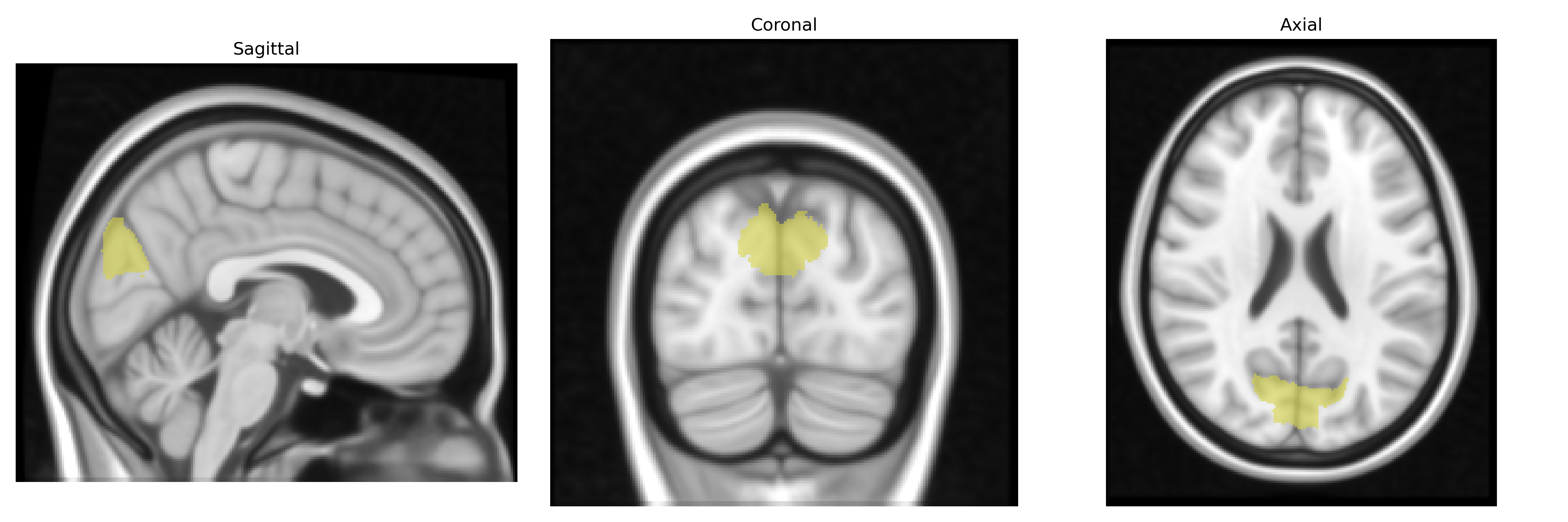
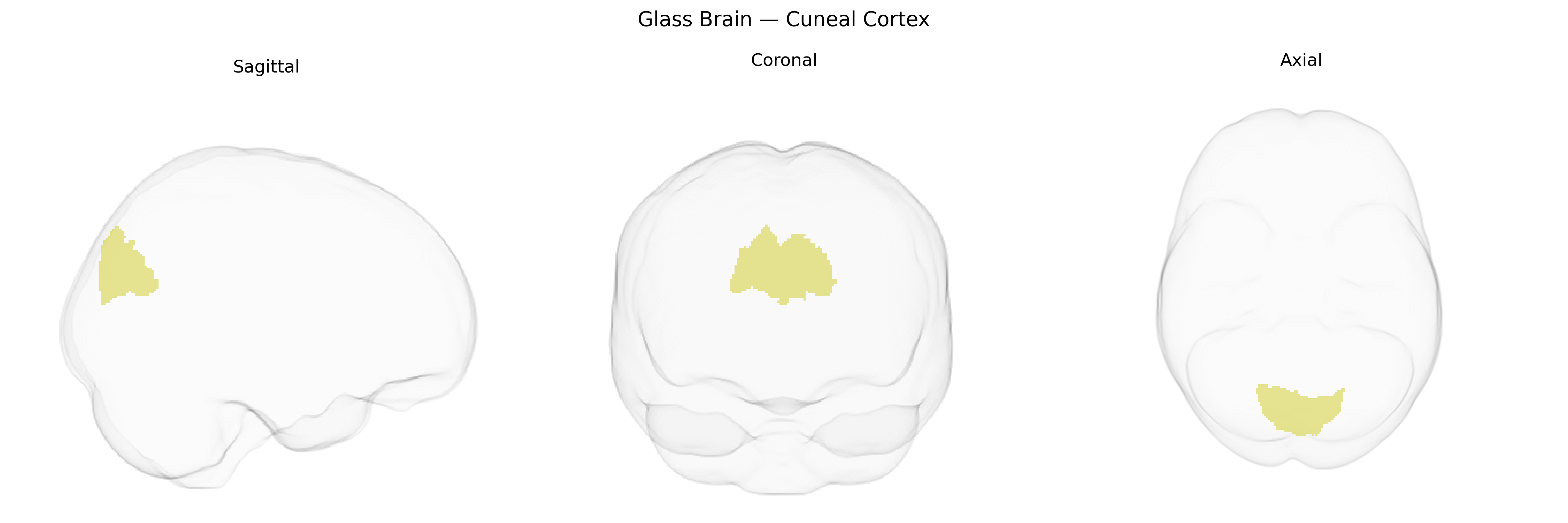
Wali Sidiqyar*, Gaurav Rudravaram*, Elyssa M. McMaster, Trent M. Schwartz, Adam M. Saunders, Kurt G. Schilling, Bennett A. Landman "Introducing SPINS: A Shared Public Visualization Library of Neuroanatomical Structures." Medical Imaging with Deep Learning- short paper
This resource is licensed under CC0 1.0 Universal (Public Domain).