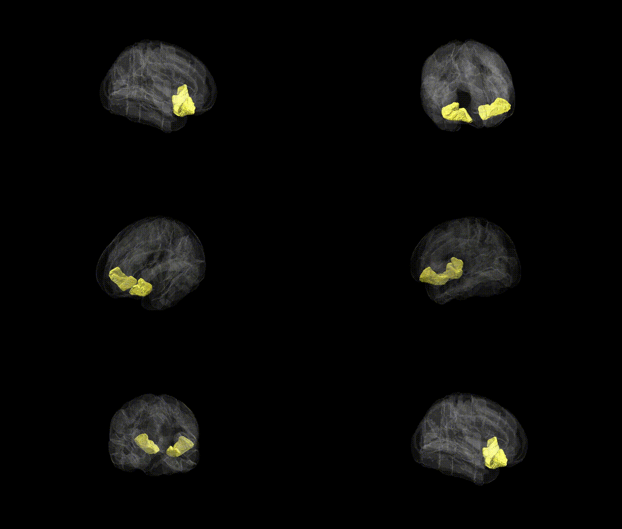
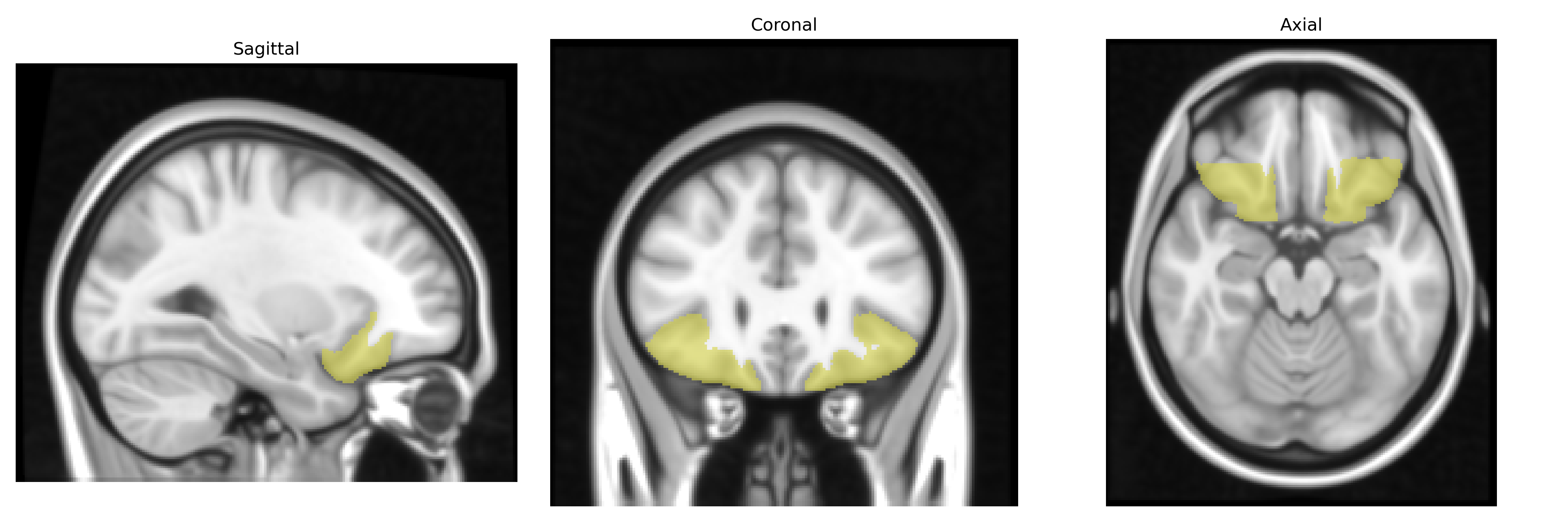
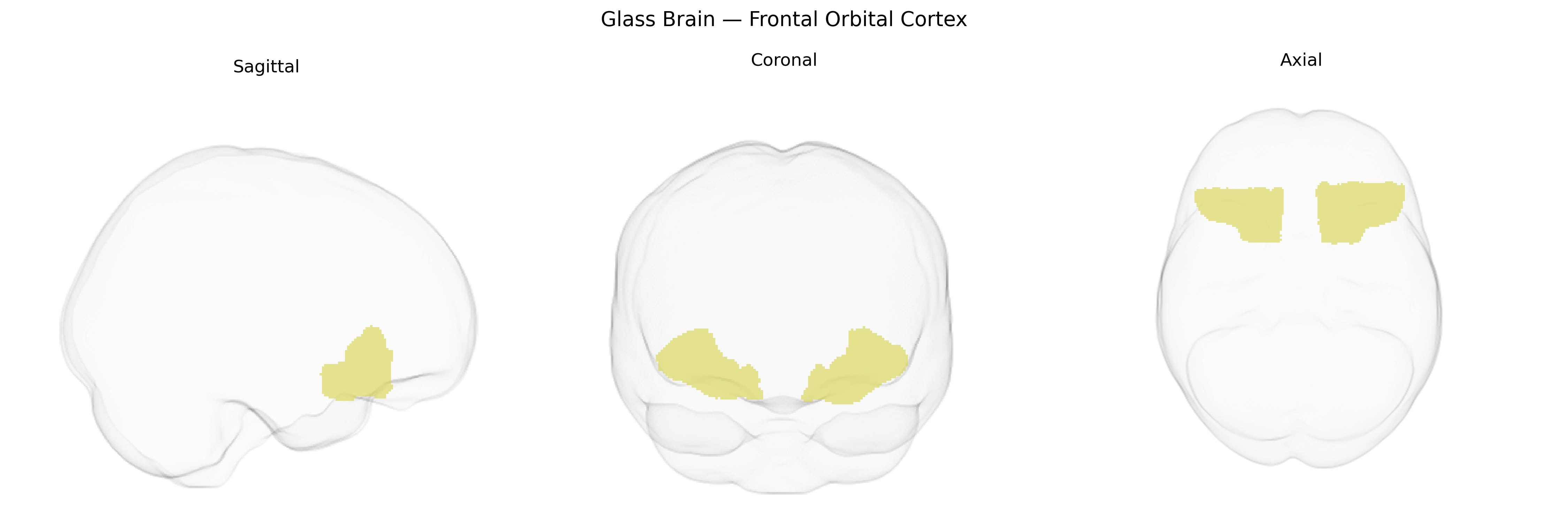
The bilateral Frontal Orbital Cortex, as defined in the Harvard-Oxford Cortical Atlas (maxprob thr25, 1 mm), corresponds to orbitofrontal regions located on the ventral surface of the frontal lobes, overlying the orbits. This cortex is composed of granular and agranular fields involved in higher-order association processing, including evaluation of reward and punishment, encoding of stimulus-value associations, decision-making under uncertainty, and modulation of affective and social behaviors. It is heavily interconnected with limbic structures (such as the amygdala and ventral striatum), medial prefrontal and anterior cingulate cortices, and sensory association areas, supporting integration of emotional, sensory, and contextual information. Lesions or dysfunction in this region have been associated with impairments in adaptive decision-making, altered reward sensitivity, disinhibition, and changes in personality. There is no direct Wikipedia article specifically titled “Frontal Orbital Cortex”; a closely related structure is the Orbitofrontal cortex.
The bilateral frontal orbital cortex (often approximating the orbitofrontal cortex in the Harvard-Oxford cortical atlas) shows genetic associations primarily through imaging–genetics and psychiatric GWAS studies implicating genes involved in synaptic function, neurodevelopment, and monoaminergic signaling. Twin and SNP-based heritability studies indicate that orbitofrontal cortical thickness and surface area are substantially heritable, with common variants in genes such as CACNA1C, GRM3, BDNF, and COMT repeatedly associated with structural and functional differences in this region, especially in the context of mood and psychotic disorders. Large-scale GWAS of cortical morphology (e.g., ENIGMA and related consortia) have identified loci near genes involved in neuronal migration and axon guidance that influence orbitofrontal thickness and surface area, while polygenic risk scores for major depressive disorder, bipolar disorder, schizophrenia, and obsessive–compulsive disorder correlate with altered volume or activity in the frontal orbital cortex. This region’s genetic correlates are also linked to traits such as impulsivity, risk-taking, addiction vulnerability (including alcohol and nicotine use), and neuroticism, with associated loci overlapping dopaminergic and serotonergic genes (e.g., DRD2, SLC6A4) and glutamatergic signaling pathways. Additionally, GWAS of Alzheimer’s disease, frontotemporal dementia, and neurodevelopmental conditions such as autism spectrum disorder and ADHD have reported orbitofrontal involvement, often mediated by variants in genes related to synaptic plasticity, immune signaling, and lipid metabolism, underscoring the frontal orbital cortex as a genetically sensitive hub for affective, reward, and decision-making circuitry.
Overview generated by GPT-4o (2026).
Region ID: 33
Hemisphere: bilateral
Atlas: HarvardOxford cort maxprob thr25 1mm
Full Quality Version: Download MP4
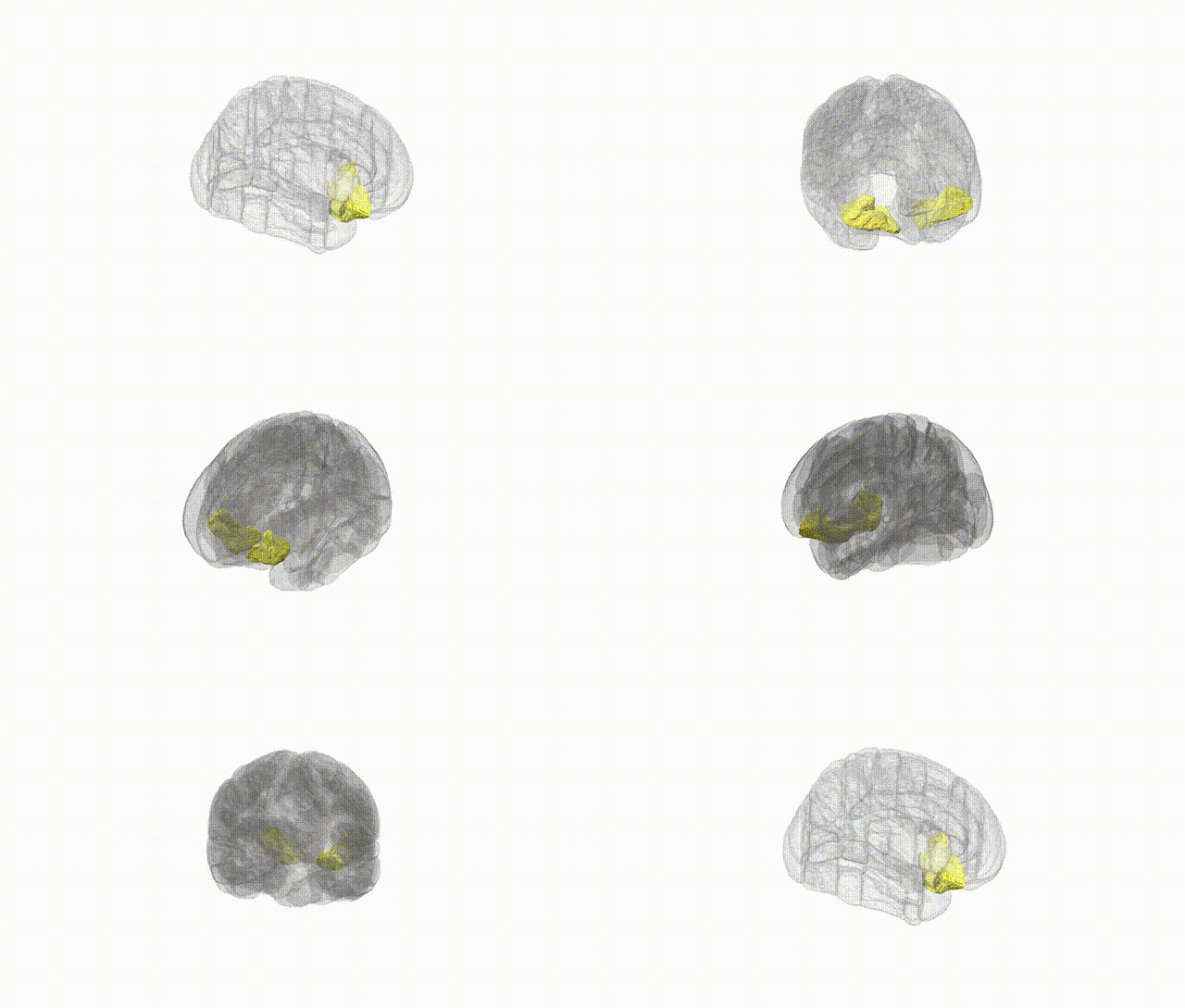
Full Quality Version: Download MP4
Wali Sidiqyar*, Gaurav Rudravaram*, Elyssa M. McMaster, Trent M. Schwartz, Adam M. Saunders, Kurt G. Schilling, Bennett A. Landman "Introducing SPINS: A Shared Public Visualization Library of Neuroanatomical Structures." Medical Imaging with Deep Learning- short paper
This resource is licensed under CC0 1.0 Universal (Public Domain).