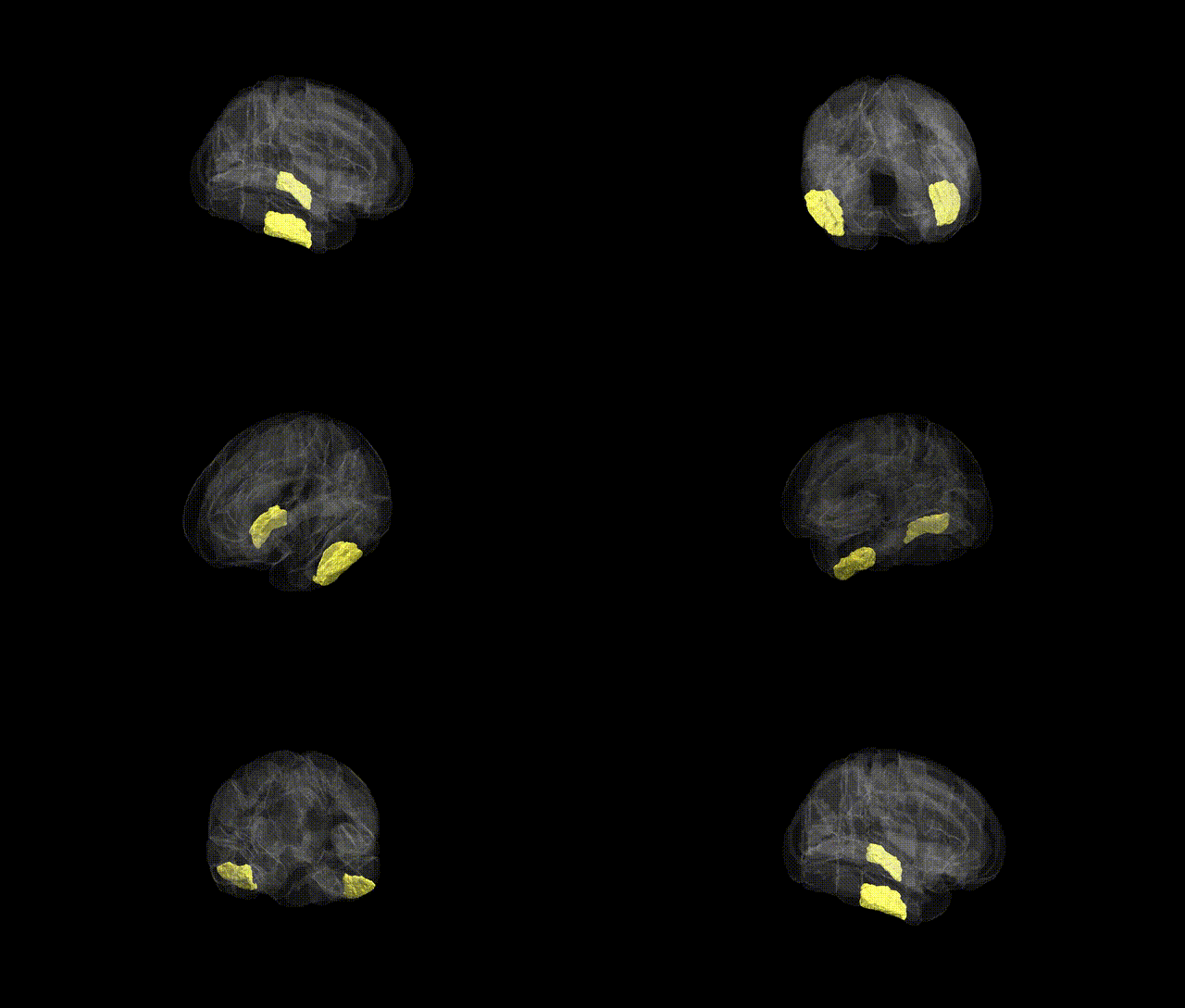
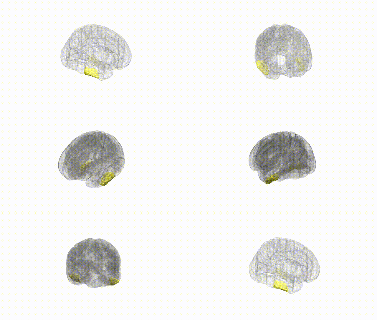
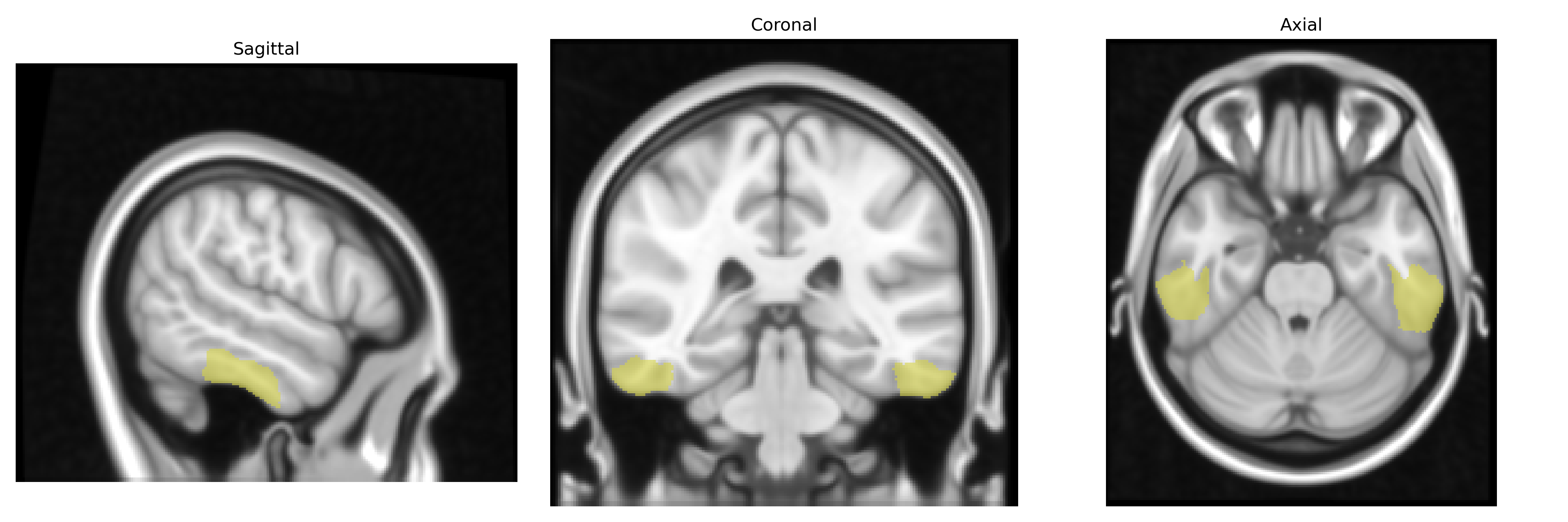
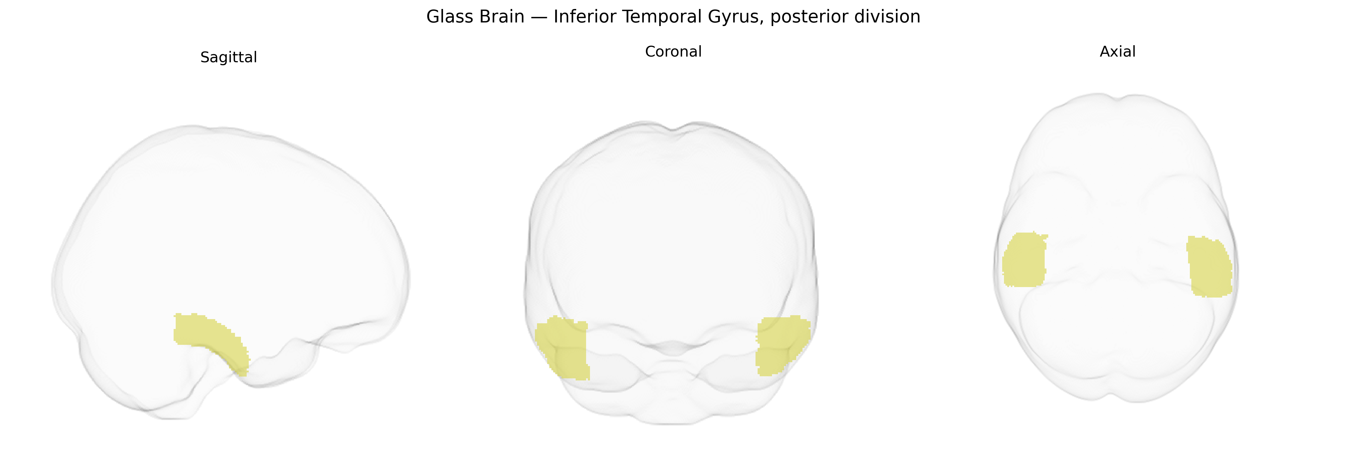
The bilateral Inferior Temporal Gyrus, posterior division (Harvard–Oxford Cortical Atlas, maxprob thr25 1 mm) refers to the caudal portion of the inferior temporal gyrus in both hemispheres, located on the ventrolateral surface of the temporal lobe, just superior to the occipitotemporal (fusiform) gyrus and inferior to the middle temporal gyrus. This region lies near the occipito-temporal junction and is supplied primarily by branches of the posterior cerebral and middle cerebral arteries. Functionally, the posterior inferior temporal cortex is implicated in high-level visual processing, including object, shape, and complex pattern recognition, and contributes to the ventral visual “what” pathway, with strong connectivity to occipital visual cortices, fusiform regions, and anterior temporal association areas. There is no dedicated Wikipedia article for the “Inferior Temporal Gyrus, posterior division”; a related structure is the Inferior temporal gyrus.
The bilateral Inferior Temporal Gyrus, posterior division (ITG-p), a high-level visual association area implicated in object recognition and semantic processing, has been indirectly associated with genetic variation primarily through large-scale imaging genetics and GWAS of brain structure and cognitive traits rather than region-specific candidate studies. Multimodal GWAS of cortical thickness and surface area (e.g., ENIGMA and UK Biobank–based analyses) have identified loci near genes such as KIAA0586, TESC, and others involved in neurodevelopment and synaptic function that influence temporal cortical morphology, with several signals encompassing inferior and lateral temporal regions that approximate the ITG-p in the Harvard-Oxford atlas. Polygenic scores for general cognitive ability, educational attainment, and reading/language traits show associations with temporal cortical measures, suggesting that distributed genetic architectures supporting semantic and visual processing extend to ITG-p structure and function. Temporal cortical GWAS have also linked variants in or near genes related to axon guidance and neuronal differentiation (e.g., DCC, CADM2) to volumetric differences that include inferior temporal territories. Disorder-focused studies, particularly in schizophrenia, autism spectrum disorder, and major depression, report genetically mediated alterations in inferior temporal and adjacent ventral visual regions, with risk loci (such as those implicating synaptic genes including GRIN2A and CACNA1C) associated with patterns of cortical thinning or surface changes that encompass the ITG-p. Furthermore, Alzheimer’s disease and frontotemporal dementia imaging genetics studies have implicated APOE and other neurodegeneration-related loci in atrophy trajectories involving temporal association cortex, including inferior temporal areas, though these findings typically reference broader temporal or temporal-occipital regions rather than a precise posterior ITG parcel. Overall, genetic associations for the bilateral ITG-p are best characterized as part of distributed temporal and ventral visual networks influenced by polygenic variation in neurodevelopmental, synaptic, and neurodegenerative pathways, with GWAS evidence coming from traits such as cognition, language, psychiatric risk, and cortical morphology rather than region-specific studies targeting this atlas-defined subdivision.
Overview generated by GPT-4o (2026).
Region ID: 15
Hemisphere: bilateral
Atlas: HarvardOxford cort maxprob thr25 1mm
Full Quality Version: Download MP4
Full Quality Version: Download MP4
Wali Sidiqyar*, Gaurav Rudravaram*, Elyssa M. McMaster, Trent M. Schwartz, Adam M. Saunders, Kurt G. Schilling, Bennett A. Landman "Introducing SPINS: A Shared Public Visualization Library of Neuroanatomical Structures." Medical Imaging with Deep Learning- short paper
This resource is licensed under CC0 1.0 Universal (Public Domain).