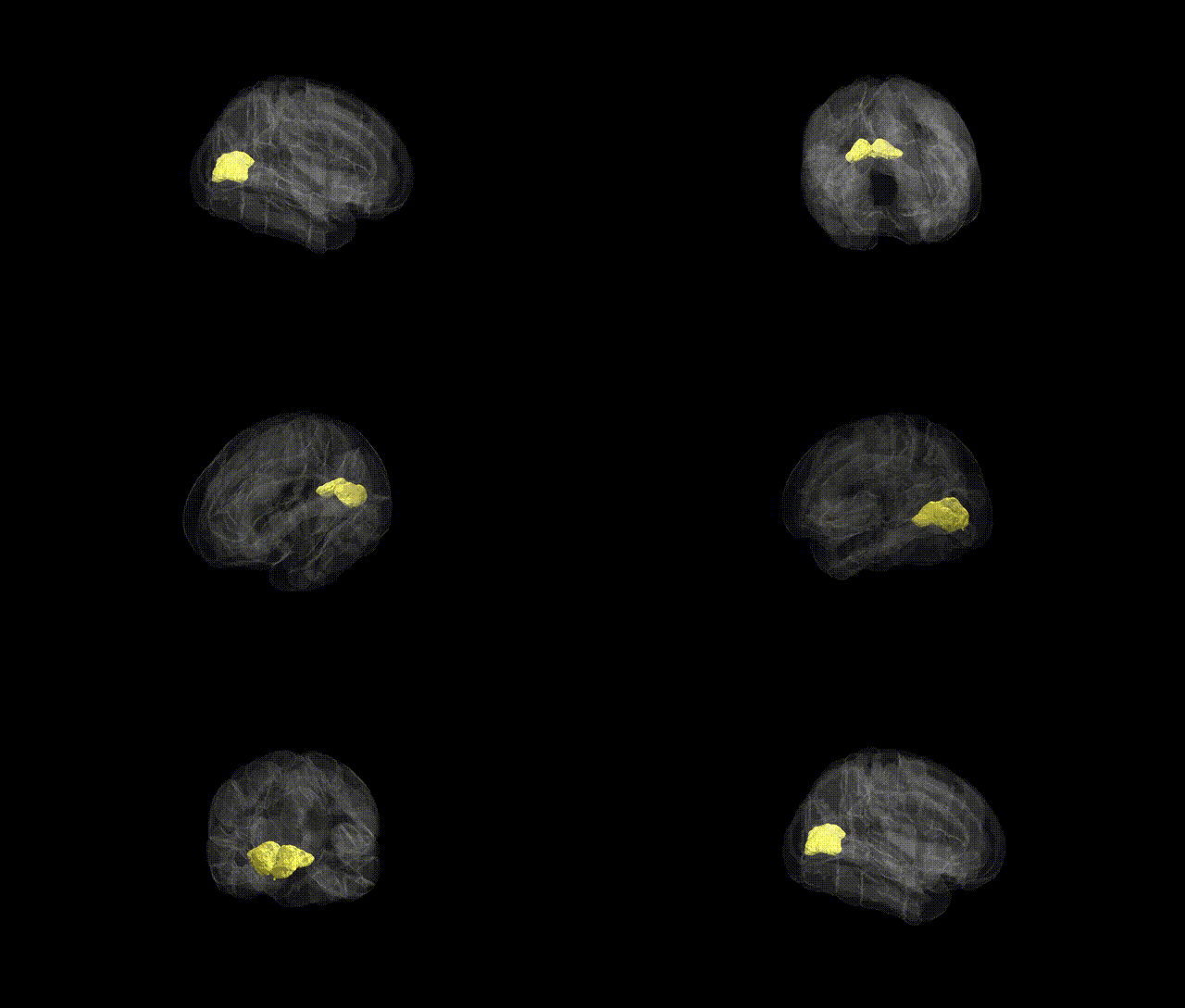
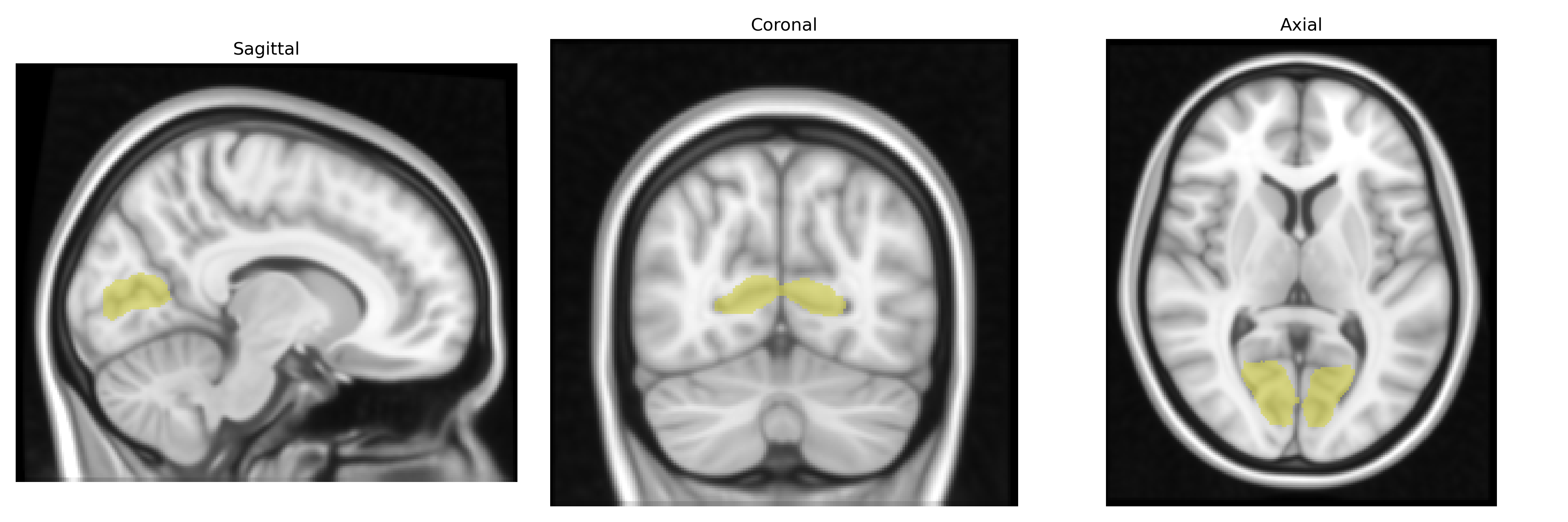
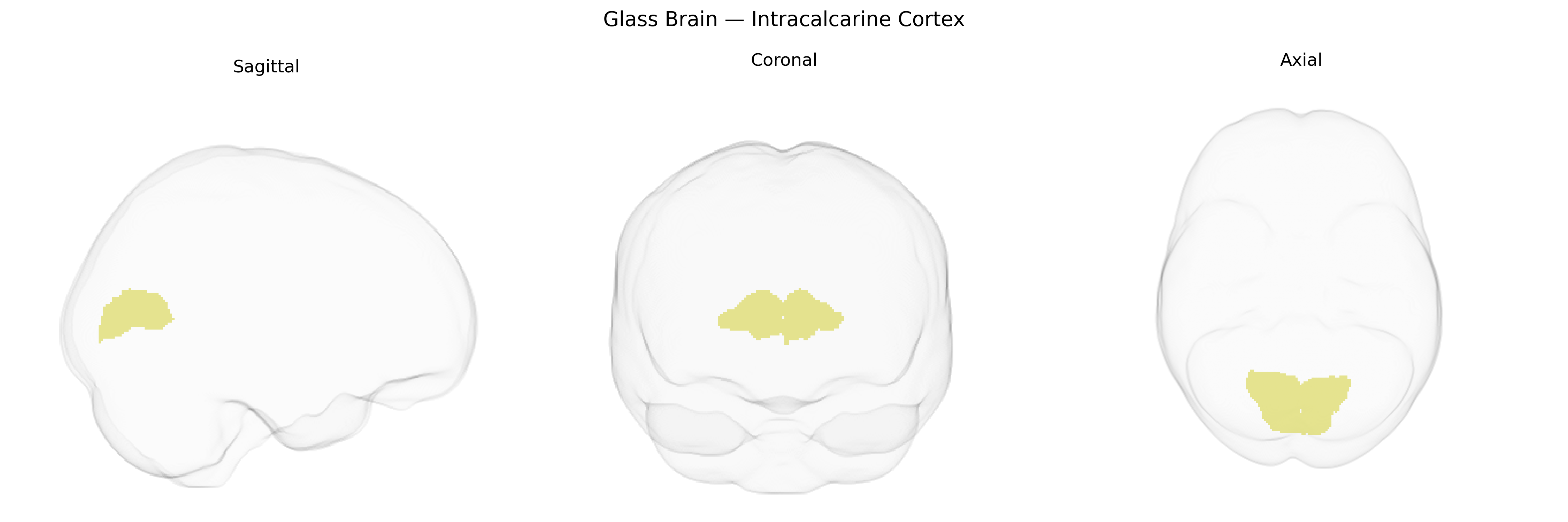
The bilateral Intracalcarine Cortex, as defined in the Harvard-Oxford cortical atlas, corresponds primarily to the cortex lining the calcarine sulcus in the medial occipital lobe and encompasses much of the primary visual cortex (V1, Brodmann area 17). This region receives dense thalamocortical input from the lateral geniculate nucleus and is organized retinotopically, with distinct representations of central and peripheral visual fields distributed along the sulcus. Neurons in the intracalcarine cortex are highly tuned to basic visual features such as orientation, spatial frequency, contrast, and motion direction, and they provide the initial cortical processing stage for visual information that is further relayed to secondary and associative visual areas. Functionally, this region is critical for conscious visual perception, including detection of edges, patterns, and simple forms, and lesions typically produce contralateral homonymous visual field defects. There is no direct Wikipedia article titled “Intracalcarine Cortex”; a closely related and encompassing structure is the primary visual cortex: Primary visual cortex.
The bilateral intracalcarine cortex, corresponding largely to primary visual cortex (V1) in the occipital lobe, has been implicated in multiple genetic and GWAS-based findings primarily through studies of brain structure, visual processing, and neuropsychiatric traits. Large-scale imaging genetics consortia such as ENIGMA and UK Biobank–based GWAS have identified common variants associated with occipital and calcarine cortical thickness, surface area, and volume, including loci near genes involved in neurodevelopment and synaptic function (e.g., variants in or near FOXO3, PAX6, and other neurodevelopmental regulators), although many signals are shared across visual and association cortices rather than being specific to the intracalcarine region. GWAS of resting-state functional connectivity and task-based activation have linked genetic variation to occipital and visual-network activity, which encompasses intracalcarine cortex, with polygenic influences overlapping those for intelligence, educational attainment, and general cognitive performance. In clinical domains, structural and functional alterations of the calcarine/intracalcarine cortex have been reported in schizophrenia, major depressive disorder, bipolar disorder, and autism spectrum disorder, with several psychiatric risk loci showing downstream effects on occipital cortical metrics in imaging–genetic analyses, though these associations are typically regionally nonspecific and reflect distributed cortical effects of polygenic risk. In rare monogenic or high-penetrance conditions affecting visual development and processing (such as some forms of congenital blindness or albinism), risk genes (e.g., those involved in retinal and optic pathway development) lead to predictable reorganization and volumetric changes in primary visual cortex, indirectly linking those genetic factors to intracalcarine morphology. Overall, genetic associations for the bilateral intracalcarine cortex are best characterized as part of broader, polygenic influences on visual system development, cortical morphology, cognitive traits, and vulnerability to neuropsychiatric disorders rather than discrete, region-exclusive GWAS signals.
Overview generated by GPT-4o (2026).
Region ID: 24
Hemisphere: bilateral
Atlas: HarvardOxford cort maxprob thr25 1mm
Full Quality Version: Download MP4
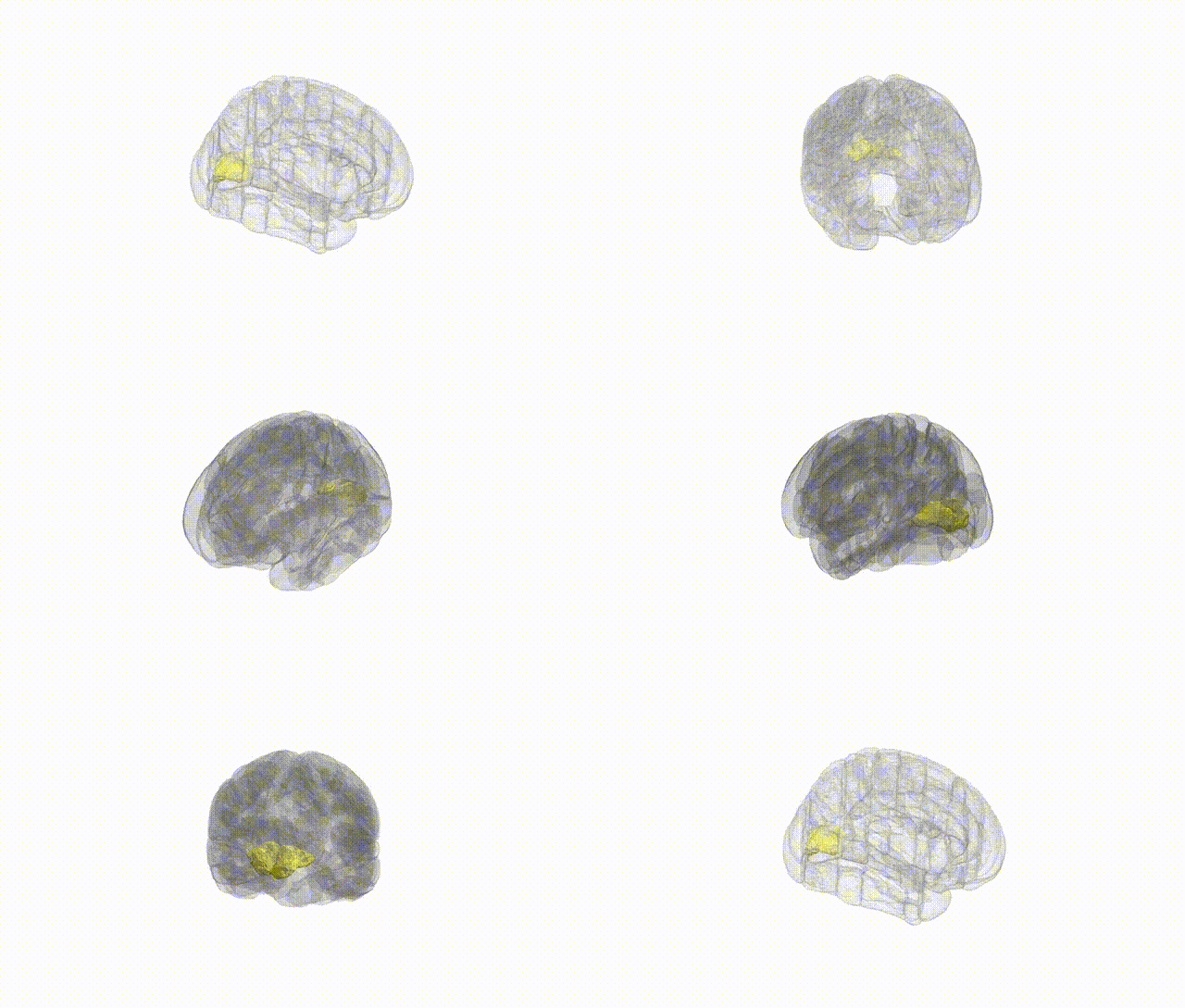
Full Quality Version: Download MP4
Wali Sidiqyar*, Gaurav Rudravaram*, Elyssa M. McMaster, Trent M. Schwartz, Adam M. Saunders, Kurt G. Schilling, Bennett A. Landman "Introducing SPINS: A Shared Public Visualization Library of Neuroanatomical Structures." Medical Imaging with Deep Learning- short paper
This resource is licensed under CC0 1.0 Universal (Public Domain).