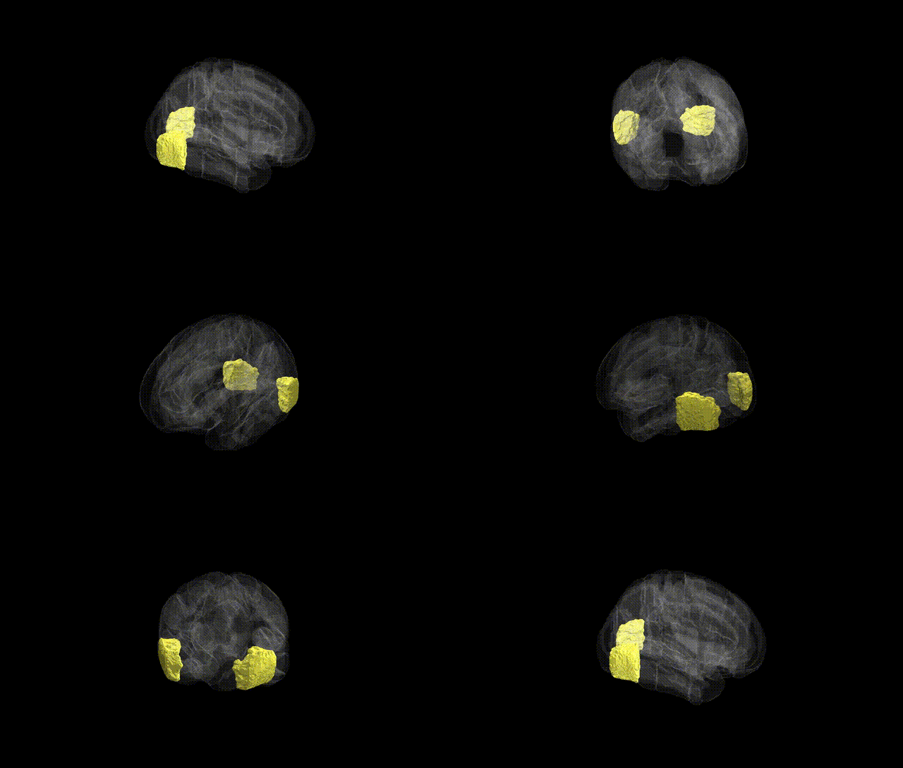
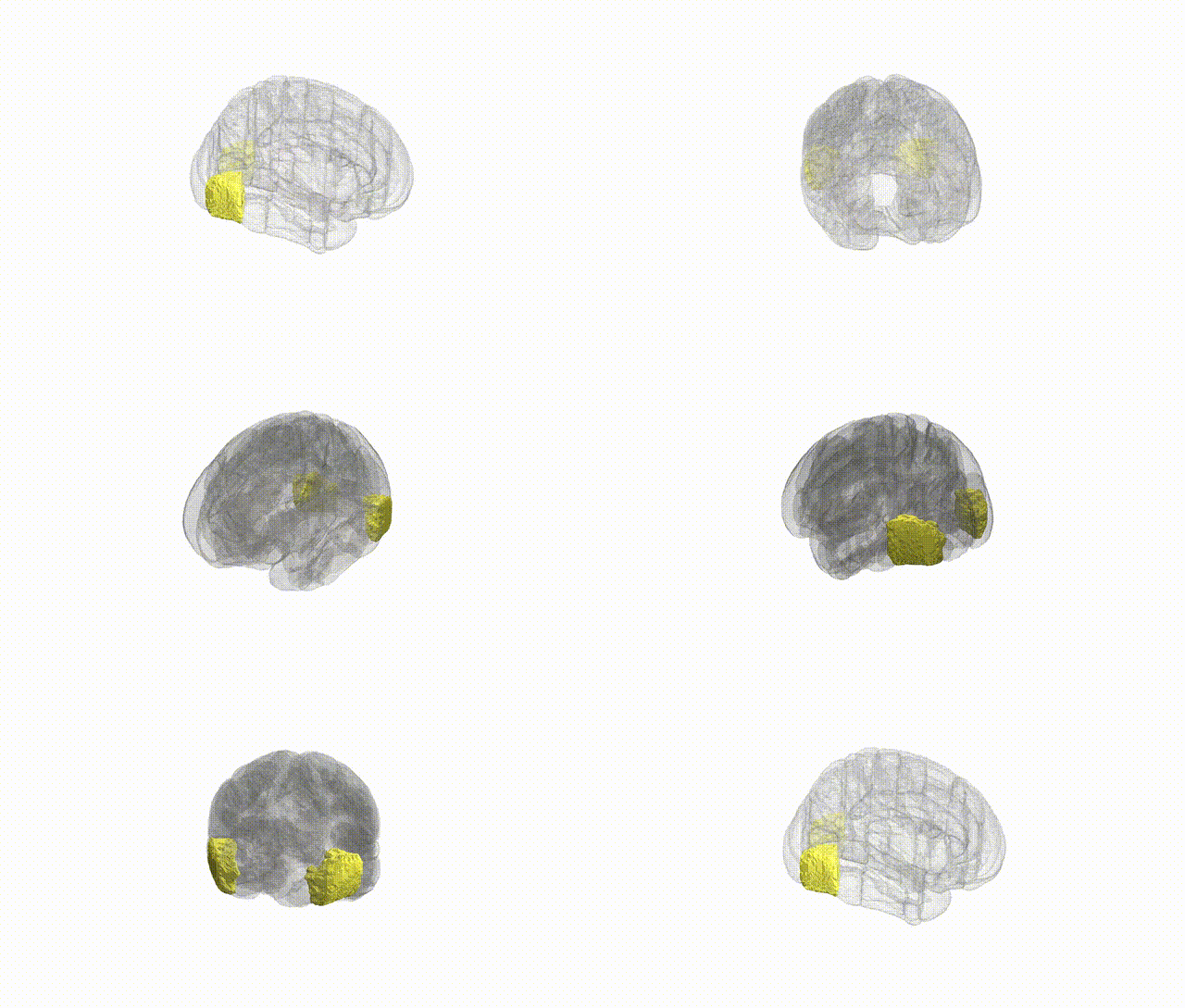
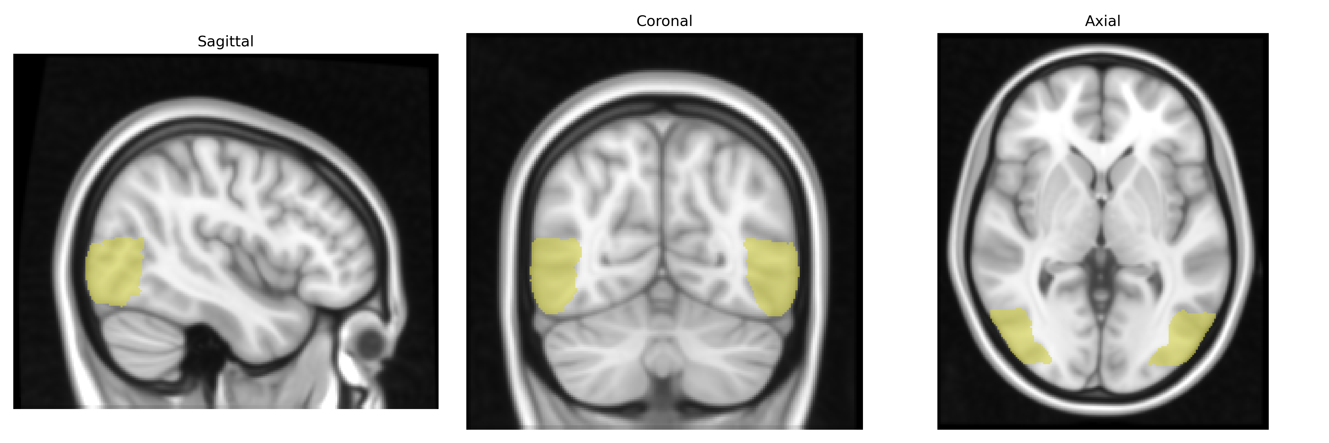
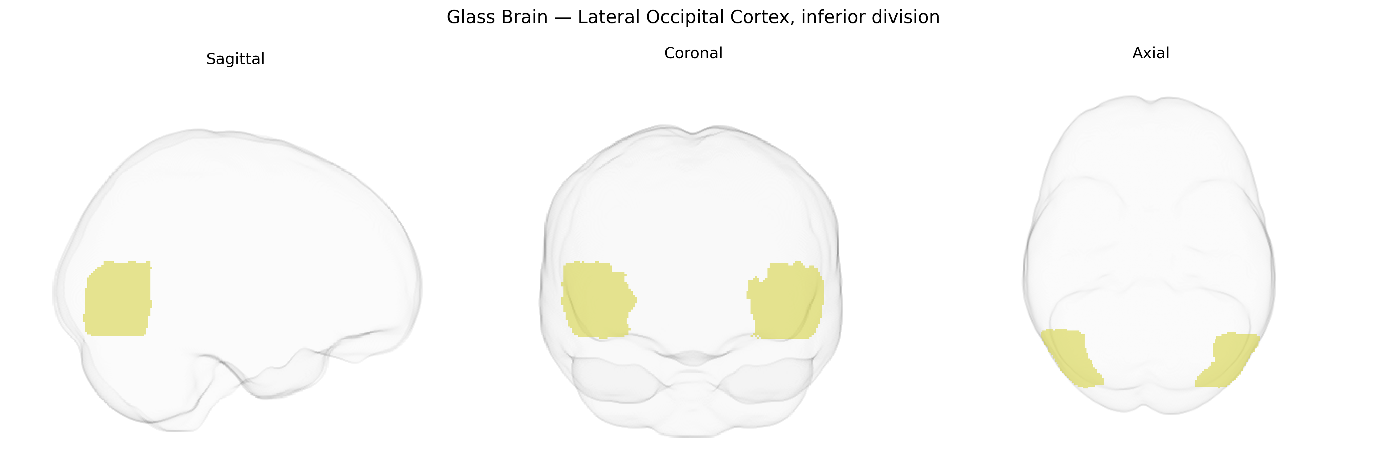
The bilateral Lateral Occipital Cortex, inferior division, as defined in the Harvard–Oxford cortical atlas (maxprob thr25, 1 mm), comprises ventrolateral occipital territories located inferior to the superior division and adjacent to the occipitotemporal border. This region forms part of the visual association cortex and is implicated in higher-order visual processing, including object recognition and shape/form analysis, integrating inputs from primary visual areas (V1/V2) and projecting to ventral visual stream structures. Functionally, it contributes to the perception of complex visual stimuli and interacts with temporal lobe regions involved in semantic and category-related processing of visual objects. There is no direct Wikipedia article for the “Lateral Occipital Cortex, inferior division”; a related structure within the same general territory is the Lateral occipital cortex.
The bilateral lateral occipital cortex, inferior division (as defined in the Harvard-Oxford atlas) has been implicated in several genetic and GWAS-based imaging-genetics studies, particularly those examining cortical thickness, surface area, and functional activation related to visual object processing. Large-scale ENIGMA and UK Biobank imaging GWAS have identified common variants near genes such as TBR1, EOMES, and other neurodevelopmental regulators that influence occipital cortical morphology, including lateral occipital regions, although these signals are often not specific to the inferior division alone. Polygenic risk scores for schizophrenia, autism spectrum disorder, and major depressive disorder have been associated with altered occipital cortical thickness or surface measures, with some reports noting lateral occipital contributions among broader visual and associative cortices. Occipital regions overlapping the lateral occipital cortex have also shown structural or functional differences linked to genetic liability for dyslexia and other neurodevelopmental conditions, and to variants associated with general cognitive ability and educational attainment, though these associations are typically modest and distributed across many cortical areas. Overall, genetic influences on the inferior lateral occipital cortex appear highly polygenic and shared with other visual and association regions, with no single disorder- or gene-specific effect uniquely and consistently localized to this subregion in current GWAS literature.
Overview generated by GPT-4o (2026).
Region ID: 23
Hemisphere: bilateral
Atlas: HarvardOxford cort maxprob thr25 1mm
Full Quality Version: Download MP4
Full Quality Version: Download MP4
Wali Sidiqyar*, Gaurav Rudravaram*, Elyssa M. McMaster, Trent M. Schwartz, Adam M. Saunders, Kurt G. Schilling, Bennett A. Landman "Introducing SPINS: A Shared Public Visualization Library of Neuroanatomical Structures." Medical Imaging with Deep Learning- short paper
This resource is licensed under CC0 1.0 Universal (Public Domain).