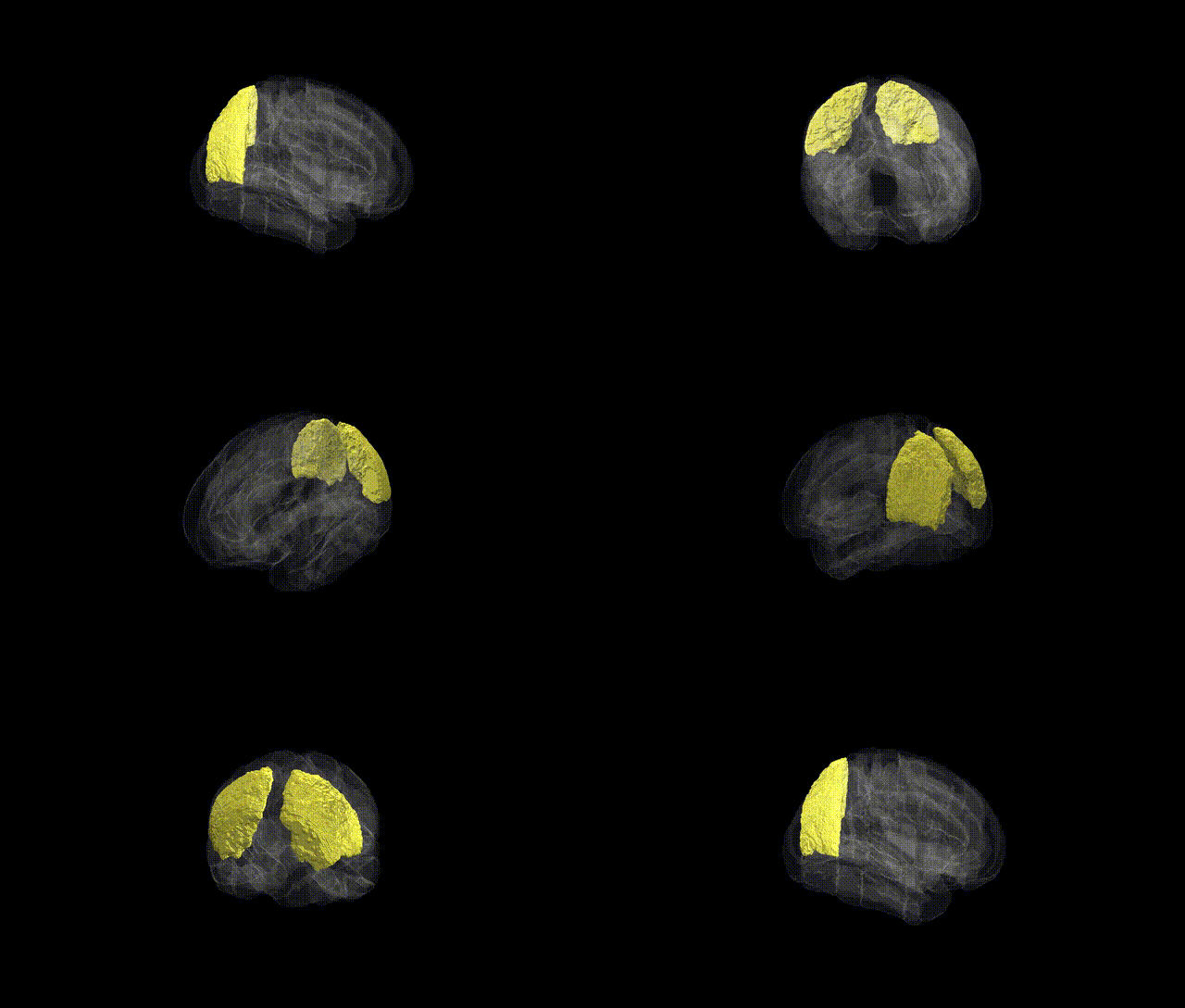
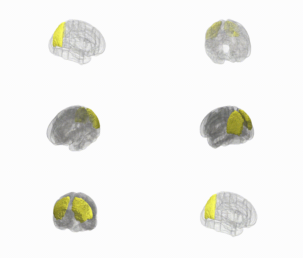
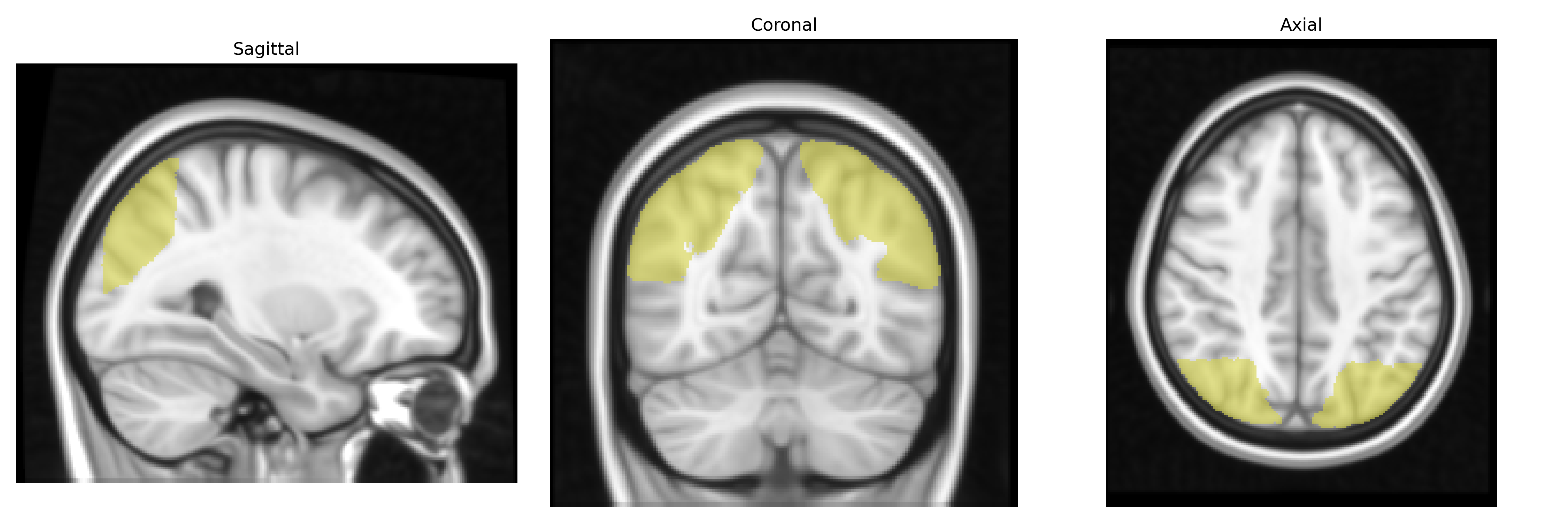
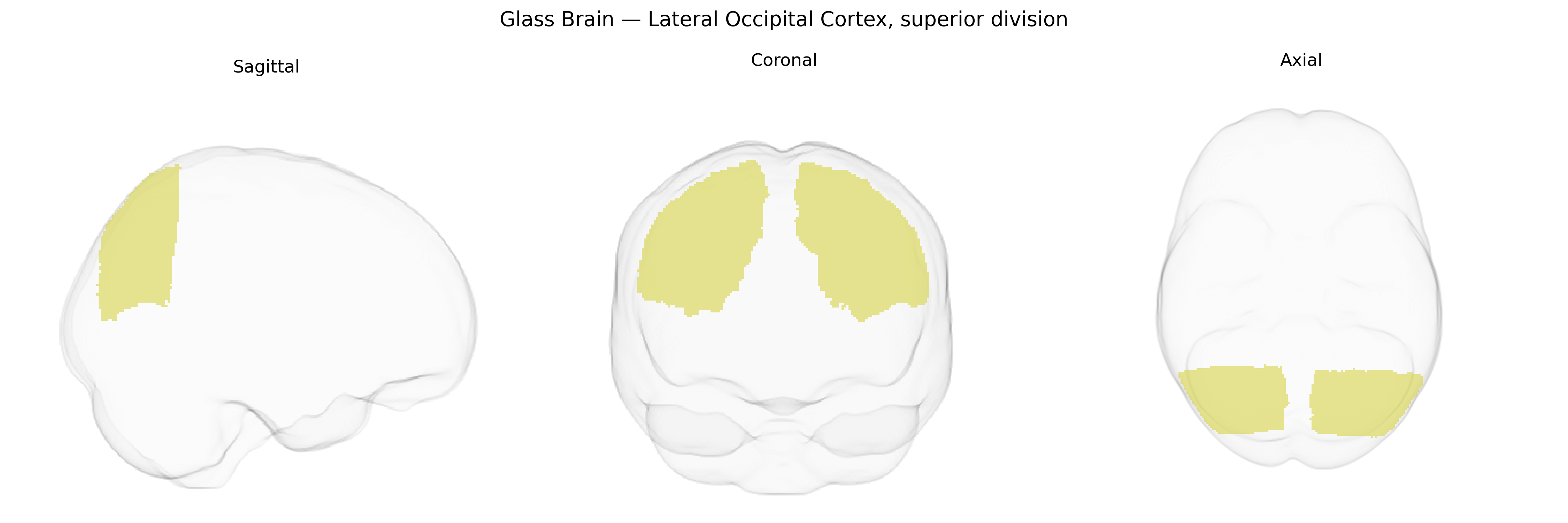
The bilateral Lateral Occipital Cortex, superior division, as defined in the Harvard-Oxford cortical atlas (maxprob thr25, 1 mm), comprises the dorsal portion of the lateral occipital cortex situated on the lateral surface of the occipital lobe, extending anteriorly toward the temporo-occipital junction and superiorly adjacent to parietal regions. This region is primarily associated with higher-order visual processing, including object recognition, shape and form analysis, and the integration of visual features into coherent percepts. It receives input from early visual areas (e.g., V1/V2) and contributes to the ventral visual stream, supporting the transformation of basic visual signals into representations relevant for perception and visually guided behavior. There is no direct Wikipedia article for the “Lateral Occipital Cortex, superior division” as named in the atlas; a closely related structure is the occipital lobe: Occipital lobe.
The bilateral lateral occipital cortex, superior division (as defined in the Harvard-Oxford atlas) has been implicated in several large-scale imaging genetics and GWAS efforts that link common variants to visual, cognitive, and neuropsychiatric traits through its structure and function. Variants in genes involved in synaptic signaling and neurodevelopment (including but not limited to BDNF, GRIN2B, and NRG3) have been associated with occipital cortical thickness, surface area, or activation patterns in visual tasks, and ENIGMA and UK Biobank imaging GWAS have reported loci near microtubule- and axon-guidance genes (e.g., MAPT region, PLCL1, and others) influencing occipital morphology and functional connectivity. The superior lateral occipital cortex, as part of the dorsal visual and object-processing streams, is frequently implicated in genetic studies of general cognitive ability, educational attainment, and reading-related traits, in which polygenic scores for intelligence and education show associations with its volume and connectivity. In neuropsychiatric genetics, schizophrenia, bipolar disorder, and major depression polygenic risk scores have been correlated with altered thickness or surface area in lateral occipital regions, and specific risk loci (such as those near ZNF804A and CACNA1C, among others) have been linked to abnormal activation during visual and working-memory tasks that recruit this region. Additionally, genetic risk for neurodevelopmental disorders, including autism spectrum disorder and dyslexia, has been associated with atypical lateral occipital structure and function, particularly in higher-order visual and face/object recognition networks, although these associations are often polygenic and not specific to this subdivision. Overall, current evidence suggests that genetic influences on synaptic development, visual-system wiring, and higher-order cognition contribute to inter-individual variability in the superior lateral occipital cortex, but no single locus or small set of genes uniquely defines this region’s genetic architecture, which remains largely polygenic and shared with broader occipito-parietal networks.
Overview generated by GPT-4o (2026).
Region ID: 22
Hemisphere: bilateral
Atlas: HarvardOxford cort maxprob thr25 1mm
Full Quality Version: Download MP4
Full Quality Version: Download MP4
Wali Sidiqyar*, Gaurav Rudravaram*, Elyssa M. McMaster, Trent M. Schwartz, Adam M. Saunders, Kurt G. Schilling, Bennett A. Landman "Introducing SPINS: A Shared Public Visualization Library of Neuroanatomical Structures." Medical Imaging with Deep Learning- short paper
This resource is licensed under CC0 1.0 Universal (Public Domain).