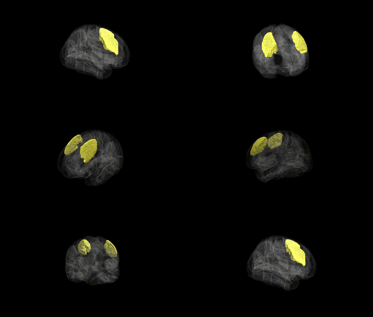
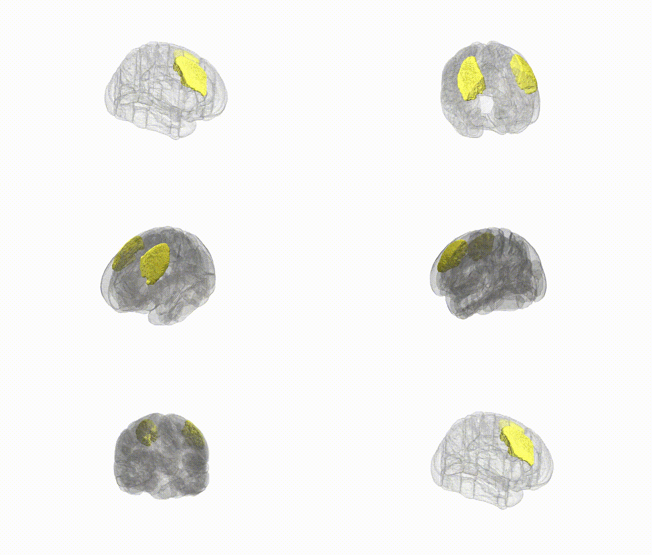
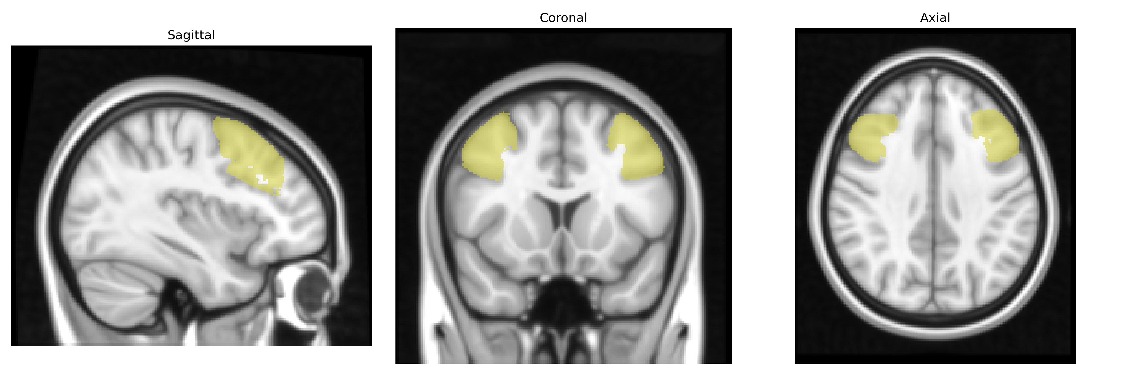
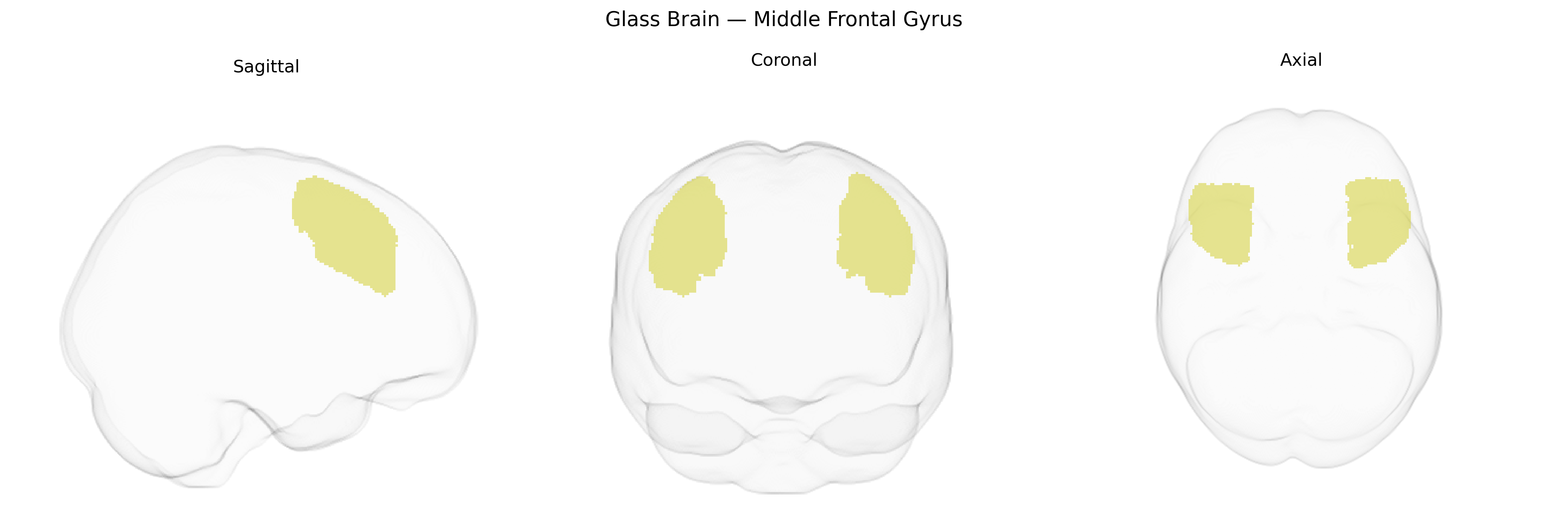
The bilateral Middle Frontal Gyrus, as defined in the Harvard–Oxford cortical maxprob threshold-25 1 mm atlas, comprises a dorsolateral segment of the frontal lobe situated between the superior and inferior frontal gyri, extending anteriorly from premotor territories toward the frontal pole. Cytoarchitectonically, it includes portions of the dorsolateral prefrontal cortex involved in higher-order executive functions such as working memory, cognitive control, planning, decision-making, and attentional regulation, as well as aspects of goal-directed behavior and abstract reasoning. This region participates in frontoparietal control networks, integrating sensory and mnemonic information to guide flexible behavior and top-down modulation of other cortical areas. There is no direct link, but the region is commonly discussed within the broader context of the Dorsolateral prefrontal cortex.
The bilateral middle frontal gyrus (MFG), as defined in the Harvard–Oxford cortical atlas, has emerged in imaging genetics and GWAS as a key association cortex region implicated in higher-order cognition, psychiatric risk, and neurodevelopment. Large-scale ENIGMA and UK Biobank studies report common variants influencing MFG cortical thickness, surface area, and activation, including loci in or near genes such as MAPT, HRK, and MIR2113, as part of broader frontal or dorsolateral prefrontal signatures. Polygenic risk scores for schizophrenia, bipolar disorder, major depressive disorder, ADHD, and autism spectrum disorder repeatedly show associations with altered MFG structure or function, often within the wider dorsolateral prefrontal–frontoparietal control network. GWAS of cognitive traits (e.g., general intelligence, working memory, educational attainment) find that variants in multiple synaptic and neurodevelopmental genes (such as CACNA1C, GRIN2B, and DISC1-related pathways) track with MFG activation during executive tasks and with its morphometry, though effects are generally small and distributed across the cortex. MFG connectivity and activation are further linked through genetic correlation and candidate-gene or polygenic analyses to risk-related traits (e.g., impulsivity, risk tolerance), substance use (particularly alcohol and nicotine), and anxiety-related phenotypes, again typically as part of broader frontal and cingulo-opercular networks. Neurodegenerative GWAS, especially in Alzheimer’s disease and frontotemporal dementia, suggest that APOE and other dementia risk loci modulate atrophy patterns including the MFG, consistent with its involvement in early executive dysfunction, but these effects are regionally nonspecific. Overall, genetic associations with the MFG reflect highly polygenic influences on frontal lobe development, synaptic signaling, and myelination, with convergent but not exclusive links to psychiatric disorders, cognitive performance, and neurodegenerative disease.
Overview generated by GPT-4o (2026).
Region ID: 4
Hemisphere: bilateral
Atlas: HarvardOxford cort maxprob thr25 1mm
Full Quality Version: Download MP4
Full Quality Version: Download MP4
Wali Sidiqyar*, Gaurav Rudravaram*, Elyssa M. McMaster, Trent M. Schwartz, Adam M. Saunders, Kurt G. Schilling, Bennett A. Landman "Introducing SPINS: A Shared Public Visualization Library of Neuroanatomical Structures." Medical Imaging with Deep Learning- short paper
This resource is licensed under CC0 1.0 Universal (Public Domain).