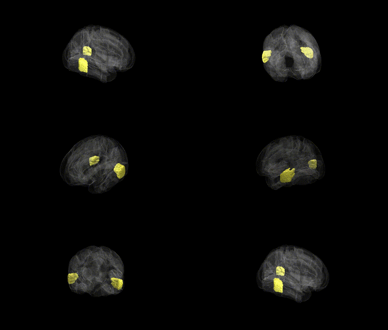
The bilateral Middle Temporal Gyrus, temporooccipital part (as defined in the Harvard-Oxford cortical maxprob atlas) corresponds to the posterior segment of the middle temporal gyrus near the temporo-occipital junction, extending along the lateral surface of the temporal lobe toward the occipital lobe. This region is implicated in higher-order visual and auditory processing, including aspects of motion perception, object recognition, and integration of visual information with semantic and language-related processes; it is also frequently engaged in reading, audiovisual integration, and multimodal association functions. Structurally, it lies lateral to the inferior temporal gyrus and inferior to the superior temporal gyrus, forming part of the broader temporal association cortex that interfaces with occipital visual areas and parietal multimodal regions. There is no direct Wikipedia article specifically for the “temporooocipital part” subdivision, but it is a subregion of the Middle Temporal Gyrus.
The bilateral middle temporal gyrus, temporooccipital part (from the Harvard-Oxford cortical atlas) corresponds largely to posterior middle temporal and lateral temporo-occipital cortex, a region repeatedly implicated in genetic studies of language, semantic processing, and higher-order visual integration. GWAS and imaging genetics work using UK Biobank and other cohorts have shown that cortical thickness and surface area in posterior temporal regions, including this area, are heritable and associated with common variants in genes involved in neurodevelopment, synaptic function, and axon guidance (for example, variants near DCC, BDNF, and several glutamatergic pathway genes have been linked to temporal-lobe morphology and cognition). Polygenic scores for educational attainment, general cognitive ability, and reading/language skills show robust associations with structure and function in this zone, consistent with its role in semantic and phonological processing. In psychiatric and neurodevelopmental GWAS, the region emerges in imaging–genetics overlays as a target of risk variants for schizophrenia and autism spectrum disorder, where temporal–occipital cortex often shows altered connectivity and activation, and for major depressive disorder, in which temporal–occipital areas are implicated in abnormal processing of social and emotional information. Additionally, rare variant and CNV studies affecting language-related genes (such as FOXP2-regulated networks) and broader cortical patterning genes (e.g., transcription factors and chromatin regulators) have been linked to atypical development or activation in posterior middle temporal cortex, further supporting a genetically influenced vulnerability of this temporooccipital region in language, social cognition, and psychosis-related phenotypes.
Overview generated by GPT-4o (2026).
Region ID: 13
Hemisphere: bilateral
Atlas: HarvardOxford cort maxprob thr25 1mm
Full Quality Version: Download MP4
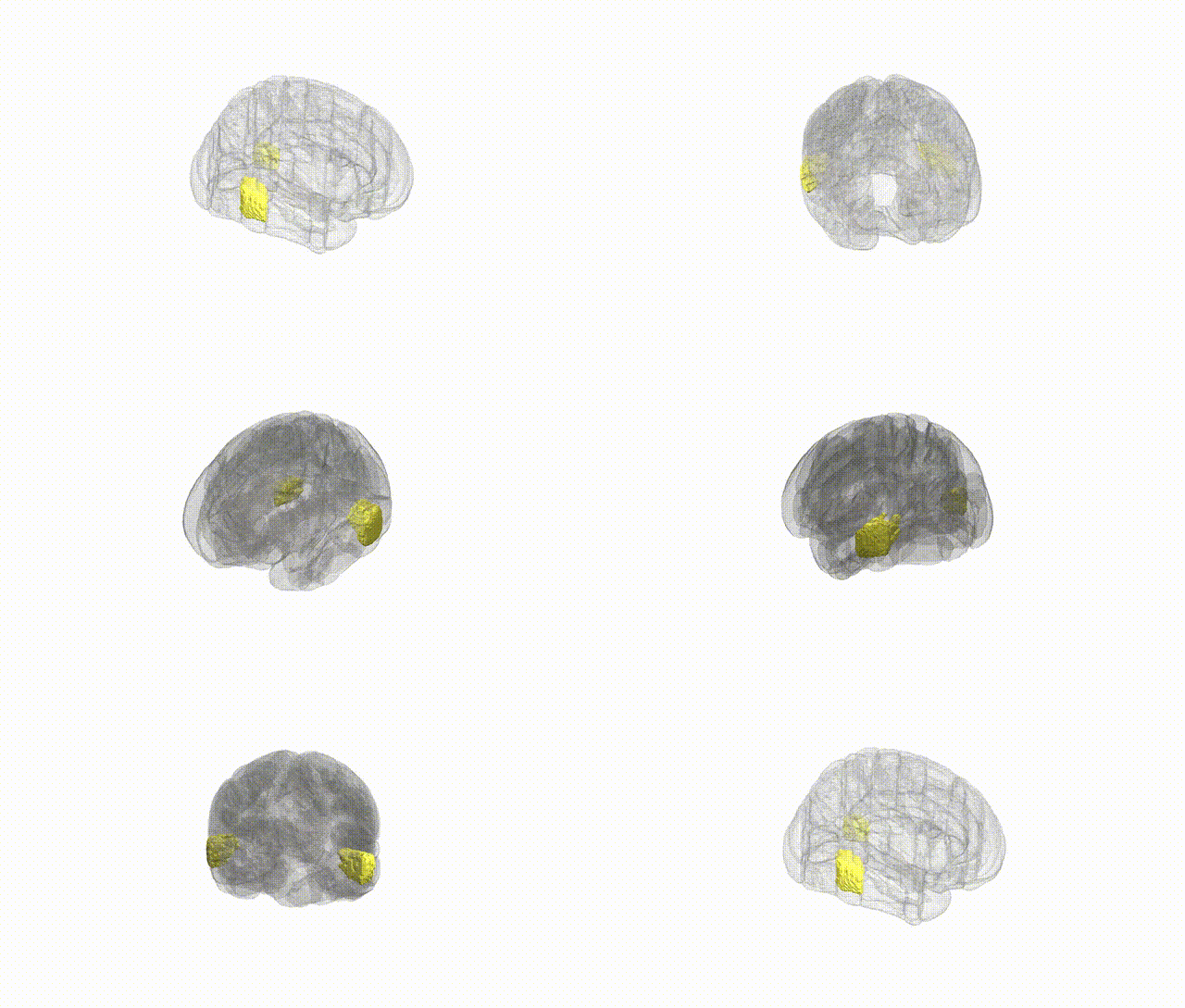
Full Quality Version: Download MP4
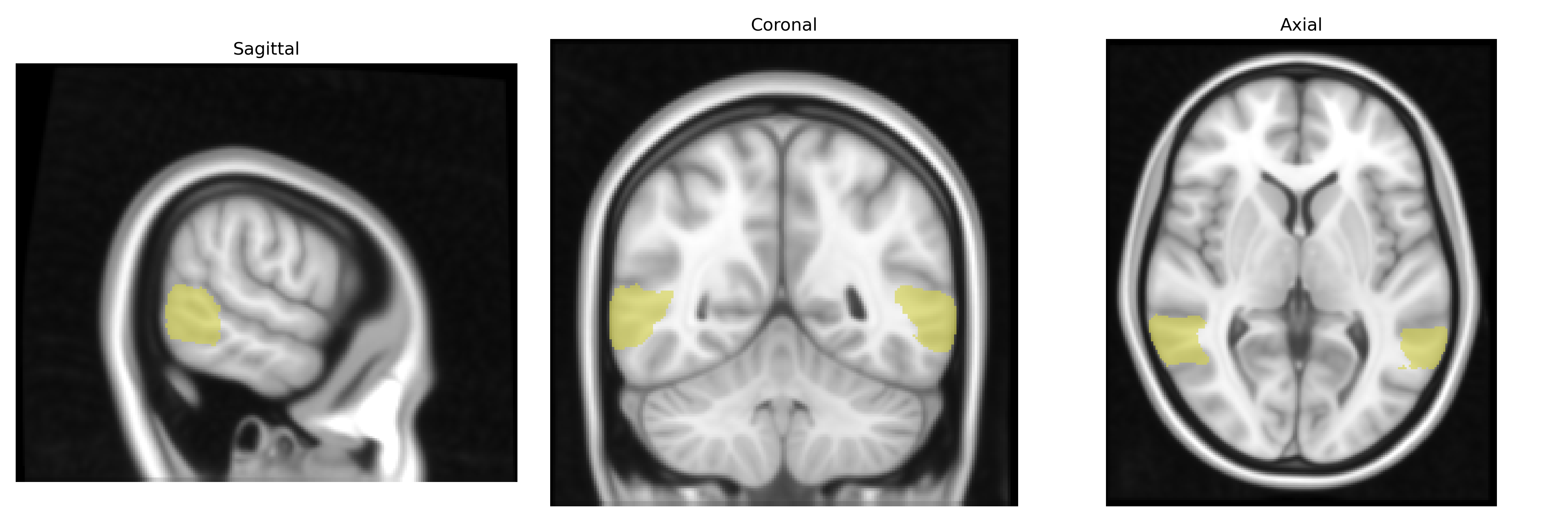
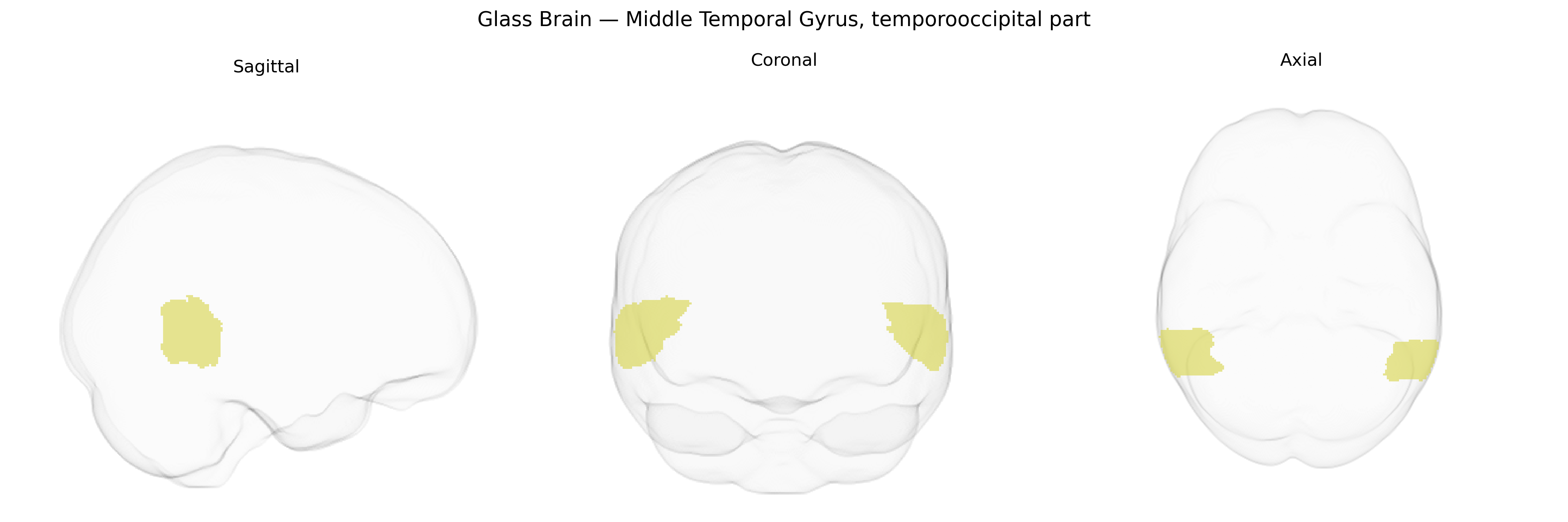
Wali Sidiqyar*, Gaurav Rudravaram*, Elyssa M. McMaster, Trent M. Schwartz, Adam M. Saunders, Kurt G. Schilling, Bennett A. Landman "Introducing SPINS: A Shared Public Visualization Library of Neuroanatomical Structures." Medical Imaging with Deep Learning- short paper
This resource is licensed under CC0 1.0 Universal (Public Domain).