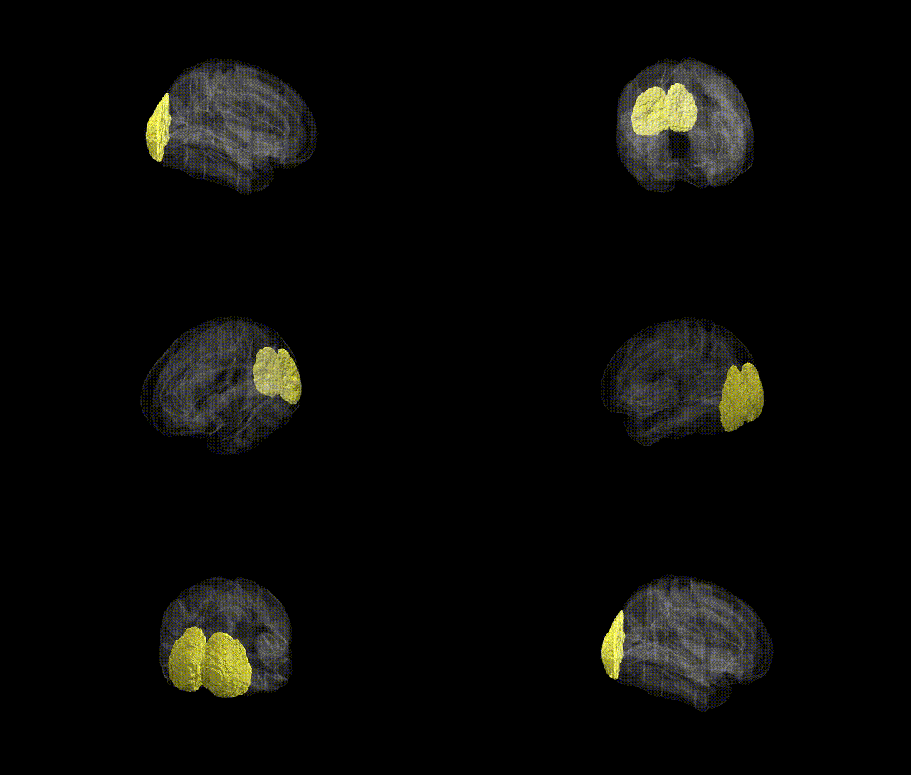
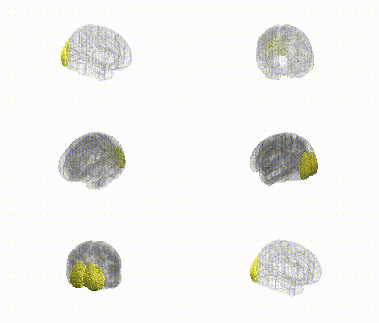
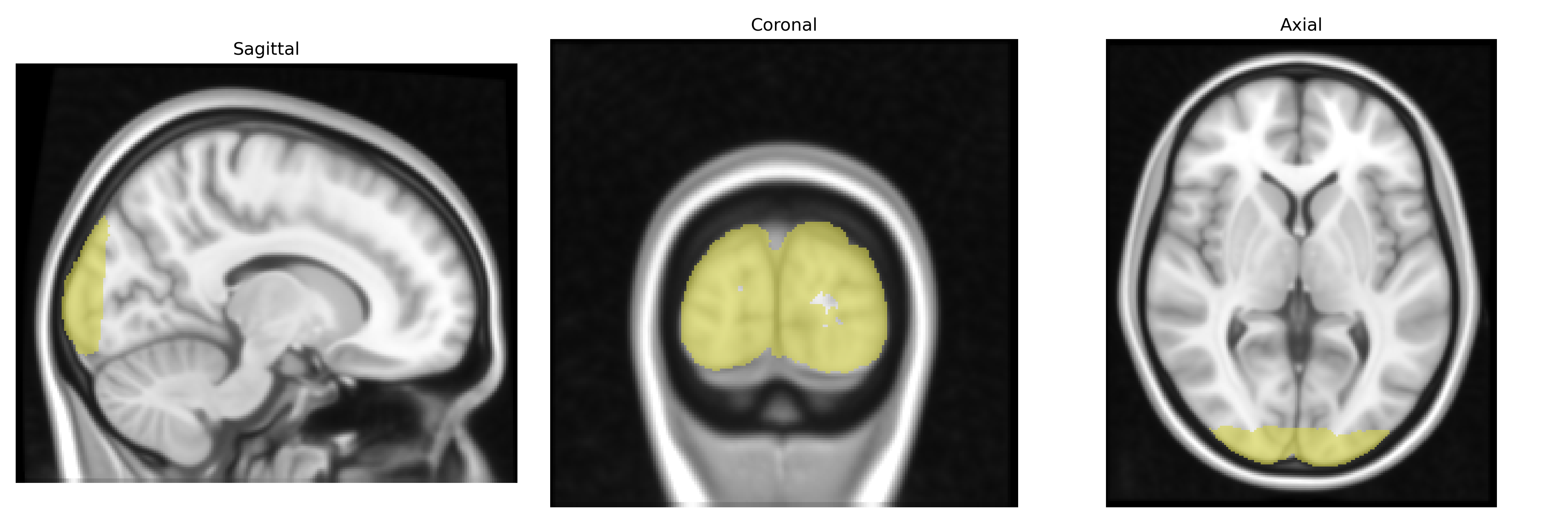
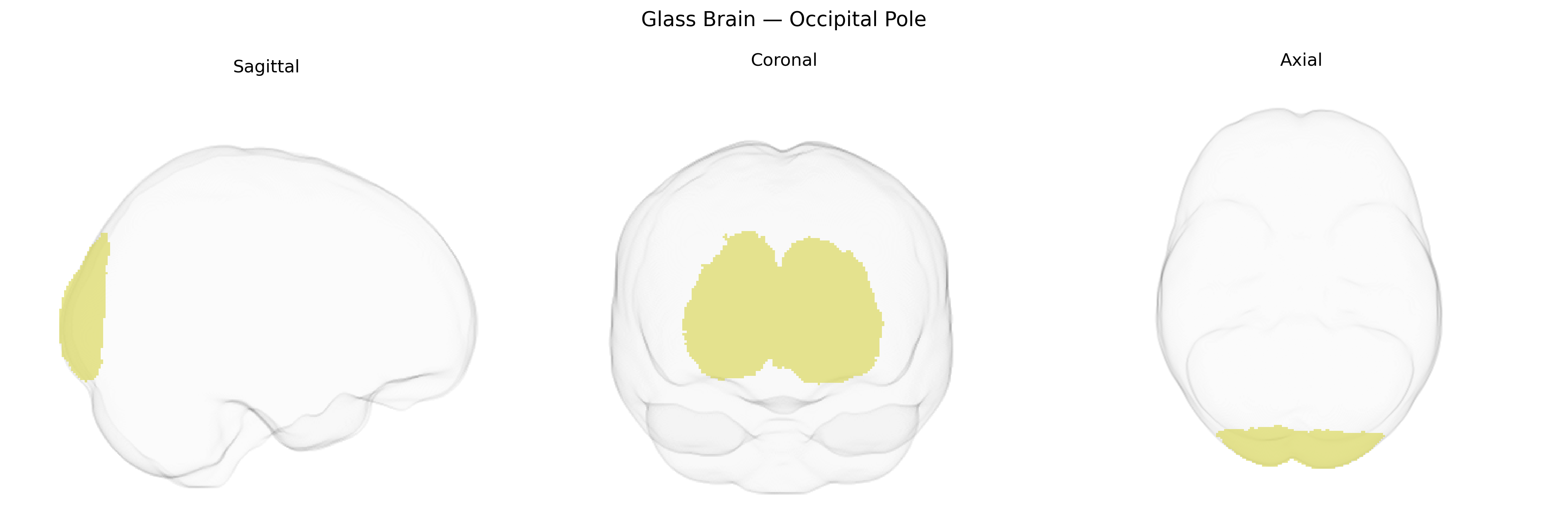
The bilateral Occipital Pole, as defined in the Harvard–Oxford cortical atlas, refers to the most posterior portion of the occipital lobes, encompassing primary and adjacent visual cortical areas that receive and process highly organized retinotopic input from the lateral geniculate nucleus of the thalamus. This region plays a central role in early-stage visual processing, including the detection of basic visual features such as orientation, spatial frequency, contrast, and simple motion, and contributes to the integration of these features into coherent visual representations that are further elaborated in higher-order occipital and temporal visual areas. Anatomically, the occipital pole lies around the posterior tip of the calcarine sulcus and is closely associated with the striate cortex (Brodmann area 17) and surrounding extrastriate regions. There is no direct Wikipedia article for “Occipital Pole”; a related structure is the Occipital lobe.
The bilateral occipital pole, corresponding to primary and early visual cortical areas in the Harvard-Oxford atlas, has been implicated in multiple genetic studies examining brain structure, function, and associated traits. GWAS of cortical thickness and surface area have identified polygenic influences on occipital regions, with variants in genes involved in neurodevelopment and synaptic function (e.g., MSRA, KCNK2, and loci near PAX6 and FGFR genes) contributing to occipital morphology and visual cortex measures. Large-scale imaging genetics consortia such as ENIGMA and UK Biobank analyses show that common variants across the genome collectively explain a substantial portion of variance in occipital cortical metrics, reflecting the highly polygenic architecture of this region. Occipital pole structure and functional activation have been indirectly linked through genetics to visual processing traits, reading and language-related phenotypes, migraine and visual aura, and neuropsychiatric conditions such as schizophrenia and major depression, where risk variants are associated with altered visual network connectivity or occipital cortical thickness, although these associations are typically modest and distributed across many brain regions rather than specific to the occipital pole alone.
Overview generated by GPT-4o (2026).
Region ID: 48
Hemisphere: bilateral
Atlas: HarvardOxford cort maxprob thr25 1mm
Full Quality Version: Download MP4
Full Quality Version: Download MP4
Wali Sidiqyar*, Gaurav Rudravaram*, Elyssa M. McMaster, Trent M. Schwartz, Adam M. Saunders, Kurt G. Schilling, Bennett A. Landman "Introducing SPINS: A Shared Public Visualization Library of Neuroanatomical Structures." Medical Imaging with Deep Learning- short paper
This resource is licensed under CC0 1.0 Universal (Public Domain).