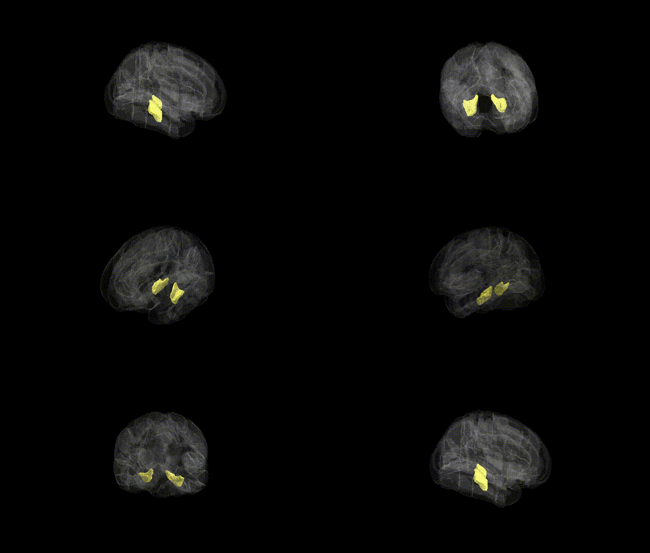
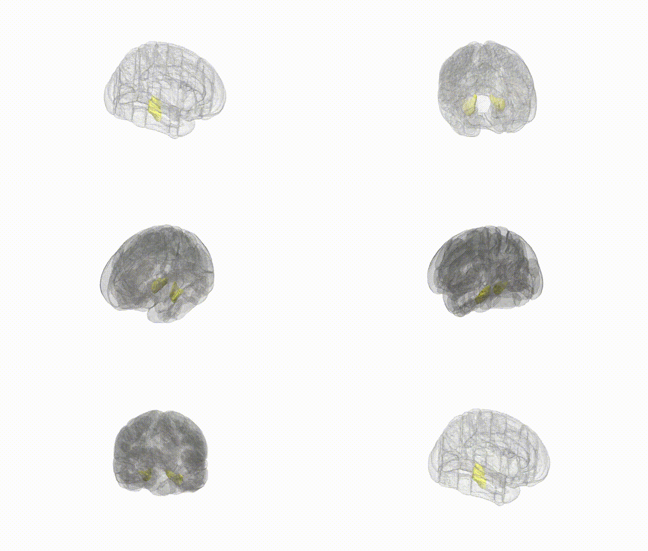
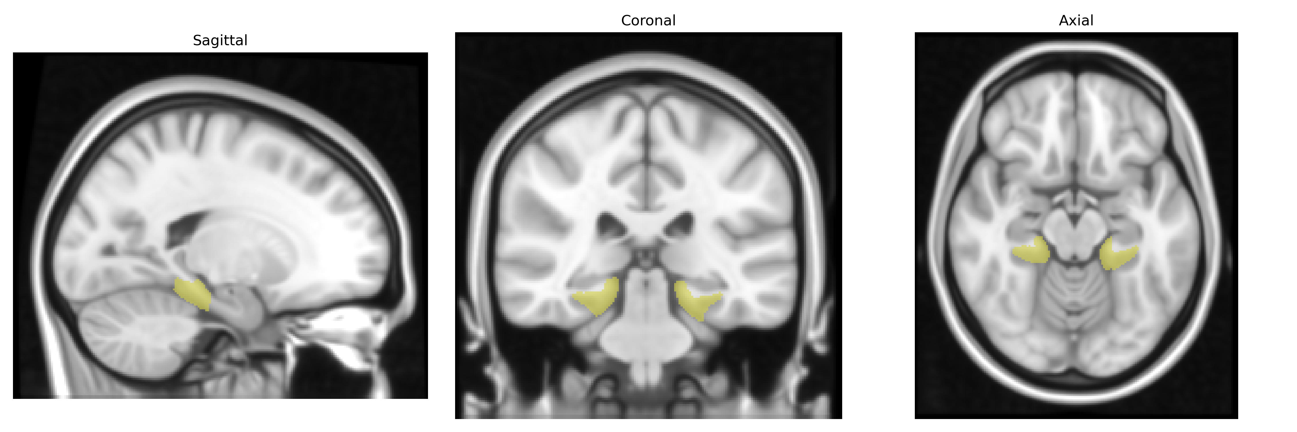
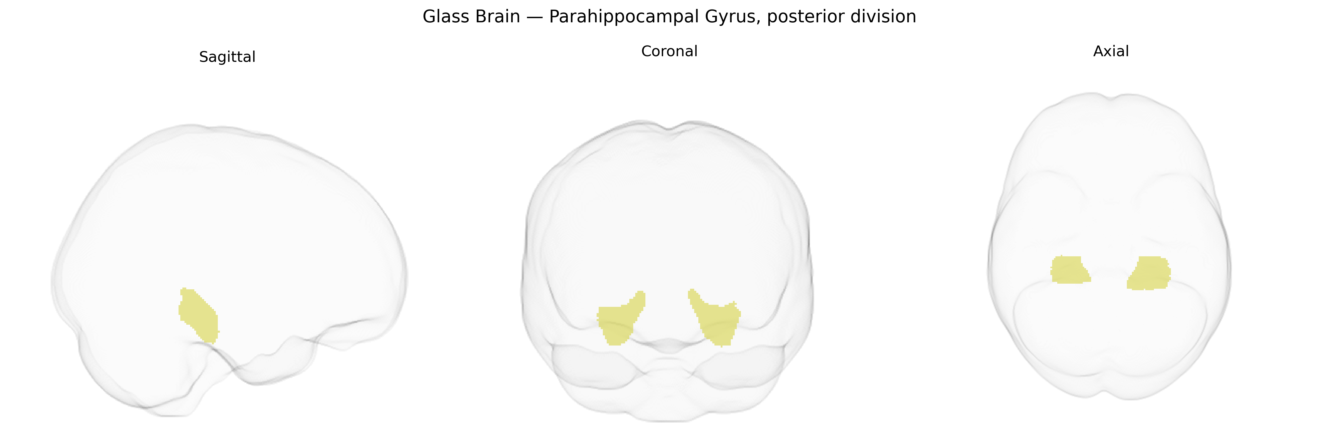
The bilateral Parahippocampal Gyrus, posterior division, as defined in the Harvard–Oxford cortical structural atlas (maxprob, thr25, 1 mm), corresponds to the posterior segment of the parahippocampal gyrus located in the medial temporal lobe, adjacent to the hippocampus and extending toward the lingual and fusiform gyri. This region is primarily associated with the encoding and retrieval of contextual and spatial information, scene processing, and aspects of episodic memory, and includes or closely overlaps the parahippocampal “place area,” which is selectively responsive to environmental scenes. Cytoarchitectonically, it forms part of the parahippocampal cortex, integrating inputs from widespread association areas and projecting to hippocampal formation structures to support navigation, memory consolidation, and the representation of visuospatial context. There is no direct Wikipedia article for the “Parahippocampal Gyrus, posterior division” as defined in the atlas; a closely related structure is the broader parahippocampal gyrus: Parahippocampal gyrus.
The bilateral parahippocampal gyrus, posterior division (a key medial temporal lobe structure involved in scene processing, contextual memory, and navigation) has been implicated in several GWAS and imaging‑genetics studies, primarily through its volume, cortical thickness, and functional connectivity rather than region‑specific SNPs. Variants in genes associated with neurodevelopment and synaptic function—such as BDNF (e.g., Val66Met), APOE (particularly ε4), TOMM40, and CLU—have been repeatedly linked to structural and functional differences in parahippocampal and adjacent medial temporal regions, often in the context of Alzheimer’s disease risk, age‑related cognitive decline, and hippocampal atrophy. Large‑scale brain MRI GWAS (e.g., ENIGMA and UK Biobank cohorts) have identified multiple loci influencing medial temporal and parahippocampal volume, including regions near WNT3, DPP4, and genes involved in neuronal signaling and axonal guidance, which correlate with memory performance, educational attainment, and general cognitive ability. Parahippocampal morphology and connectivity show genetic correlations with neuropsychiatric conditions such as schizophrenia, major depressive disorder, and PTSD, where polygenic risk scores for these disorders predict alterations in medial temporal lobe structure and activity. Additionally, GWAS of spatial navigation, episodic memory, and scene recognition traits—though still limited—indicate that common variants affecting glutamatergic transmission, neurogenesis, and tau/amyloid pathways contribute to individual differences in parahippocampal function, situating this region as a convergent locus for genetic influences on memory, Alzheimer’s disease susceptibility, and affective psychopathology.
Overview generated by GPT-4o (2026).
Region ID: 35
Hemisphere: bilateral
Atlas: HarvardOxford cort maxprob thr25 1mm
Full Quality Version: Download MP4
Full Quality Version: Download MP4
Wali Sidiqyar*, Gaurav Rudravaram*, Elyssa M. McMaster, Trent M. Schwartz, Adam M. Saunders, Kurt G. Schilling, Bennett A. Landman "Introducing SPINS: A Shared Public Visualization Library of Neuroanatomical Structures." Medical Imaging with Deep Learning- short paper
This resource is licensed under CC0 1.0 Universal (Public Domain).