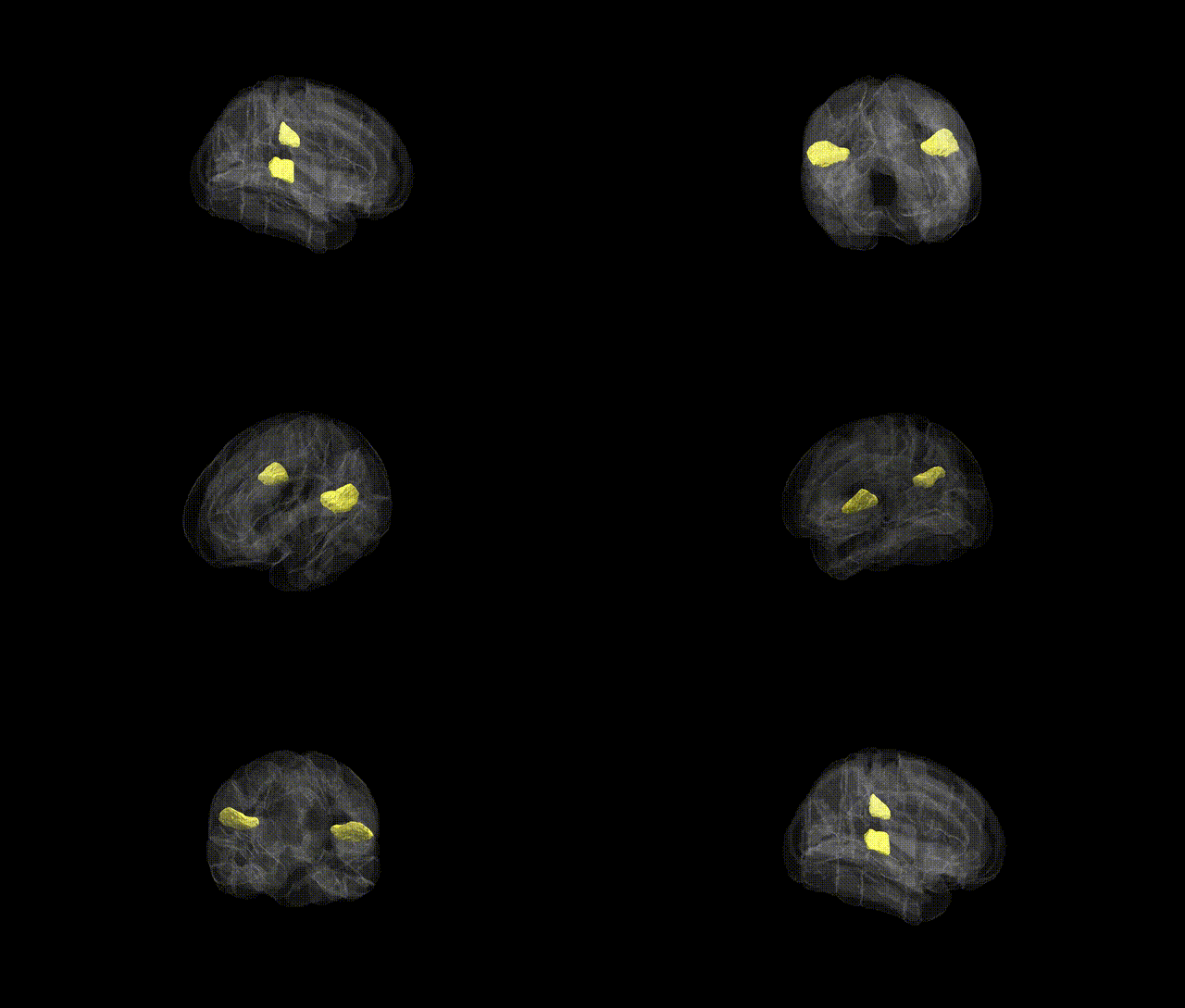
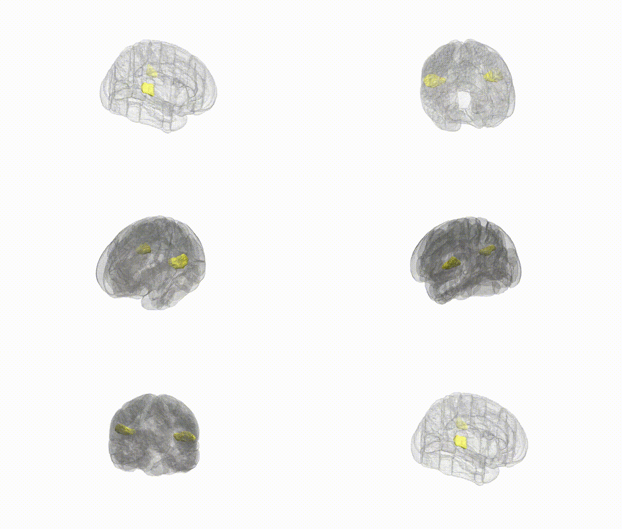
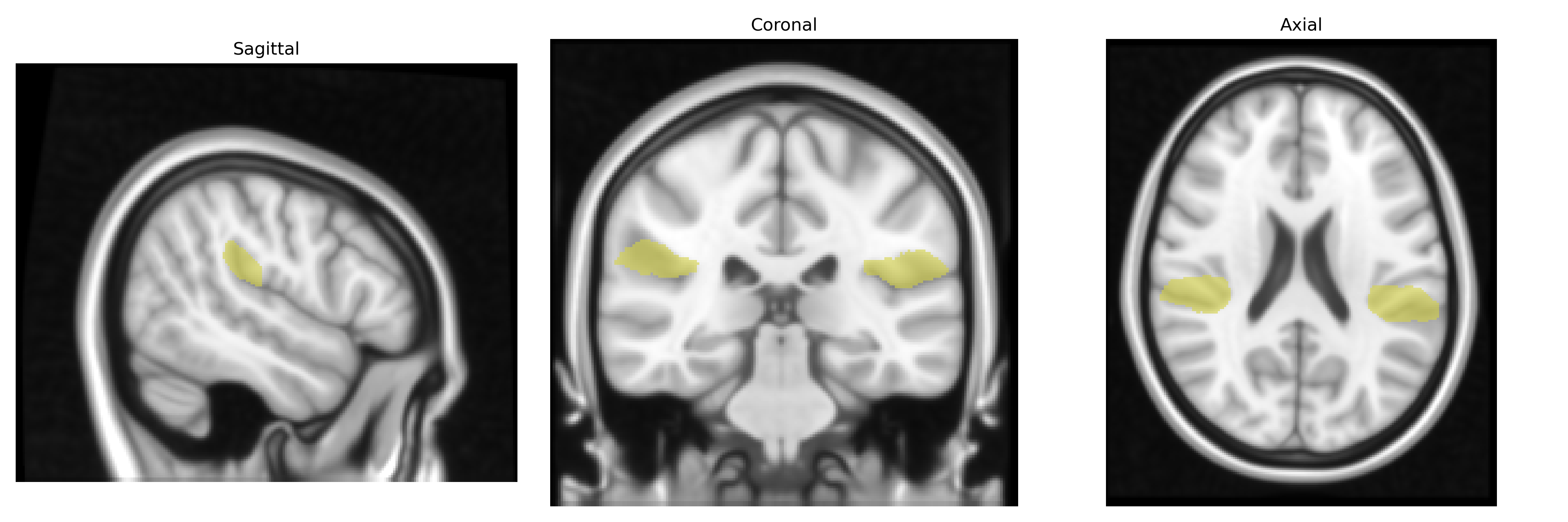
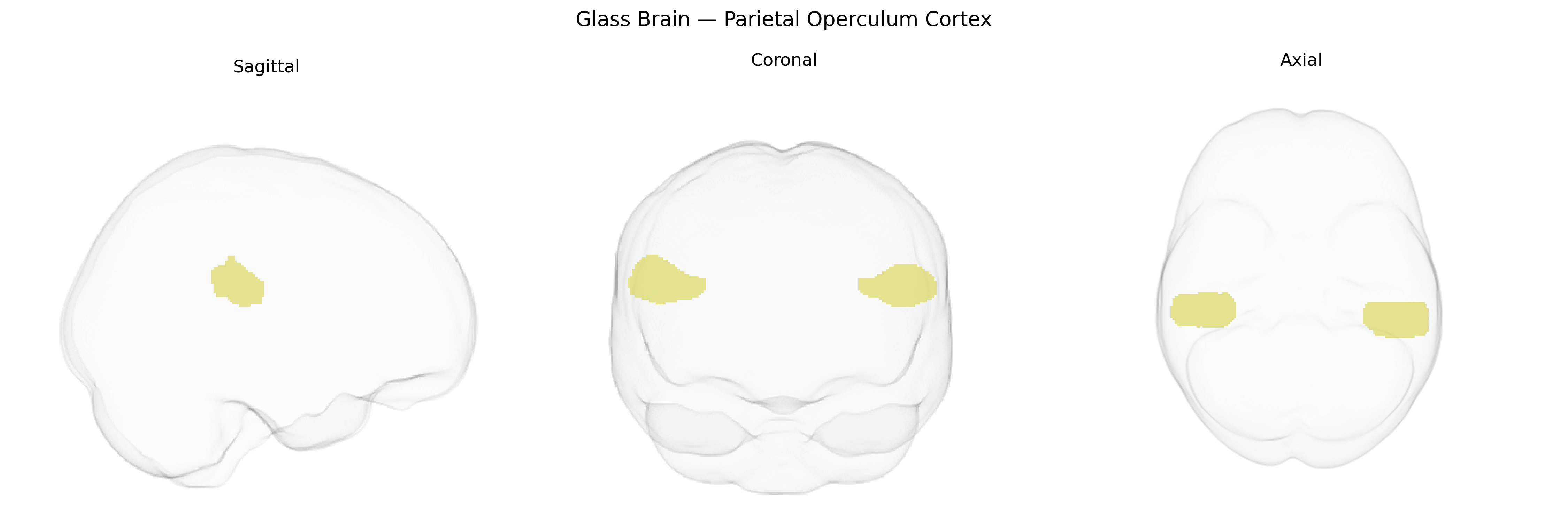
The bilateral Parietal Operculum Cortex, as defined in the Harvard–Oxford cortical structural atlas (maxprob thr25, 1 mm), comprises the opercular portion of the inferior parietal lobe overlying the insula and forming part of the lateral wall of the Sylvian (lateral) fissure. Structurally, it includes cortex adjacent to the supramarginal gyrus and is closely associated with the secondary somatosensory area (S2), participating in higher-order processing of tactile and nociceptive information, sensorimotor integration, and aspects of body representation. Functionally, this region is implicated in bilateral somatosensory perception, integration of multimodal sensory inputs, and may contribute to aspects of pain perception and tactile object recognition. There is no direct Wikipedia article specifically for “Parietal Operculum Cortex”; however, it is part of the broader opercular cortex associated with the Operculum.
The bilateral parietal operculum cortex, as defined in the Harvard-Oxford Cortical Atlas, overlaps structurally and functionally with secondary somatosensory and sensorimotor integration areas, and genetic associations identified through GWAS and imaging–genetics studies often implicate variants affecting cortical thickness, surface area, and functional connectivity in this region. Large-scale neuroimaging GWAS (e.g., ENIGMA, UK Biobank) have reported associations between parietal operculum morphology and variants in genes involved in neurodevelopment, synaptic organization, and axon guidance (such as loci near MAPT, TBR1, and genes in Wnt and calcium-signaling pathways), although findings are typically regionally broad and not specific to the operculum alone. Functionally, this region has been linked via imaging–genetic correlations to pain sensitivity, somatosensory perception, and motor learning traits, and it appears in polygenic models of cognitive performance and educational attainment that relate distributed cortical patterns, including opercular parietal areas, to common variant load. Clinically, altered structure or activation in the parietal operculum has been observed in genetic risk contexts for epilepsy, autism spectrum disorder, and schizophrenia, but these associations arise from genome-wide polygenic effects on network-level organization rather than single, region-specific causal genes, and no disorder is currently defined by a unique, robust GWAS signal localized exclusively to the parietal operculum cortex.
Overview generated by GPT-4o (2026).
Region ID: 43
Hemisphere: bilateral
Atlas: HarvardOxford cort maxprob thr25 1mm
Full Quality Version: Download MP4
Full Quality Version: Download MP4
Wali Sidiqyar*, Gaurav Rudravaram*, Elyssa M. McMaster, Trent M. Schwartz, Adam M. Saunders, Kurt G. Schilling, Bennett A. Landman "Introducing SPINS: A Shared Public Visualization Library of Neuroanatomical Structures." Medical Imaging with Deep Learning- short paper
This resource is licensed under CC0 1.0 Universal (Public Domain).