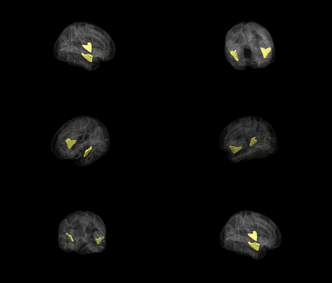
The bilateral Planum Polare is a cortical region located anterior to Heschl’s gyrus on the superior temporal plane, forming part of the anterior auditory association cortex within the temporal lobe. It lies on the dorsal surface of the superior temporal gyrus, extending into the transverse temporal structures, and is involved in higher-order auditory processing, including analysis of complex sounds and aspects of music and speech. Functionally, the Planum Polare contributes to the integration of spectral and temporal features of auditory stimuli and is thought to participate in predictive and contextual processing of sounds within broader auditory networks. There is no direct Wikipedia article for Planum Polare; a related structure is the superior temporal gyrus: Superior temporal gyrus.
The bilateral Planum Polare, an anterior temporal lobe region involved in higher-order auditory, language, and semantic processing, has been implicated indirectly in genetic studies through its overlap with temporal association cortex rather than via region-specific GWAS signals. Large neuroimaging genetics consortia (e.g., ENIGMA, UK Biobank) have identified common variants in genes such as HMGA2, IGF1, KIAA0586, and others that influence global and temporal lobe cortical thickness and surface area, within which Planum Polare territory is included, but these effects are typically reported at lobar or parcel-aggregated scales rather than for this atlas-defined region alone. GWAS of language-related traits and reading abilities, involving loci near genes such as CNTNAP2, FOXP2, DCDC2, and KIAA0319, and disorders like developmental language disorder and dyslexia, have shown associations with structural and functional measures in the superior and middle temporal cortices, including anterior temporal regions encompassing Planum Polare, though the genetic effects are spatially broad and not anatomically specific to this label. Similarly, variants linked to schizophrenia, bipolar disorder, and autism spectrum disorder (for example, in CACNA1C, GRIN2A, NRXN1, and other synaptic and neurodevelopmental genes) have been associated with altered cortical thickness, gyrification, or activation patterns in temporal association areas that include Planum Polare, but current GWAS and imaging-genetic frameworks do not isolate robust, replicated SNP-level associations uniquely to the Planum Polare region from the Harvard-Oxford Cortical Atlas.
Overview generated by GPT-4o (2026).
Region ID: 44
Hemisphere: bilateral
Atlas: HarvardOxford cort maxprob thr25 1mm
Full Quality Version: Download MP4
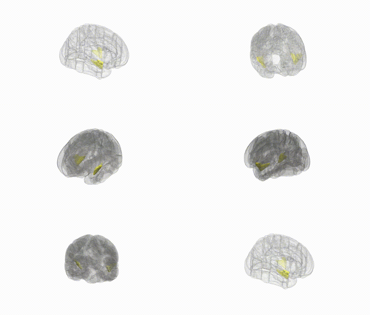
Full Quality Version: Download MP4
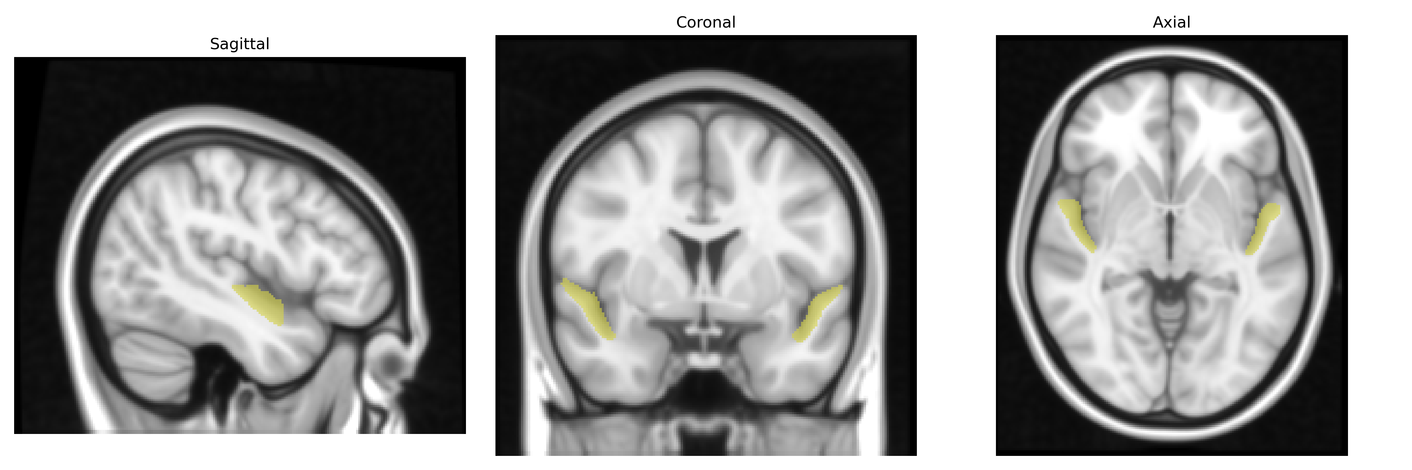
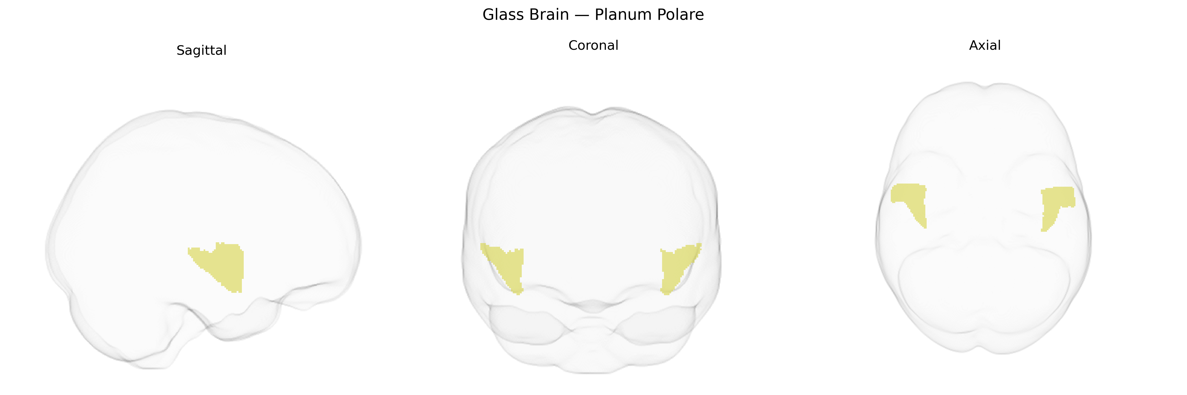
Wali Sidiqyar*, Gaurav Rudravaram*, Elyssa M. McMaster, Trent M. Schwartz, Adam M. Saunders, Kurt G. Schilling, Bennett A. Landman "Introducing SPINS: A Shared Public Visualization Library of Neuroanatomical Structures." Medical Imaging with Deep Learning- short paper
This resource is licensed under CC0 1.0 Universal (Public Domain).