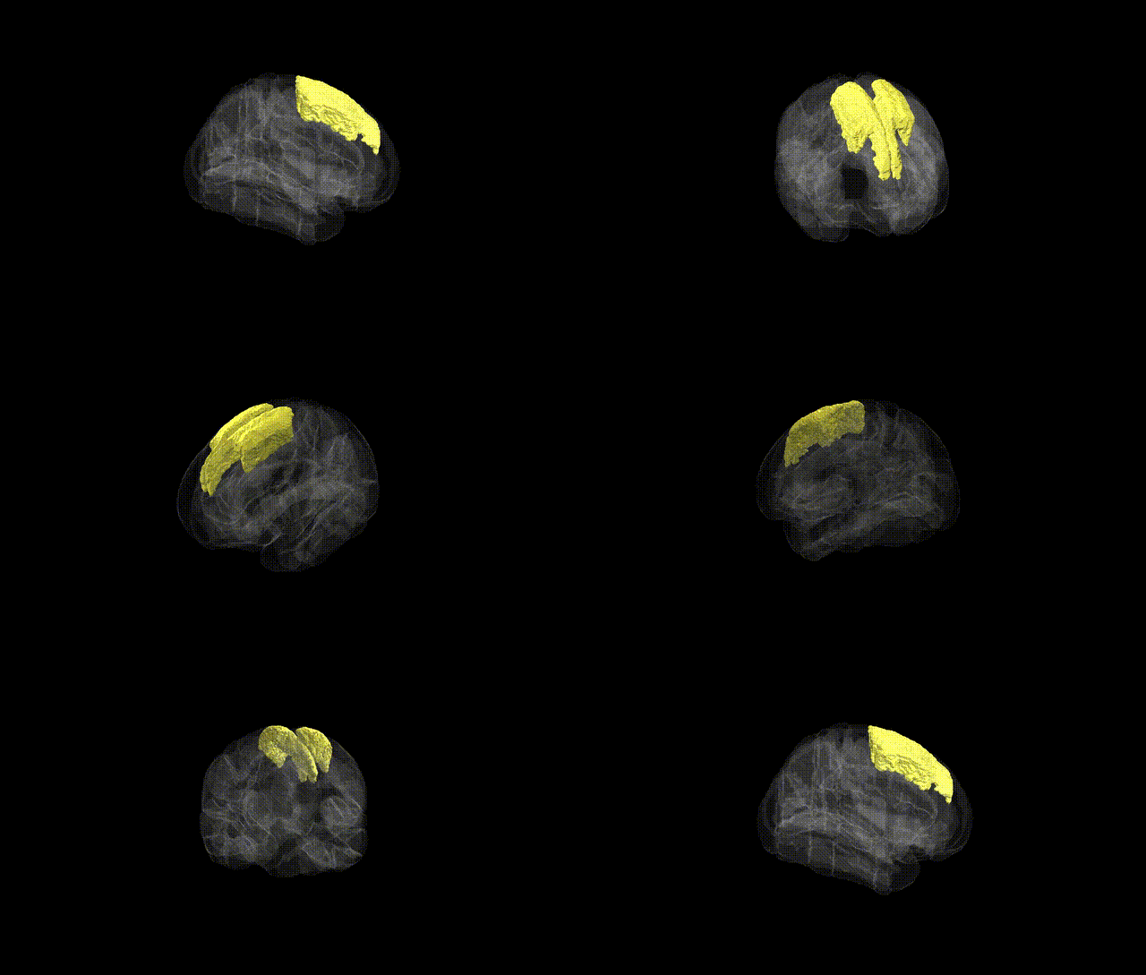
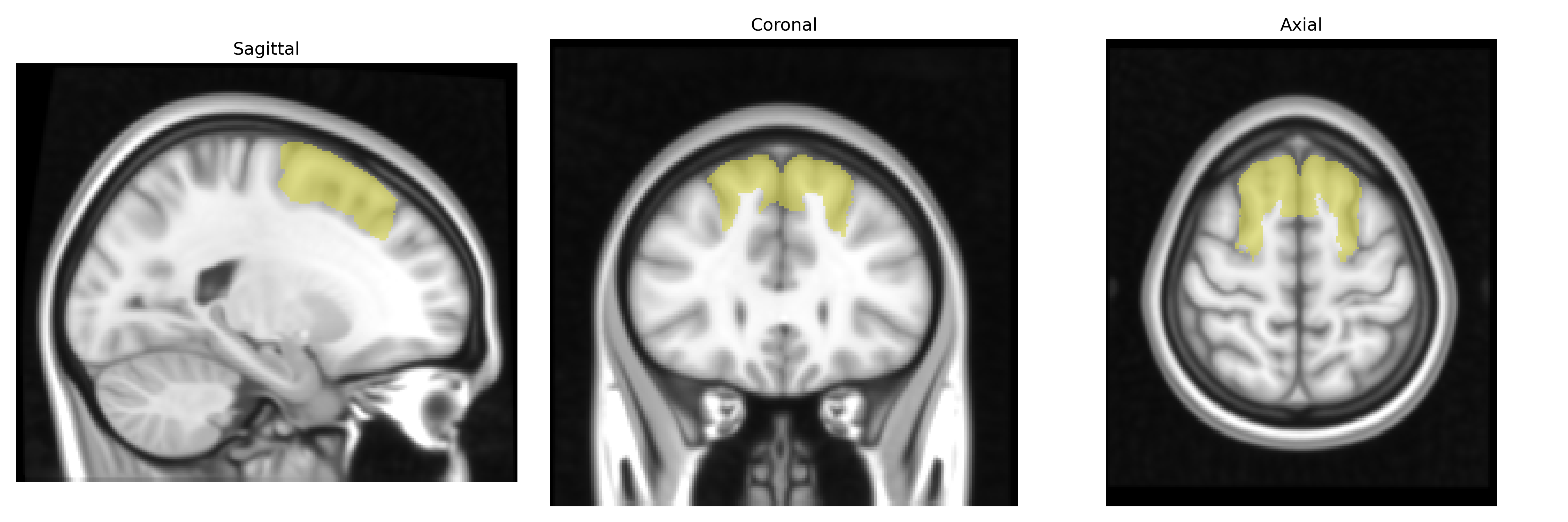
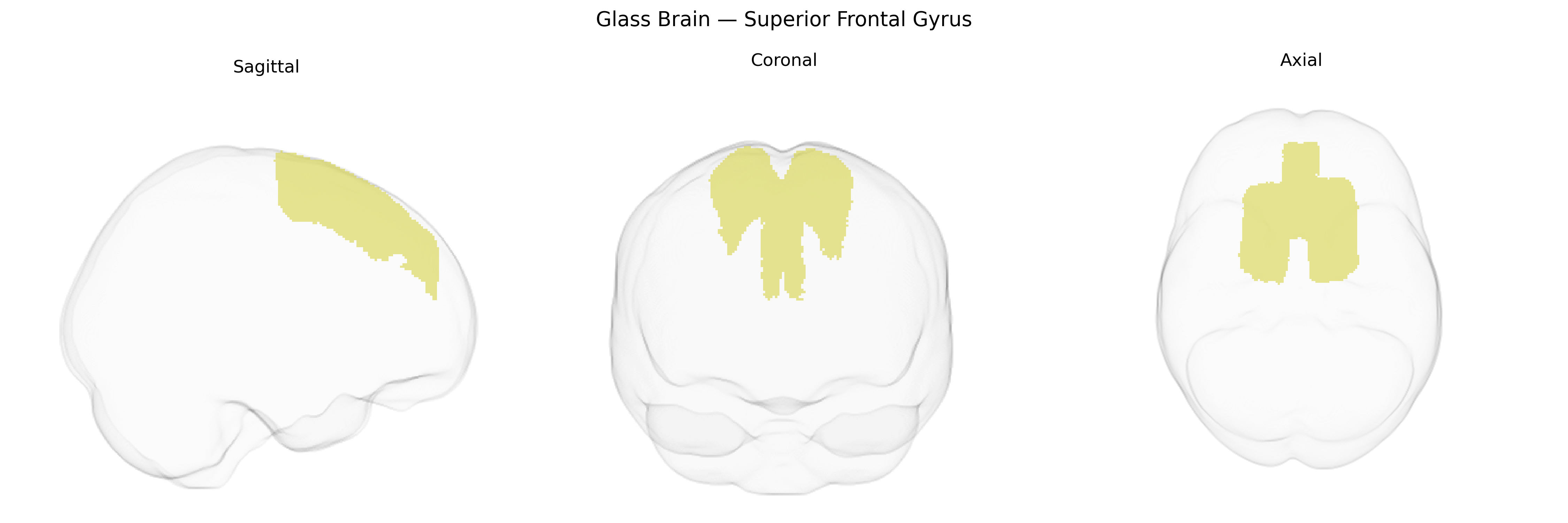
The bilateral Superior Frontal Gyrus, as defined in the Harvard-Oxford Cortical Structural Atlas (maxprob thr25, 1 mm), is a dorsal frontal lobe structure extending along the superior aspect of the frontal cortex, situated medial to the middle frontal gyrus and anterior to the precentral gyrus. It participates in higher-order cognitive and executive functions, including working memory, attention, self-referential processing, and aspects of motor planning, with functional connectivity to medial prefrontal, cingulate, and parietal association areas. Cytoarchitectonically, it encompasses portions of premotor and prefrontal territories, with anterior regions more associated with abstract, integrative processing and posterior regions linked to motor-related functions. The region shows hemispheric symmetry in gross anatomy but may exhibit lateralized specializations depending on task demands and functional network engagement. Superior frontal gyrus
The bilateral Superior Frontal Gyrus (SFG), corresponding to the Harvard–Oxford cortical maxprob thr25 1 mm atlas label, has been implicated in multiple genetic and GWAS findings, largely through imaging genetics and disorder-focused studies rather than single-gene associations specific to this region. Heritability analyses from twin and family studies show that SFG volume, thickness, and surface area are moderately to highly heritable, with large consortia such as ENIGMA and UK Biobank demonstrating polygenic influences on its structure and function. GWAS of cortical morphology have identified common variants near genes involved in neurodevelopment, synaptic signaling, and cell adhesion (e.g., loci near HMGA2, IGF1, and other regulatory genes) that affect frontal cortical thickness and surface area, often including the superior frontal territory. Functionally, the SFG—central to cognitive control, self-referential processing, and components of the default mode and executive networks—has been linked via imaging-genetic studies to polygenic risk scores for schizophrenia, major depressive disorder, bipolar disorder, ADHD, and autism, where risk variants are associated with altered SFG activation or morphology. In Alzheimer’s disease and frontotemporal dementia, genetic risk (e.g., APOE ε4 and MAPT haplotypes) correlates with frontal cortical atrophy patterns that frequently include the SFG, while GWAS of traits such as general cognitive ability, educational attainment, and neuroticism show distributed polygenic effects on frontal regions encompassing this gyrus. Overall, current evidence supports a highly polygenic, pleiotropic architecture in which many common variants influencing neurodevelopment, synaptic function, and psychiatric risk contribute to structural and functional variation in the bilateral SFG, rather than a small number of region-specific genetic determinants.
Overview generated by GPT-4o (2026).
Region ID: 3
Hemisphere: bilateral
Atlas: HarvardOxford cort maxprob thr25 1mm
Full Quality Version: Download MP4
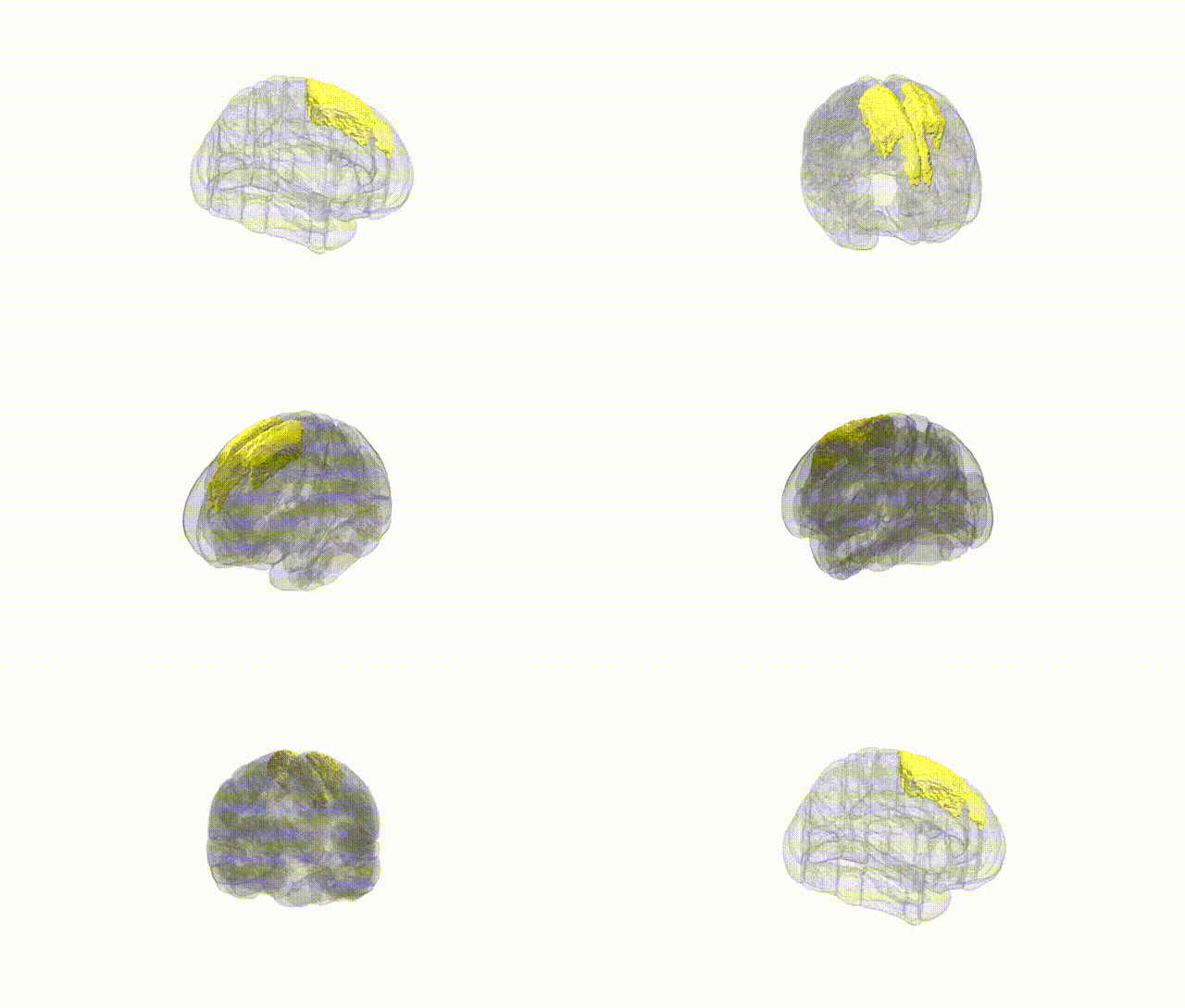
Full Quality Version: Download MP4
Wali Sidiqyar*, Gaurav Rudravaram*, Elyssa M. McMaster, Trent M. Schwartz, Adam M. Saunders, Kurt G. Schilling, Bennett A. Landman "Introducing SPINS: A Shared Public Visualization Library of Neuroanatomical Structures." Medical Imaging with Deep Learning- short paper
This resource is licensed under CC0 1.0 Universal (Public Domain).