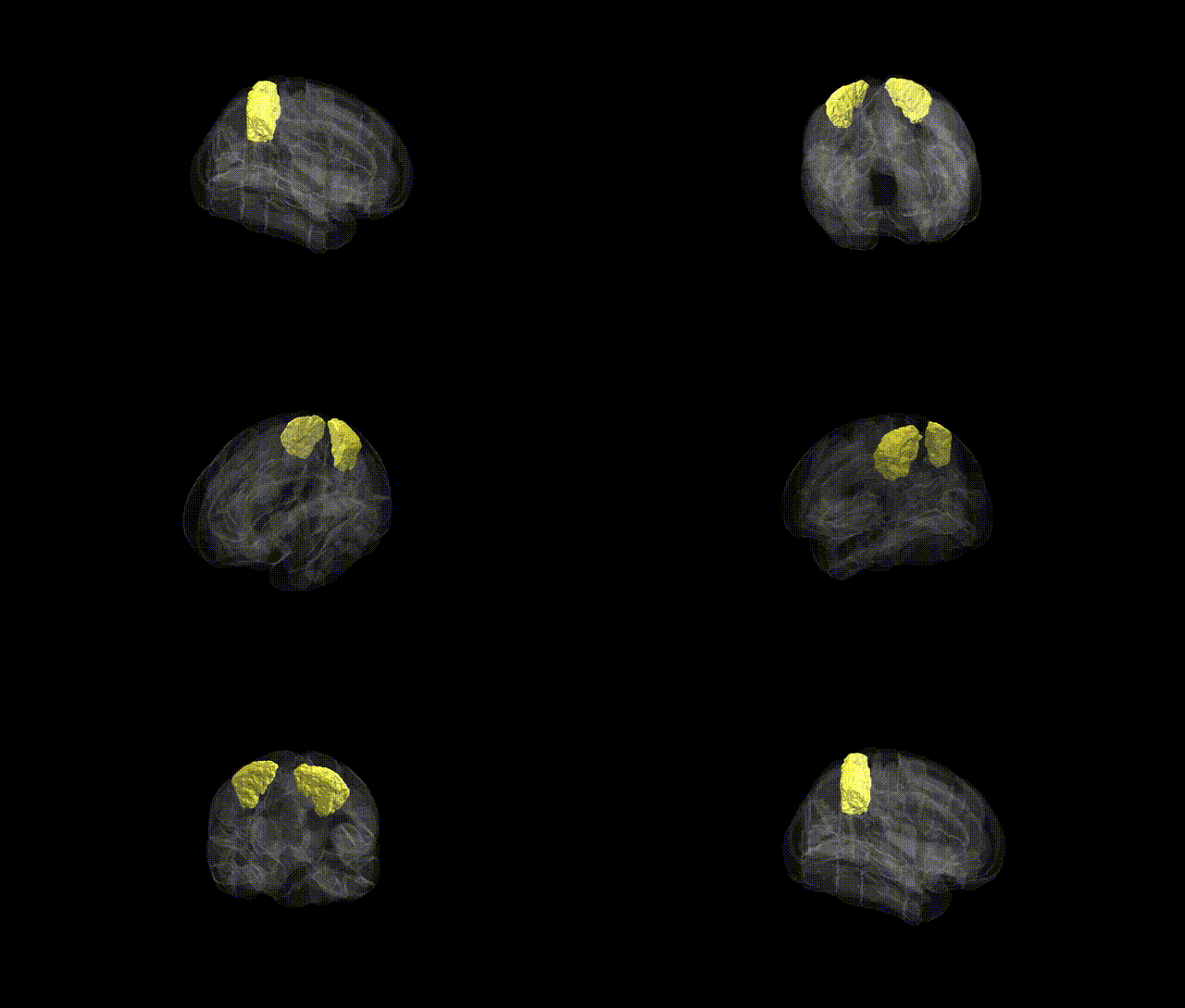
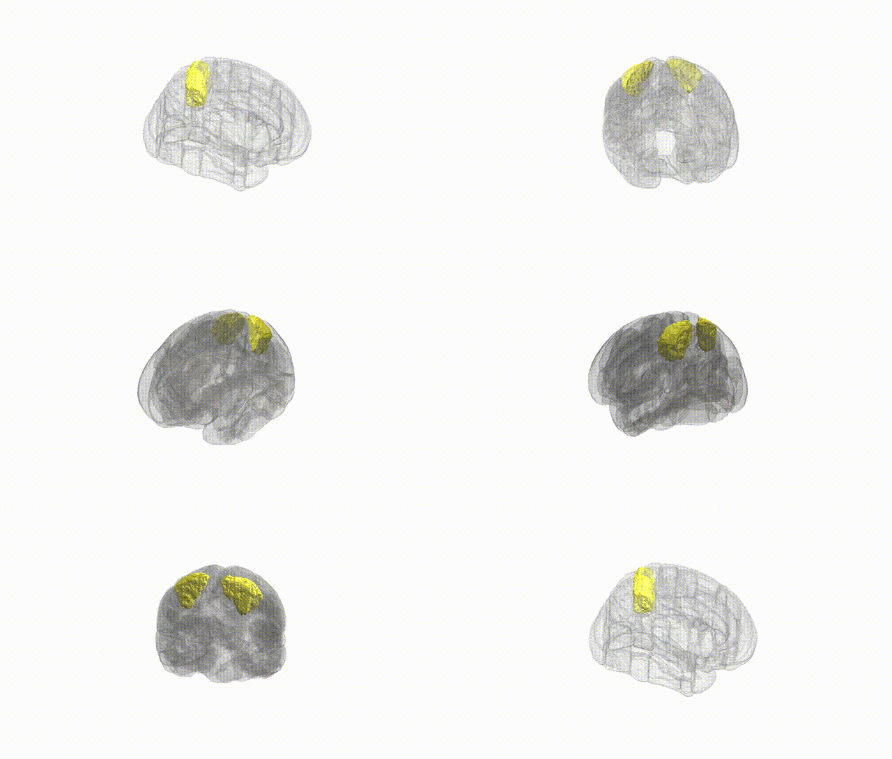
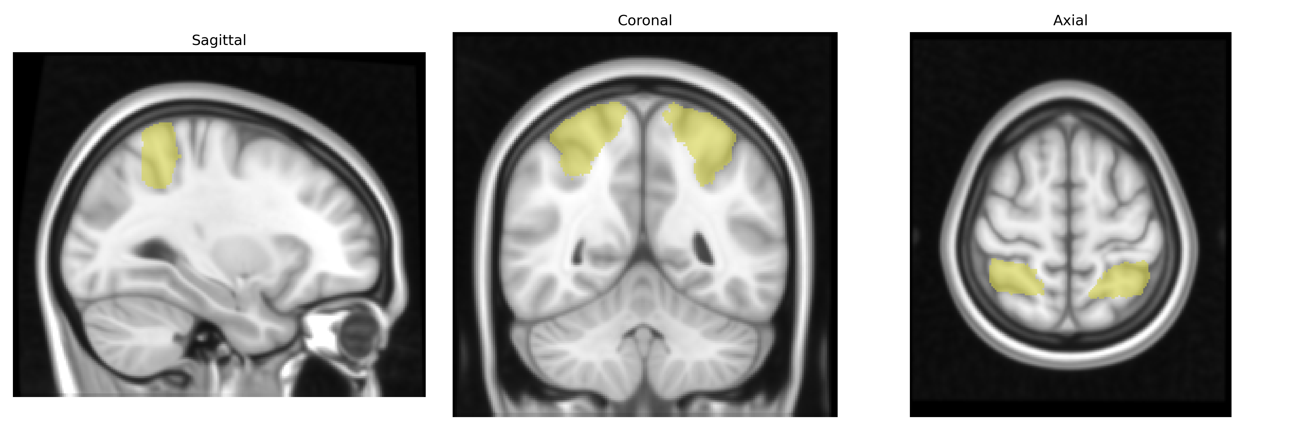
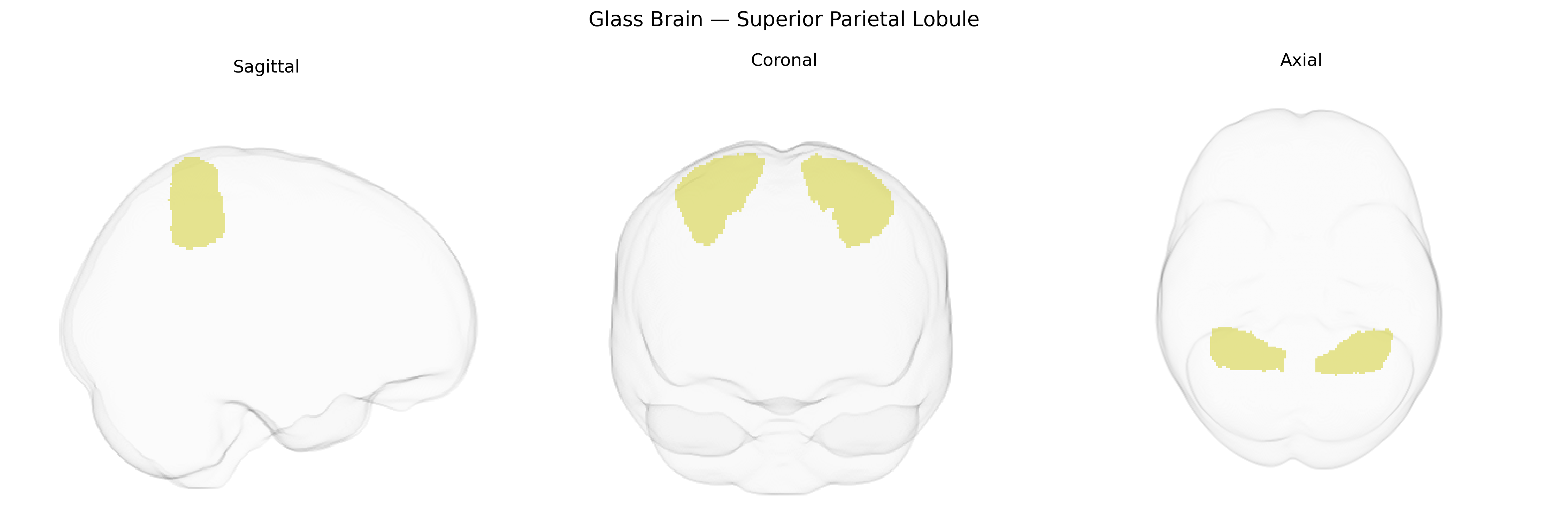
The bilateral Superior Parietal Lobule (SPL), as defined in the Harvard-Oxford cortical structural atlas (maxprob, thr25, 1 mm), is a dorsally positioned parietal association region located posterior to the postcentral gyrus and superior to the intraparietal sulcus, extending medially toward the precuneus. It is primarily involved in higher-order somatosensory integration, visuospatial processing, and the coordination of attention and action, contributing to functions such as spatial orientation, body schema representation, and the online control of reaching and grasping movements. The SPL receives convergent input from primary and secondary somatosensory cortices and visual areas, and projects to premotor and other parietal association regions, thereby forming part of a dorsal “where/how” pathway critical for sensorimotor transformation and visuomotor integration. Related article: Superior parietal lobule.
The bilateral Superior Parietal Lobule (SPL), as defined in the Harvard-Oxford cortical maxprob atlas, has been implicated in multiple genetic and GWAS findings linking common and rare variants to individual differences in visuospatial attention, working memory, sensorimotor integration, and higher-order cognitive function. Large-scale brain-morphometry GWAS (e.g., ENIGMA, UK Biobank) have identified polygenic influences on SPL cortical thickness, surface area, and volume, with loci in genes involved in neurodevelopment, synaptic signaling, and axon guidance (such as variants near HMGA2, microtubule- and cytoskeleton-related genes, and several intergenic regulatory regions) contributing to parietal structure. SPL activity and connectivity show heritable patterns associated with general intelligence and educational attainment, aligning with GWAS loci in genes related to neuronal differentiation and cortical patterning. Parietal abnormalities involving SPL are reported in neurodevelopmental and psychiatric disorders, including autism spectrum disorder, attention-deficit/hyperactivity disorder, and schizophrenia; risk variants in genes governing synaptic plasticity and excitatory–inhibitory balance (for example, in glutamatergic and GABAergic pathways) are indirectly linked to altered SPL structure and function in these conditions. Additionally, SPL-related networks show genetic correlations with traits such as handedness, motor coordination, and risk-taking behavior, reflecting shared polygenic architectures between SPL morphology/function and complex behavioral phenotypes.
Overview generated by GPT-4o (2026).
Region ID: 18
Hemisphere: bilateral
Atlas: HarvardOxford cort maxprob thr25 1mm
Full Quality Version: Download MP4
Full Quality Version: Download MP4
Wali Sidiqyar*, Gaurav Rudravaram*, Elyssa M. McMaster, Trent M. Schwartz, Adam M. Saunders, Kurt G. Schilling, Bennett A. Landman "Introducing SPINS: A Shared Public Visualization Library of Neuroanatomical Structures." Medical Imaging with Deep Learning- short paper
This resource is licensed under CC0 1.0 Universal (Public Domain).