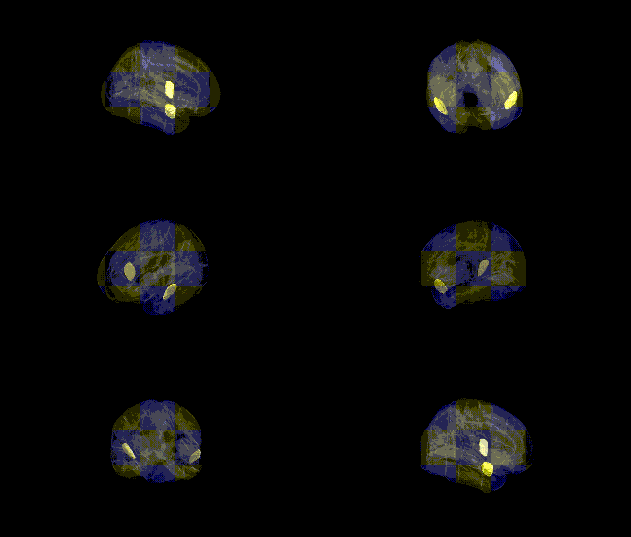
The bilateral Superior Temporal Gyrus, anterior division (from the Harvard–Oxford Cortical Structural Atlas, maxprob thr25 1 mm) comprises the rostral segment of the superior temporal gyrus in both hemispheres, bordering the temporal pole anteriorly and extending posteriorly toward mid–superior temporal regions. It is part of the lateral temporal neocortex and is supplied predominantly by branches of the middle cerebral artery. Cytoarchitectonically, this region contains higher-order auditory and multimodal associative cortex, integrating complex auditory inputs (such as speech and environmental sounds) with semantic, social, and emotional information, and participating in language comprehension networks in the dominant hemisphere as well as in voice and socio-emotional processing bilaterally. The anterior superior temporal gyrus maintains strong structural and functional connectivity with the temporal pole, middle and inferior temporal gyri, inferior parietal lobule, insula, and prefrontal association cortex, and is implicated in disorders affecting language, social cognition, and auditory perception. Superior temporal gyrus
The bilateral anterior division of the Superior Temporal Gyrus (STG), as defined in the Harvard–Oxford cortical atlas, has been implicated in several genetically influenced traits and disorders, primarily through imaging genetics and GWAS of brain structure and function rather than region-specific single-locus findings. Large-scale neuroimaging GWAS (e.g., ENIGMA consortium, UK Biobank) have identified multiple common variants influencing temporal lobe cortical thickness and surface area, including loci near genes such as KIAA0586, PAX8, TCF4, and variants in regulatory regions affecting synaptic and neurodevelopmental pathways, with some associations extending to the superior temporal cortex. The anterior STG, a key node for language, social cognition, and auditory processing, shows volumetric and functional alterations in schizophrenia, autism spectrum disorder, and bipolar disorder, with polygenic risk scores for these conditions predicting STG morphology and activity; candidate and genome-wide studies implicate genes involved in glutamatergic signaling (e.g., GRIN2A), synaptic scaffolding (e.g., DLG4), and neurodevelopment (e.g., CNTNAP2, FOXP2-related networks) in circuits encompassing this region. GWAS of language-related traits (speech sound disorder, reading and verbal ability) and social communication have reported associations in genes that modulate temporal lobe structure and connectivity, though not always localized specifically to the anterior STG. Additionally, risk variants for Alzheimer’s disease (e.g., APOE ε4 and loci near CLU, BIN1) and frontotemporal dementia influence atrophy patterns and functional disruption in temporal cortices, including the STG, reflecting genetic contributions to vulnerability of this region in neurodegeneration. Overall, genetic influences on the anterior STG appear highly polygenic, mediated by neurodevelopmental, synaptic, and plasticity-related pathways, and are most clearly observed in multivariate GWAS of cortical morphology and connectivity rather than in single-trait, region-specific association studies.
Overview generated by GPT-4o (2026).
Region ID: 9
Hemisphere: bilateral
Atlas: HarvardOxford cort maxprob thr25 1mm
Full Quality Version: Download MP4
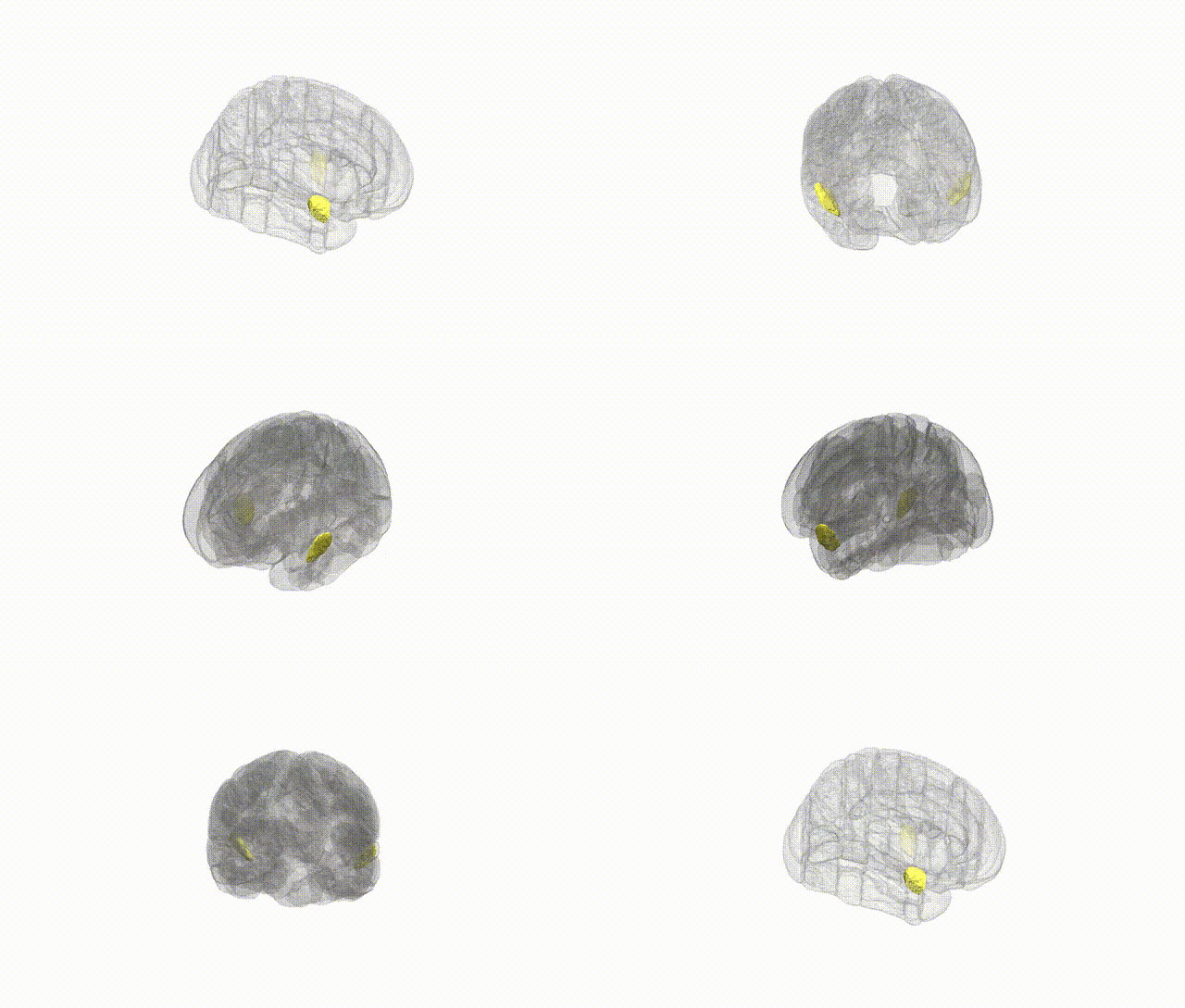
Full Quality Version: Download MP4
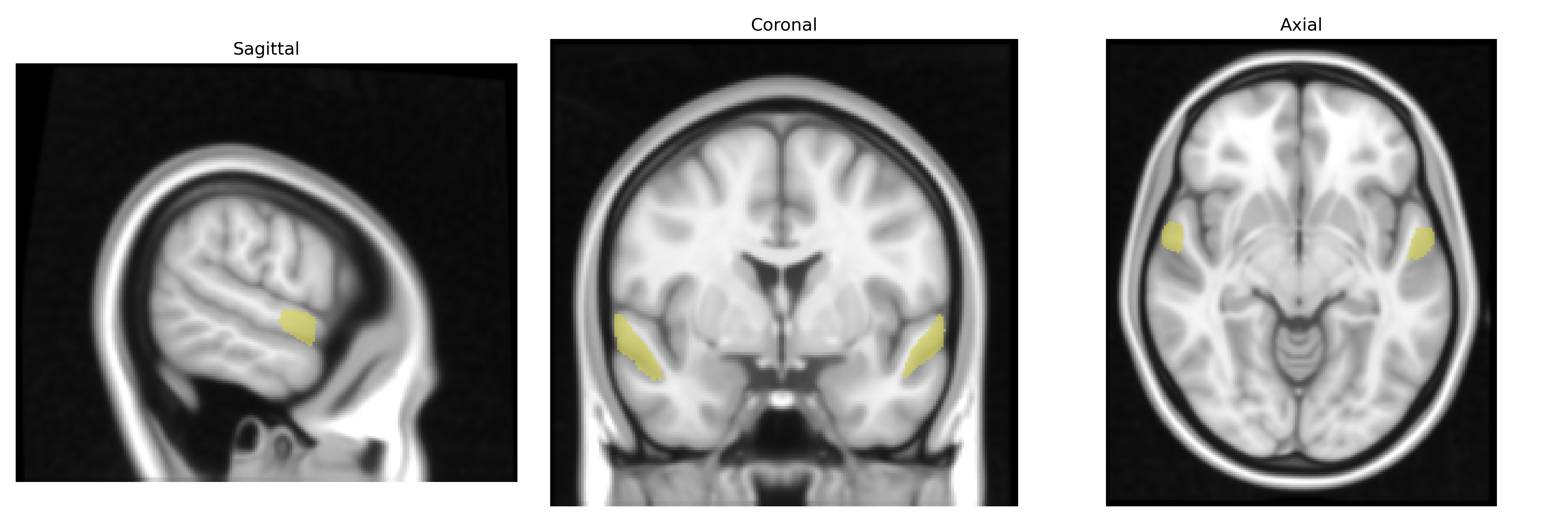
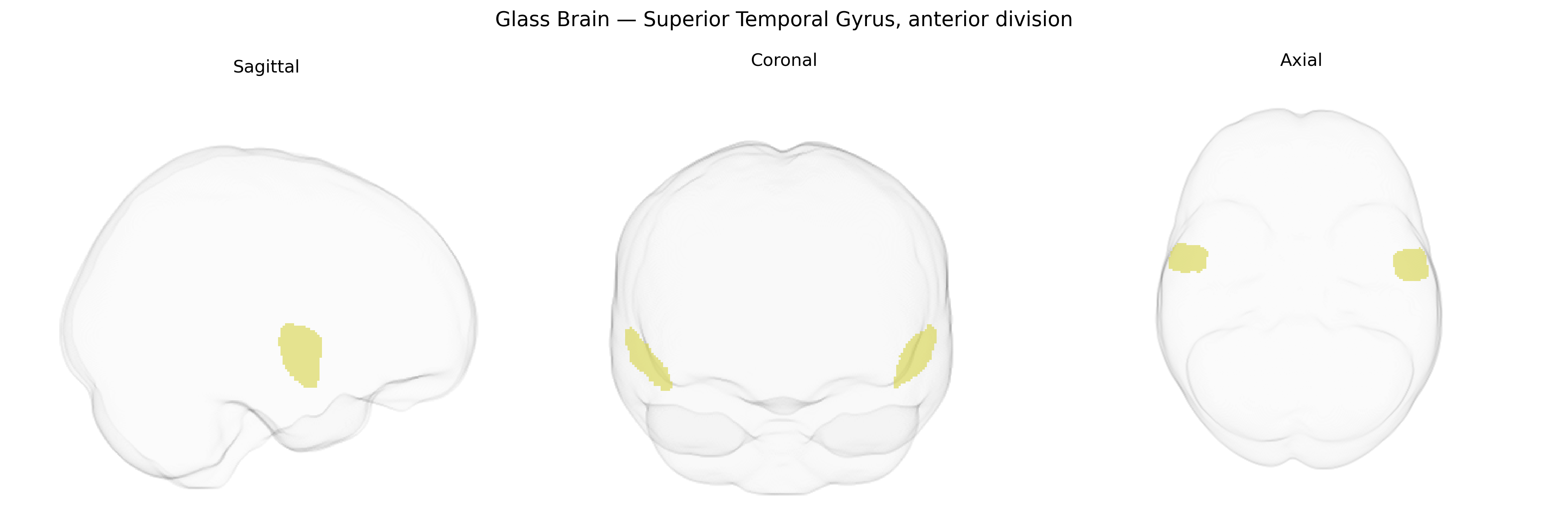
Wali Sidiqyar*, Gaurav Rudravaram*, Elyssa M. McMaster, Trent M. Schwartz, Adam M. Saunders, Kurt G. Schilling, Bennett A. Landman "Introducing SPINS: A Shared Public Visualization Library of Neuroanatomical Structures." Medical Imaging with Deep Learning- short paper
This resource is licensed under CC0 1.0 Universal (Public Domain).