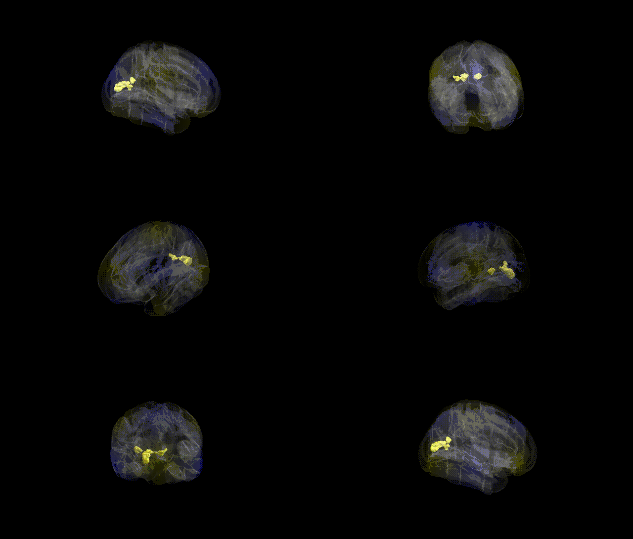
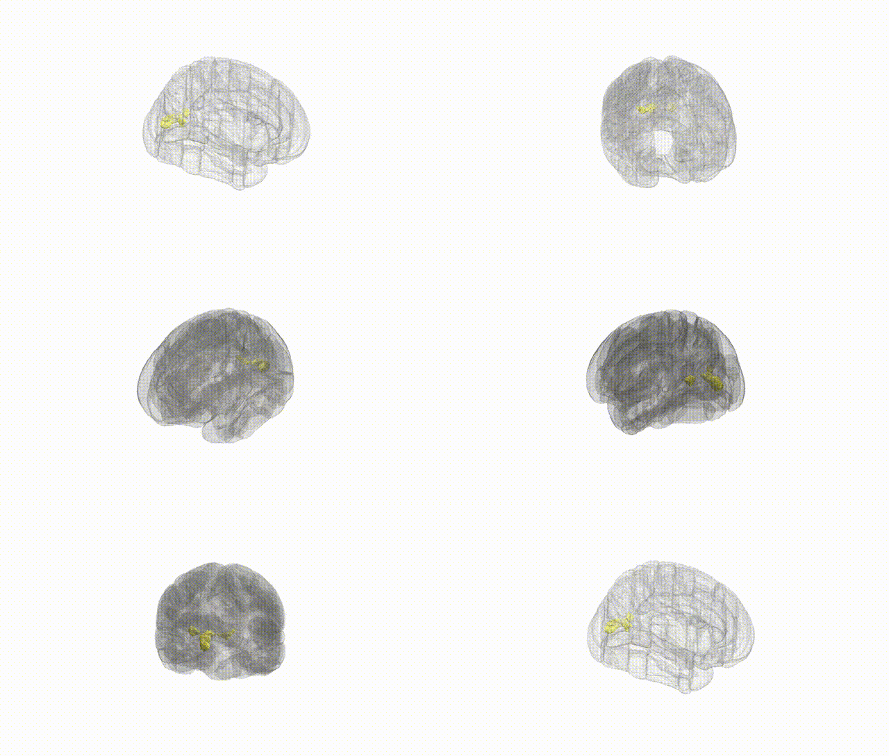
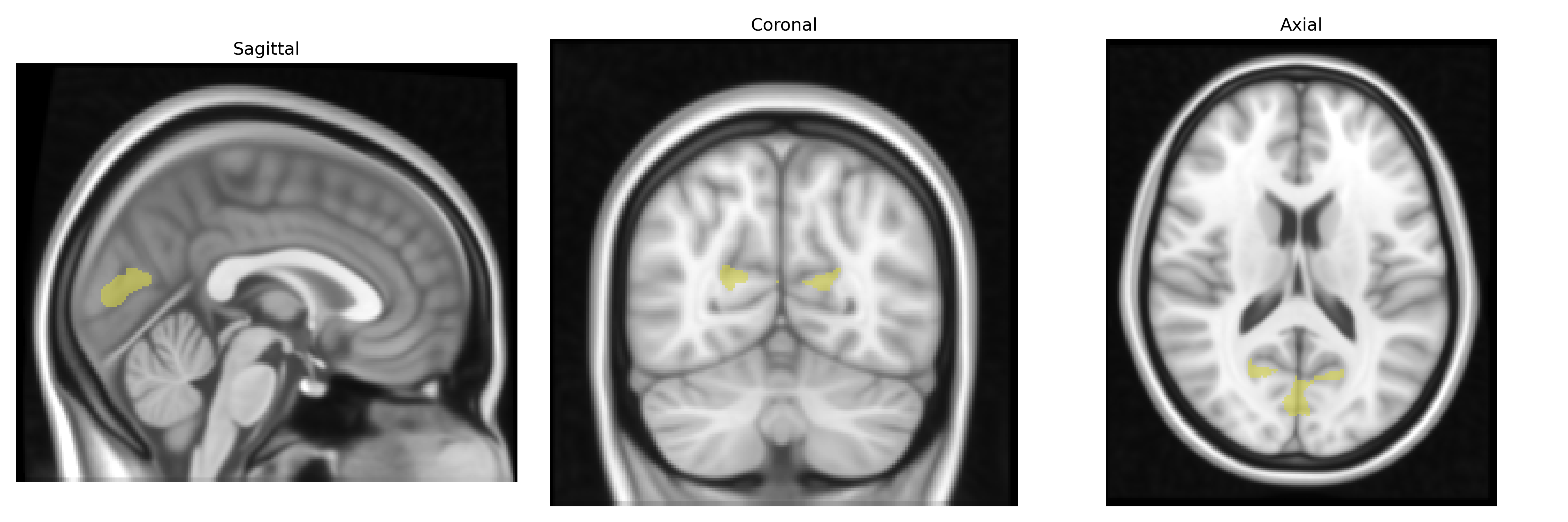
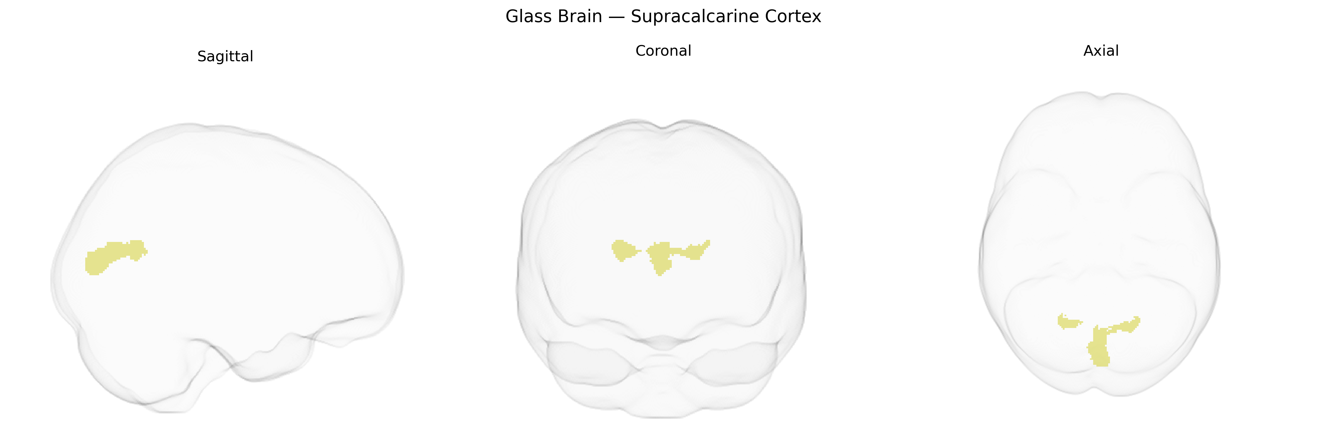
The bilateral Supracalcarine Cortex, as defined in the Harvard-Oxford cortical structural atlas, refers to cortical tissue located superior to the calcarine sulcus in the occipital lobe, adjacent to and functionally related to primary visual cortex (V1, Brodmann area 17). This region is part of the early visual processing network and participates in the initial analysis of visual input, particularly aspects of basic visual features such as luminance, contrast, and spatial patterns, with strong retinotopic organization. Although “Supracalcarine Cortex” itself does not have a dedicated Wikipedia entry, it is anatomically and functionally related to the Primary Visual Cortex, situated along the calcarine fissure.
The bilateral supracalcarine cortex, corresponding largely to primary visual areas around the calcarine sulcus in the Harvard–Oxford cortical atlas, has been implicated in genetic studies primarily through its role in visual processing, structural variation, and functional connectivity rather than via region-specific GWAS hits labeled “supracalcarine.” Large-scale imaging genetics consortia such as ENIGMA and UK Biobank have identified heritable variation in occipital and calcarine cortical thickness, surface area, and volume, with associated loci often involving genes related to neurodevelopment (e.g., microtubule and axon guidance pathways) and synaptic function, though these findings usually reference broader occipital or calcarine regions rather than the supracalcarine label specifically. Polygenic risk scores for neuropsychiatric disorders—including schizophrenia, major depressive disorder, and autism spectrum disorder—have been associated with altered occipital/calarcarine structure or activation patterns in some studies, suggesting that genetic liability to these conditions can influence supracalcarine cortex morphology or function as part of distributed brain effects. Visual system GWAS, such as those on visual acuity, photoreceptor and retinal traits, and migraine with aura, occasionally report downstream associations with occipital activation and connectivity, including the calcarine region, implying that genetic variation affecting visual pathways can shape supracalcarine cortex function indirectly. However, as of current literature, there are no widely replicated GWAS findings that single out the supracalcarine cortex by name as a primary locus; instead, genetic associations are inferred from broader visual cortex measures, multimodal imaging–genetics analyses, and polygenic effects on brain networks that encompass this region.
Overview generated by GPT-4o (2026).
Region ID: 47
Hemisphere: bilateral
Atlas: HarvardOxford cort maxprob thr25 1mm
Full Quality Version: Download MP4
Full Quality Version: Download MP4
Wali Sidiqyar*, Gaurav Rudravaram*, Elyssa M. McMaster, Trent M. Schwartz, Adam M. Saunders, Kurt G. Schilling, Bennett A. Landman "Introducing SPINS: A Shared Public Visualization Library of Neuroanatomical Structures." Medical Imaging with Deep Learning- short paper
This resource is licensed under CC0 1.0 Universal (Public Domain).