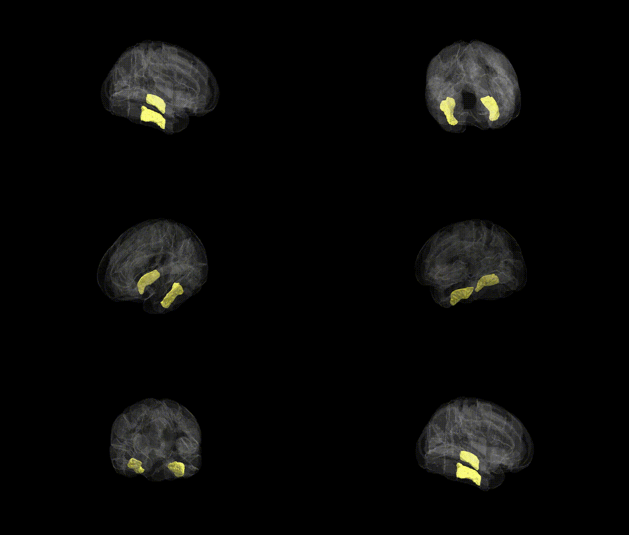
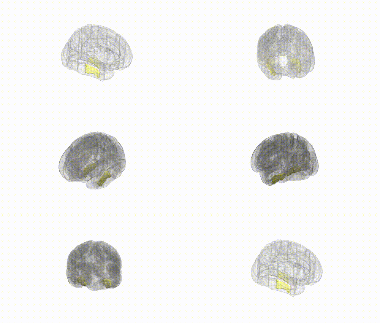
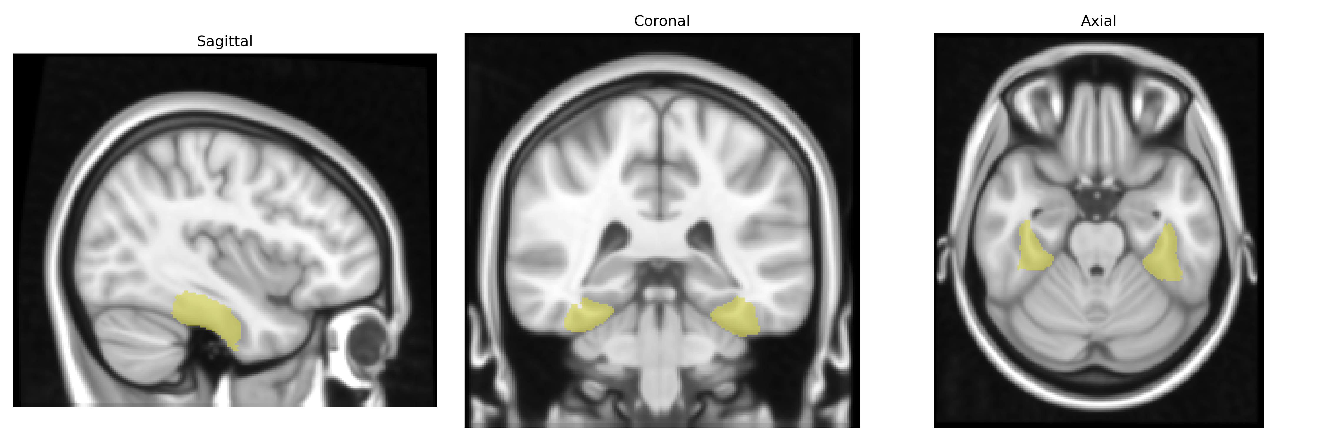
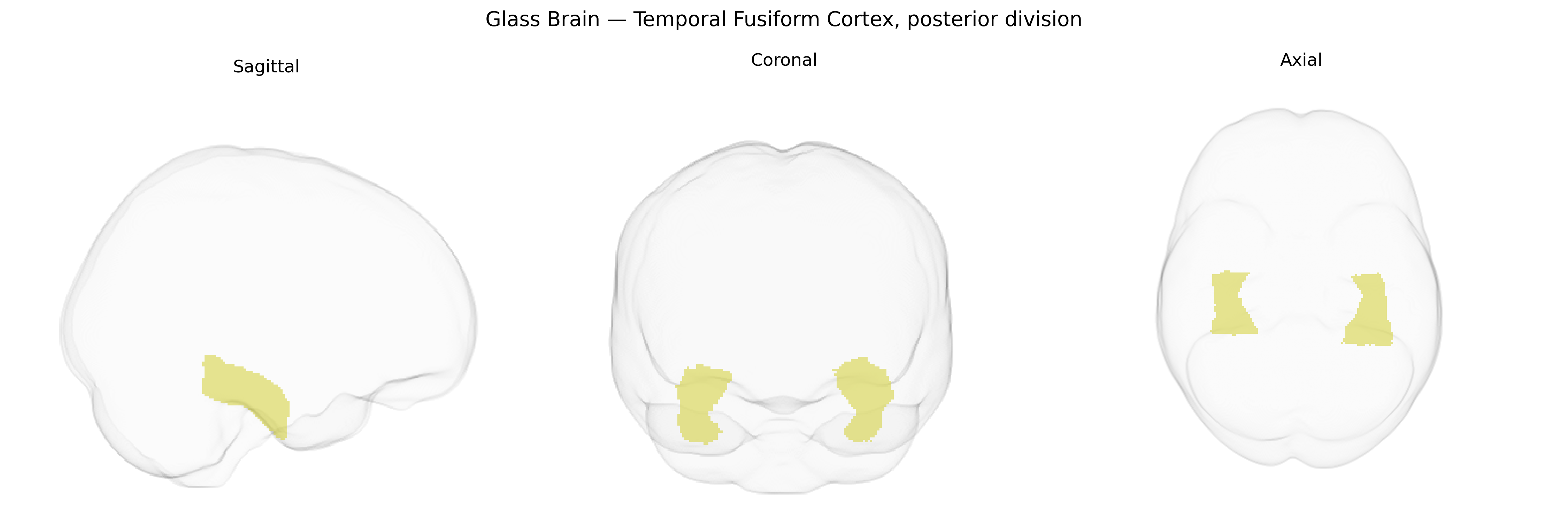
The bilateral Temporal Fusiform Cortex, posterior division, as defined in the Harvard–Oxford cortical maxprob atlas (25% threshold, 1 mm), corresponds to the posterior segment of the fusiform gyrus within the ventral temporal lobe, situated between the inferior temporal gyrus laterally and the parahippocampal gyrus medially. This region lies along the ventral visual processing stream and is implicated in high-level object and face perception, including contributions to category-selective processing of complex visual stimuli. Cytoarchitectonically, it overlaps portions of what has been described as occipitotemporal (fusiform) cortex, with dense reciprocal connectivity to occipital visual areas, inferior temporal cortex, and limbic structures involved in memory and semantic processing. There is no direct Wikipedia article specifically for “Temporal Fusiform Cortex, posterior division”; a closely related structure is the Fusiform gyrus.
The bilateral temporal fusiform cortex, posterior division (often overlapping with posterior fusiform gyrus and parts of the ventral temporal visual stream) shows genetic associations primarily through imaging–genetics and GWAS of cortical morphology and functional activation. Large-scale neuroimaging GWAS (e.g., ENIGMA and UK Biobank) have identified common variants in genes related to neurodevelopment, axon guidance, and synaptic plasticity (including loci near genes such as MAPT, TBR1, and KIAA0586 in broader temporal and ventral occipito‑temporal regions) that influence fusiform cortical thickness, surface area, and gyrification. Polygenic risk for neurodevelopmental and psychiatric disorders—autism spectrum disorder, schizophrenia, and major depressive disorder—has been associated with structural and functional alterations in posterior fusiform/ventral temporal cortex, particularly in face- and word-selective areas, suggesting shared genetic architectures influencing social cognition and high-level visual processing. GWAS of face perception and recognition have implicated variants near genes involved in occipito‑temporal development (for example, loci near PRDM13 and NEGR1 in broader visual and temporal cortex) that modulate activation of fusiform face-selective regions, and familial/rare-variant studies of congenital prosopagnosia have reported linkage and mutations in genes affecting posterior fusiform and adjacent ventral temporal organization, although specific risk alleles remain incompletely characterized. Overall, current evidence supports a polygenic, pleiotropic pattern in which variants influencing cortical development, microstructure, and connectivity contribute to individual differences and disorder-related changes in the posterior temporal fusiform cortex rather than a small set of region-specific “fusiform genes.”
Overview generated by GPT-4o (2026).
Region ID: 38
Hemisphere: bilateral
Atlas: HarvardOxford cort maxprob thr25 1mm
Full Quality Version: Download MP4
Full Quality Version: Download MP4
Wali Sidiqyar*, Gaurav Rudravaram*, Elyssa M. McMaster, Trent M. Schwartz, Adam M. Saunders, Kurt G. Schilling, Bennett A. Landman "Introducing SPINS: A Shared Public Visualization Library of Neuroanatomical Structures." Medical Imaging with Deep Learning- short paper
This resource is licensed under CC0 1.0 Universal (Public Domain).