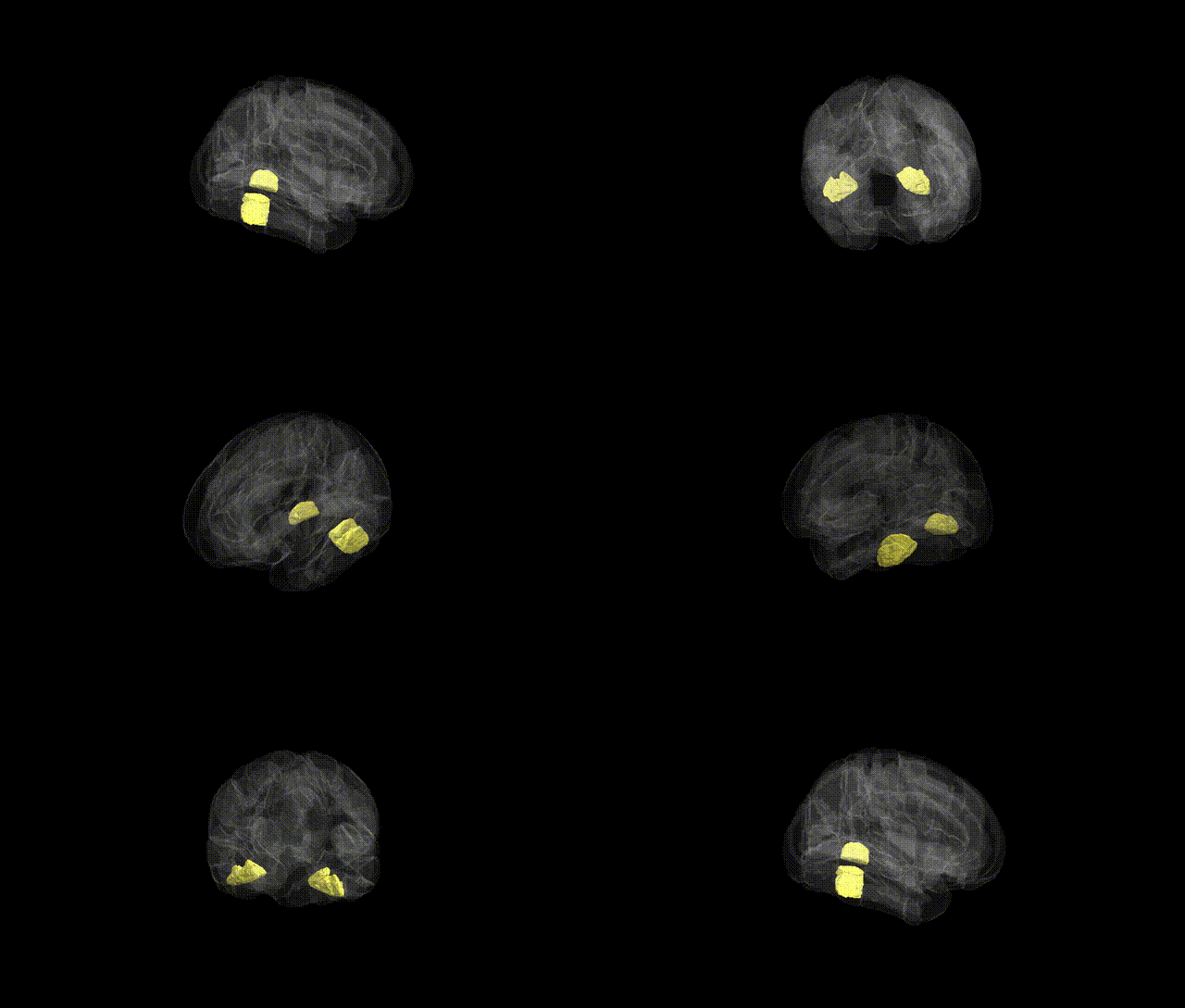
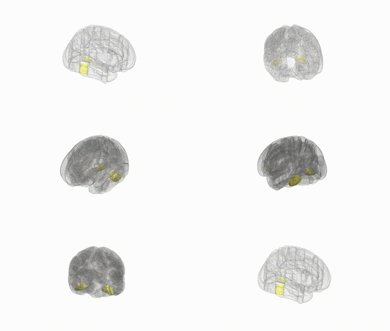
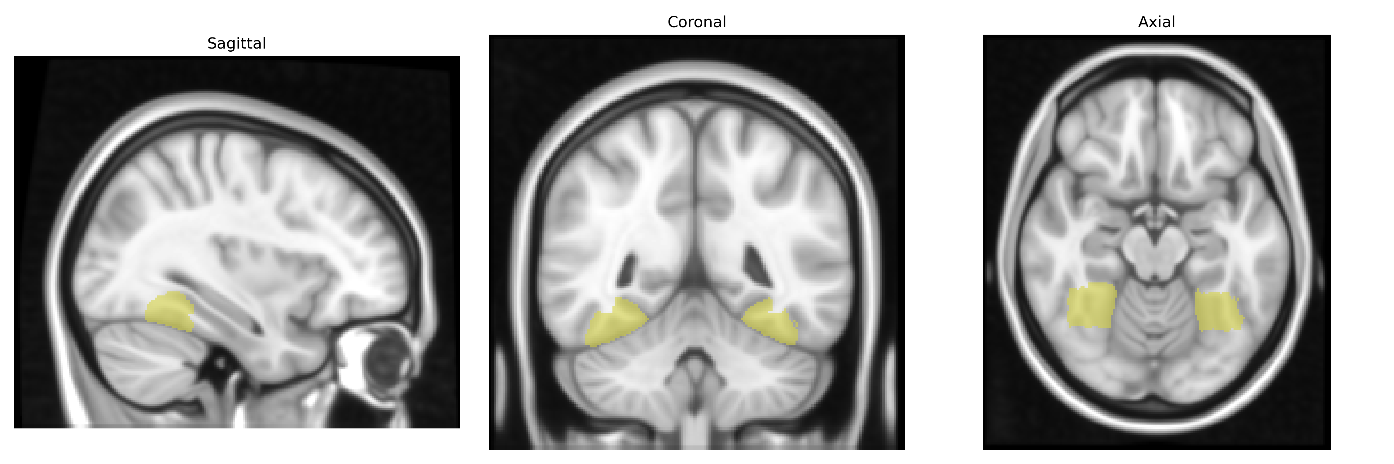
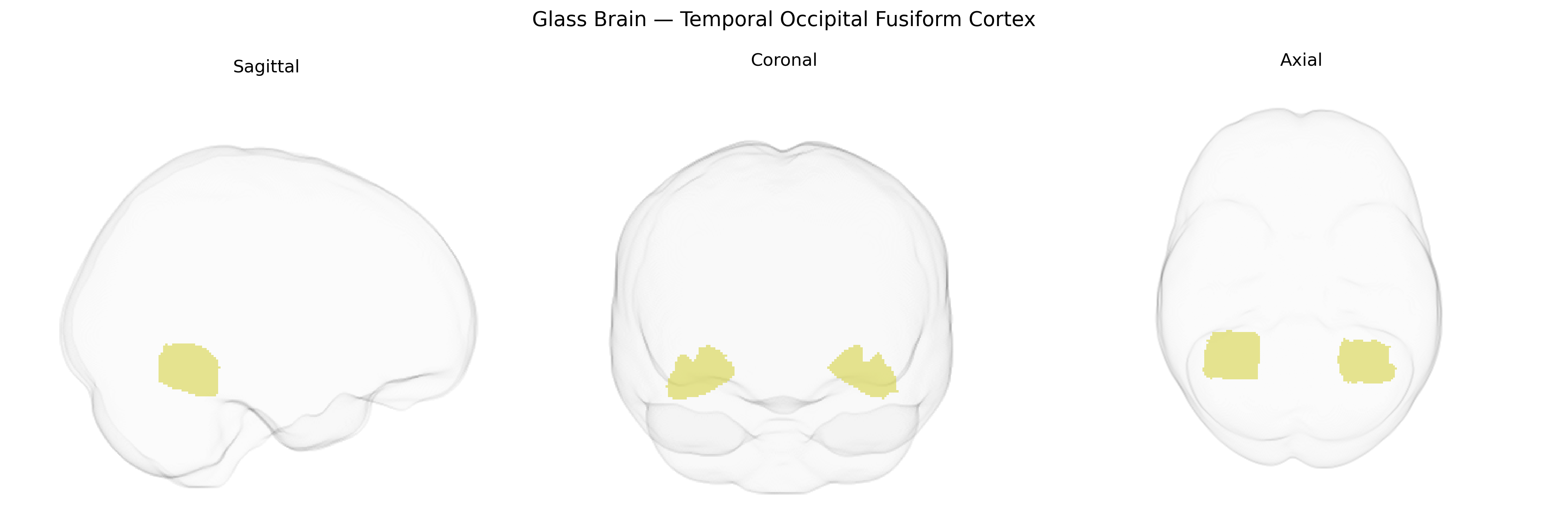
The bilateral Temporal Occipital Fusiform Cortex is a ventral temporal lobe region located at the junction of the temporal and occipital lobes, encompassing portions of the fusiform gyrus involved in high-level visual processing. In the Harvard-Oxford Cortical Atlas, this area corresponds to a fusiform extension that supports functions such as object recognition, processing of complex visual patterns, and category-selective perception (e.g., faces, words), with hemispheric specializations and extensive connections to occipital visual cortices and anterior temporal structures. There is no direct link for this exact atlas label; a closely related structure is the Fusiform gyrus.
The bilateral temporal occipital fusiform cortex, encompassing regions implicated in high-level visual processing such as face and word recognition, has been linked through imaging‑genetics and GWAS-based studies to multiple neurodevelopmental, psychiatric, and cognitive traits, although precise gene–region mappings remain incomplete. Polygenic influences on fusiform volume and cortical thickness have emerged from large-scale brain MRI GWAS (e.g., ENIGMA, UK Biobank), with signals implicating genes involved in neurodevelopment, synaptic function, and axonal guidance, including loci near genes such as HMGA2, IGF1, and various microtubule- and CAM-related genes, although these associations are typically reported for broader temporal or occipital regions rather than the HarvardOxford temporal occipital fusiform label specifically. Functionally, genetic risk for autism spectrum disorder and schizophrenia has been associated with altered fusiform structure and activation during face and social processing tasks, including reduced fusiform volume or atypical fusiform response to faces in carriers of higher polygenic risk scores for these disorders. Variants related to dyslexia and reading ability, including those in DCDC2, KIAA0319, and other language/reading-associated loci, have been tied to altered structure and activation in ventral occipitotemporal regions encompassing fusiform “visual word form” areas, linking genetic influences on reading to this cortex. Additional associations include familial and genetic contributions to prosopagnosia (developmental face blindness), where linkage and candidate gene work (e.g., MCTP2 and other synaptic genes) converge on face-selective fusiform regions, and genetic correlations between fusiform measures and general cognitive ability, educational attainment, and intracranial volume from multivariate GWAS. Overall, the genetic architecture of the temporal occipital fusiform cortex appears highly polygenic, overlapping with broader temporal and occipital cortical genetics, with convergent evidence tying this region’s structure and function to polygenic risk for neurodevelopmental and psychiatric disorders, literacy and visual expertise traits, and general brain and cognitive development, though region-specific GWAS with the exact HarvardOxford-defined fusiform parcel are still limited and under refinement.
Overview generated by GPT-4o (2026).
Region ID: 39
Hemisphere: bilateral
Atlas: HarvardOxford cort maxprob thr25 1mm
Full Quality Version: Download MP4
Full Quality Version: Download MP4
Wali Sidiqyar*, Gaurav Rudravaram*, Elyssa M. McMaster, Trent M. Schwartz, Adam M. Saunders, Kurt G. Schilling, Bennett A. Landman "Introducing SPINS: A Shared Public Visualization Library of Neuroanatomical Structures." Medical Imaging with Deep Learning- short paper
This resource is licensed under CC0 1.0 Universal (Public Domain).