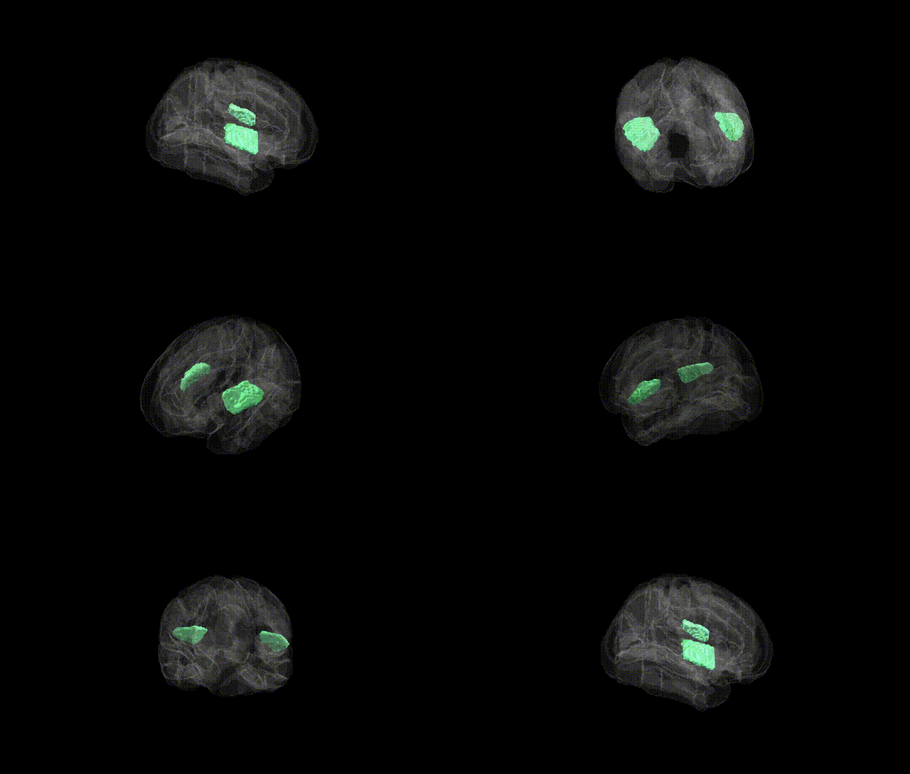
The bilateral Central Opercular Cortex, as defined in the Harvard–Oxford Cortical Structural Atlas (maxprob thr25, 2 mm), comprises portions of the opercular region of the frontal and parietal lobes that overlie the insula and form part of the peri-Sylvian cortex. This region contributes to sensorimotor integration for orofacial structures, speech articulation, and aspects of language processing, and is closely associated with the broader opercular areas that participate in complex motor planning and somatosensory processing. Anatomically, it lies adjacent to primary somatosensory and motor cortices and forms a structural and functional interface between neocortical sensorimotor fields and deeper insular circuitry. There is no direct link for “Central Opercular Cortex”; a related structure encompassing this region is the Operculum.
The bilateral Central Opercular Cortex, part of the perisylvian opercular region implicated in speech articulation, orofacial sensorimotor integration, and language processing, has been indirectly linked to several genetic associations through neuroimaging and GWAS of cortical structure and language-related traits rather than region-specific studies from the Harvard-Oxford atlas itself. Large-scale GWAS of cortical thickness and surface area (e.g., ENIGMA) have implicated loci near genes such as MAPT, KIAA0586, and microtubule and synaptic pathway genes that broadly affect perisylvian and frontal opercular morphology, including opercular subdivisions. Language and speech GWAS have associated variants in FOXP2, CNTNAP2, DCDC2, and KIAA0319 with functional and structural differences in opercular and inferior frontal language networks, which encompass the central opercular area. Neuroimaging genetics in developmental speech and language disorders, stuttering, and dyslexia have repeatedly shown altered activation and structure in opercular regions, with genetic risk loci for these conditions (e.g., in GNPTAB/GNPTG for stuttering, and multiple polygenic risk variants for dyslexia) correlating with atypical perisylvian and opercular organization. Additionally, polygenic risk for schizophrenia, autism spectrum disorder, and major depressive disorder has been associated with distributed cortical changes involving the operculum, and GWAS of smoking, obesity, and general cognitive ability report cortical morphometric effects in opercular language–sensorimotor networks, indicating that the Central Opercular Cortex participates in genetically influenced circuits underlying language, motor control, and neuropsychiatric vulnerability, even though no major GWAS has yet isolated this atlas-defined region as a primary, standalone genetic target.
Overview generated by GPT-4o (2026).
Region ID: 42
Hemisphere: bilateral
Atlas: HarvardOxford cort maxprob thr25 2mm
Full Quality Version: Download MP4
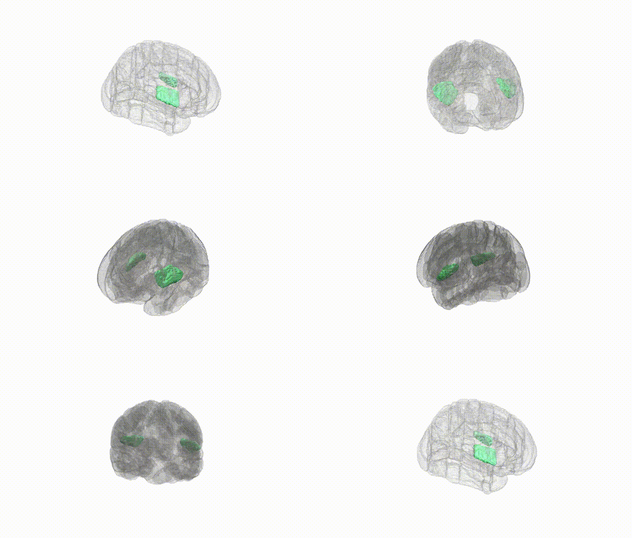
Full Quality Version: Download MP4
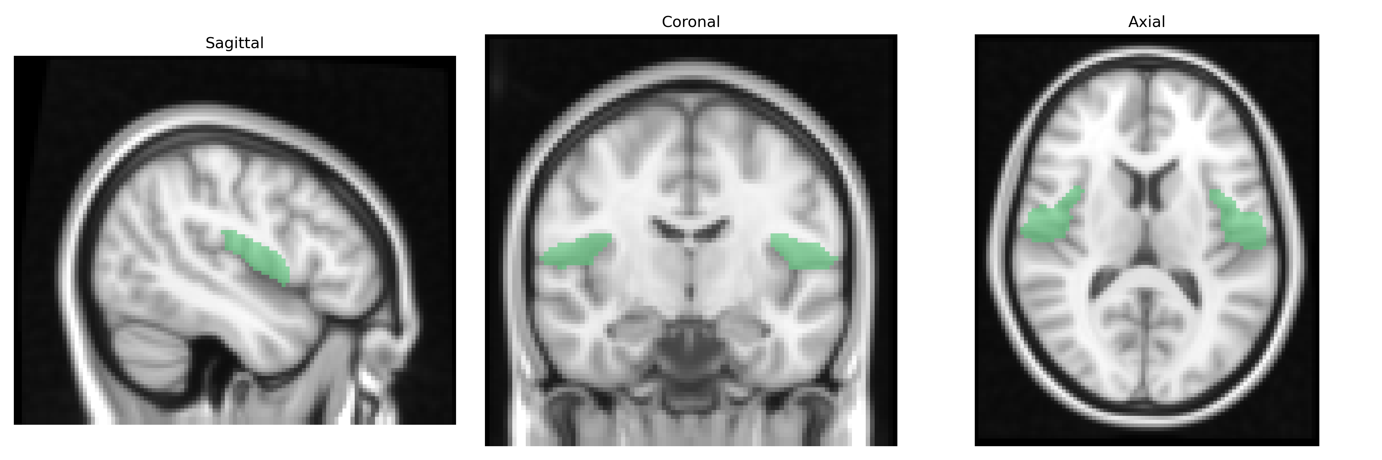
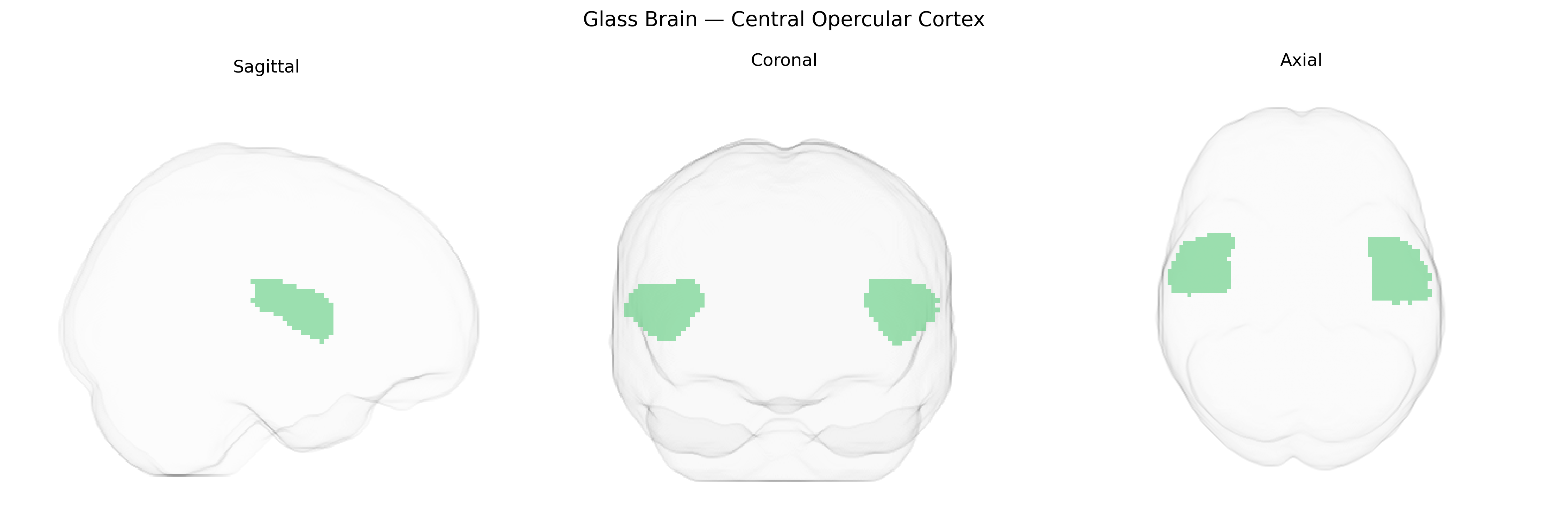
Wali Sidiqyar*, Gaurav Rudravaram*, Elyssa M. McMaster, Trent M. Schwartz, Adam M. Saunders, Kurt G. Schilling, Bennett A. Landman "Introducing SPINS: A Shared Public Visualization Library of Neuroanatomical Structures." Medical Imaging with Deep Learning- short paper
This resource is licensed under CC0 1.0 Universal (Public Domain).