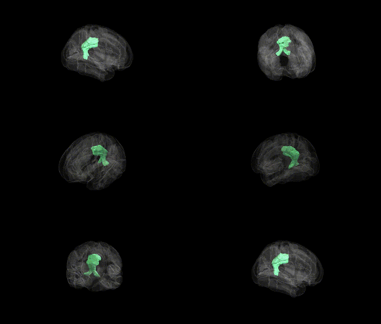
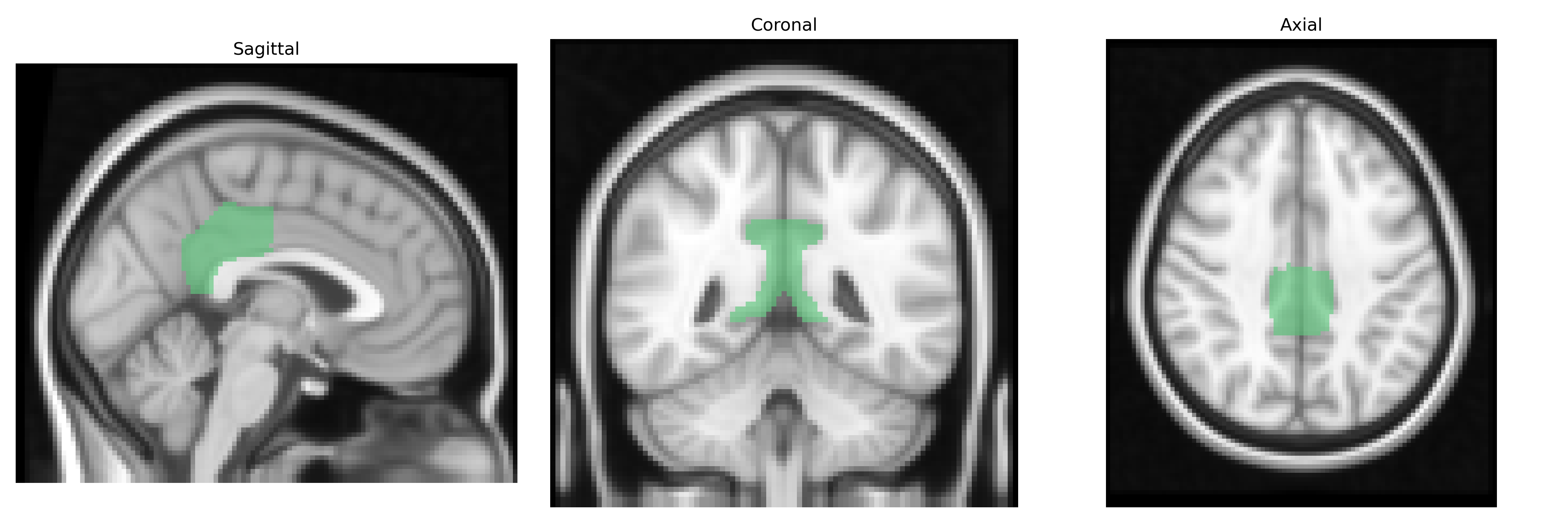
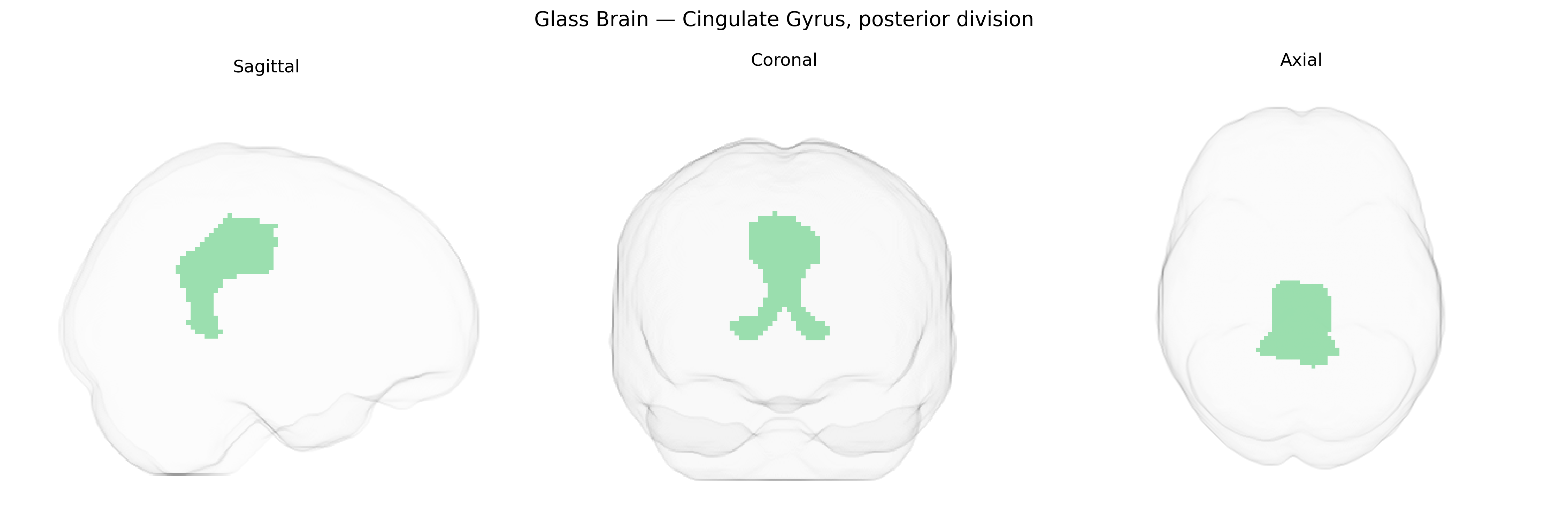
The bilateral cingulate gyrus, posterior division, as defined in the Harvard-Oxford Cortical Structural Atlas (maxprob thr25, 2 mm), encompasses the posterior segment of the cingulate cortex located on the medial surface of the cerebral hemispheres, extending from the midcingulate region toward the precuneus and retrosplenial cortex. This region is part of the limbic system and is strongly associated with the default mode network, contributing to internally directed cognition, autobiographical memory, visuospatial imagery, and integration of sensory and emotional information. It receives multimodal inputs from parietal, medial temporal, and thalamic nuclei and projects to widespread cortical and subcortical targets, supporting roles in attention, environmental monitoring, and context-dependent behavioral responses. There is no direct Wikipedia page specifically for the “posterior division” of the cingulate gyrus in this atlas; a related article is the broader Cingulate gyrus.
The bilateral posterior cingulate gyrus (PCG), as approximated by the Harvard–Oxford “Cingulate Gyrus, posterior division,” has been implicated in multiple genetic association studies, particularly through imaging genetics and large-scale GWAS linking cortical morphology and connectivity to specific loci and polygenic architectures. Variants in genes involved in synaptic plasticity, myelination, and neurodevelopment (e.g., BDNF, NRG1, DISC1, and APOE in the context of Alzheimer’s disease) have been associated with posterior cingulate volume, thickness, metabolism, or functional connectivity, consistent with the region’s vulnerability in neurodegenerative and psychiatric conditions. GWAS of cortical thickness and surface area (e.g., ENIGMA and UK Biobank–based consortia) have identified genome-wide significant loci near genes such as HMGA2, MIR2113, and those within Wnt and calcium-signaling pathways that influence cingulate and adjacent medial parietal cortex structure, while polygenic risk scores for schizophrenia, major depressive disorder, bipolar disorder, ADHD, autism spectrum disorder, and Alzheimer’s disease show associations with altered posterior cingulate morphology or default mode network connectivity. Additionally, genetic variants related to metabolic and cardiovascular traits (including APOE ε4 and loci implicated in lipid metabolism and vascular risk) have been linked to posterior cingulate hypometabolism and atrophy, particularly in preclinical and clinical Alzheimer’s disease, and GWAS of cognitive traits (general intelligence, memory performance, and educational attainment) suggest that overlapping polygenic architectures contribute to inter-individual differences in posterior cingulate structure and function, though precise, region-specific causal genes remain incompletely resolved.
Overview generated by GPT-4o (2026).
Region ID: 30
Hemisphere: bilateral
Atlas: HarvardOxford cort maxprob thr25 2mm
Full Quality Version: Download MP4
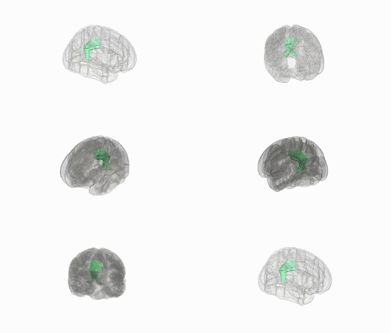
Full Quality Version: Download MP4
Wali Sidiqyar*, Gaurav Rudravaram*, Elyssa M. McMaster, Trent M. Schwartz, Adam M. Saunders, Kurt G. Schilling, Bennett A. Landman "Introducing SPINS: A Shared Public Visualization Library of Neuroanatomical Structures." Medical Imaging with Deep Learning- short paper
This resource is licensed under CC0 1.0 Universal (Public Domain).