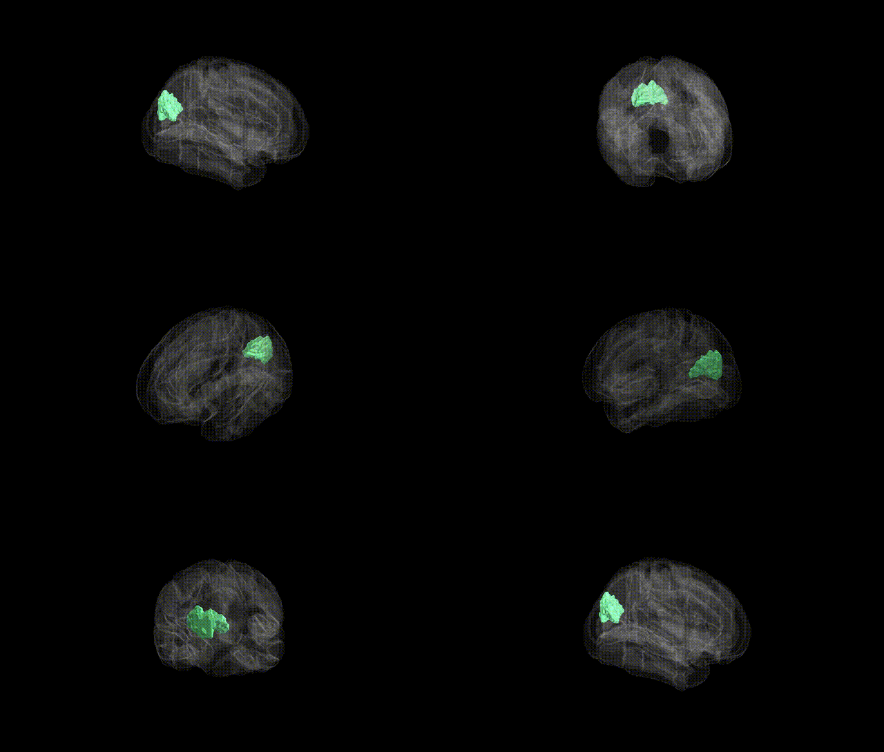
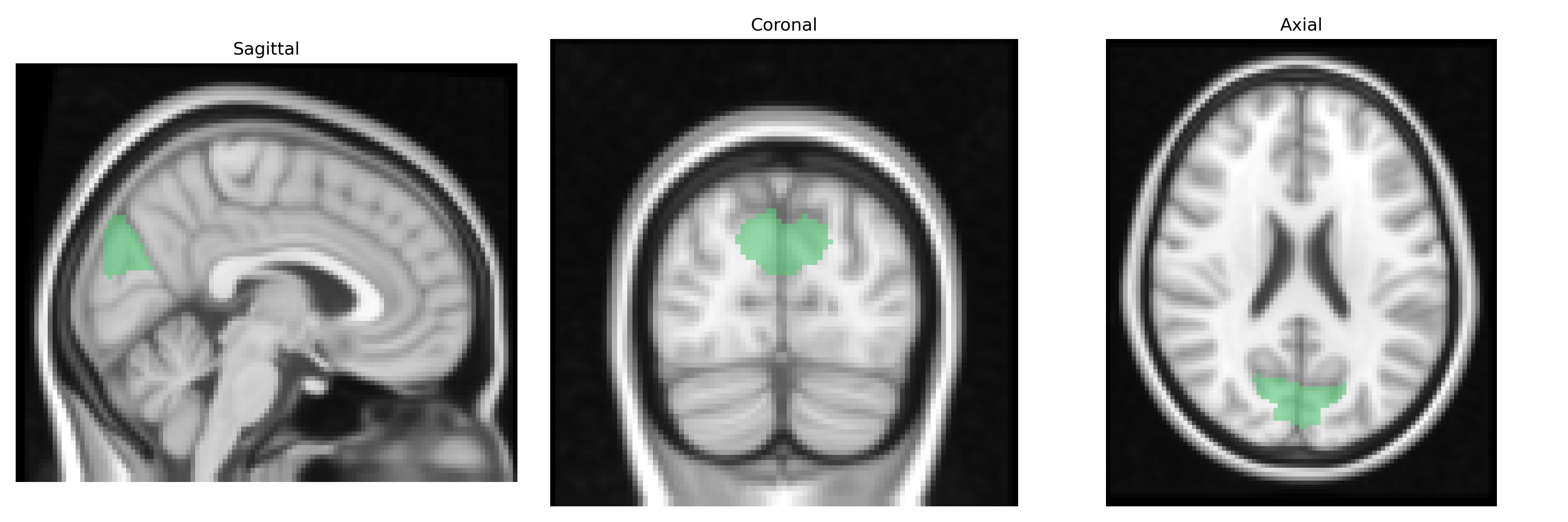
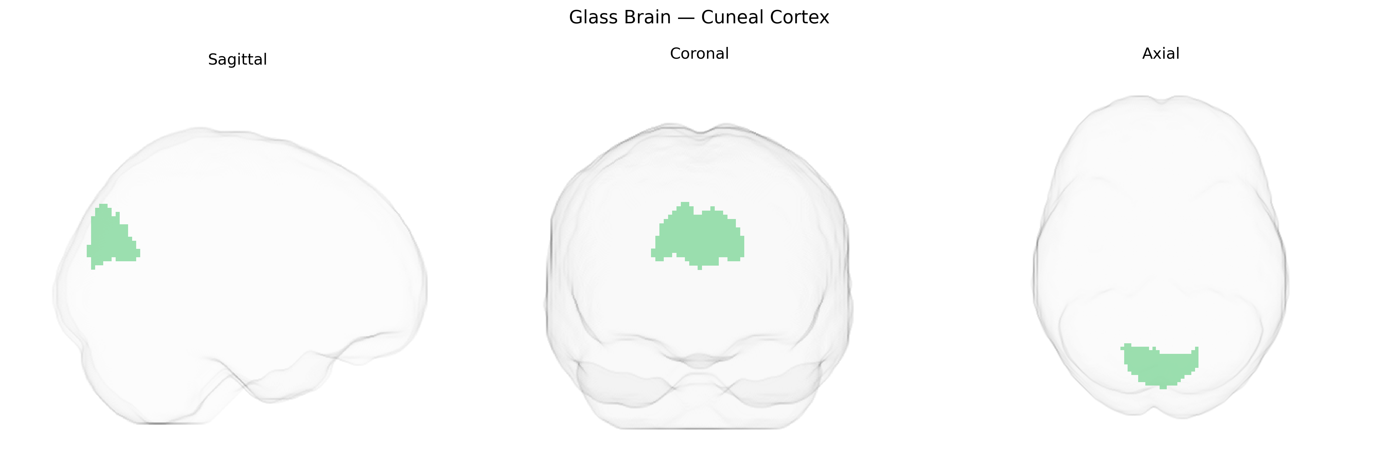
The bilateral cuneal cortex is a medial occipital lobe region corresponding largely to the cuneus, situated between the parieto-occipital sulcus and the calcarine fissure, and forming part of the primary and secondary visual cortices. It is supplied predominantly by branches of the posterior cerebral artery and is organized retinotopically, with lower visual field representations mapped dorsally. Functionally, the cuneal cortex participates in early-stage visual processing, including basic feature analysis (e.g., orientation, spatial frequency), integration of binocular input, and contributions to visuospatial attention and eye-movement–related visual modulation. It is interconnected with other occipital visual areas, dorsal parietal regions, and subcortical visual relays, supporting both low-level sensory processing and higher-order visuoperceptual functions. No direct Wikipedia article exists for “cuneal cortex”; see the related structure Cuneus.
The bilateral cuneal cortex, a primary visual and associative occipital region as defined in the Harvard-Oxford cortical atlas, has been implicated by imaging genetics and GWAS-based neuroimaging studies in several heritable traits and disorders, although typically as part of broader occipital or visual network findings rather than as an isolated locus. Large-scale brain morphology GWAS (e.g., ENIGMA, UK Biobank–based studies) have reported common variants in genes involved in neurodevelopment, axon guidance, and synaptic signaling (such as variants near or within MAPT, HMGA2, and other neurodevelopmental genes) associated with occipital cortical thickness, surface area, or volume that include the cuneus. Functional imaging genetics studies have linked cuneal cortex activation and structure to variants influencing visual processing, attentional control, and default mode/visual network coupling, and polygenic risk scores for schizophrenia, major depressive disorder, and bipolar disorder show subtle associations with altered occipital, including cuneal, morphology and connectivity. GWAS of neuropsychiatric and neurodegenerative conditions have not identified the cuneal cortex as a primary, region-specific genetic target, but convergent evidence from multimodal studies suggests that genetic risk factors for disorders with visual-perceptual or attentional components (such as schizophrenia, migraine with aura, and certain forms of epilepsy) may manifest partly through structural or functional differences in occipital regions encompassing the cuneal cortex.
Overview generated by GPT-4o (2026).
Region ID: 32
Hemisphere: bilateral
Atlas: HarvardOxford cort maxprob thr25 2mm
Full Quality Version: Download MP4
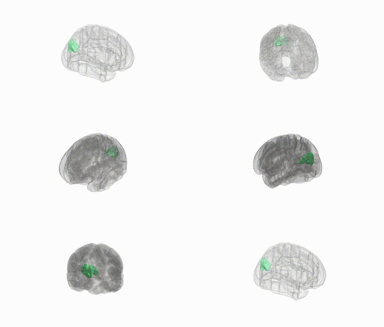
Full Quality Version: Download MP4
Wali Sidiqyar*, Gaurav Rudravaram*, Elyssa M. McMaster, Trent M. Schwartz, Adam M. Saunders, Kurt G. Schilling, Bennett A. Landman "Introducing SPINS: A Shared Public Visualization Library of Neuroanatomical Structures." Medical Imaging with Deep Learning- short paper
This resource is licensed under CC0 1.0 Universal (Public Domain).